

System and Method for Performing Single Photon Emission Computed Tomography (Spect) with a Focal-Length Cone-Beam Collimation
a single-photon emission and computed tomography technology, applied in tomography, instruments, applications, etc., can solve the problems of inability or inability to achieve a centrally-peaked sensitivity function, spect imaging of deep brain structures may be compromised by a loss of photons, and achieve precise estimates of striatal activity concentrations , the effect of improving detection and activity quantification
- Summary
- Abstract
- Description
- Claims
- Application Information
AI Technical Summary
Benefits of technology
Problems solved by technology
Method used
Image

Examples
Embodiment Construction
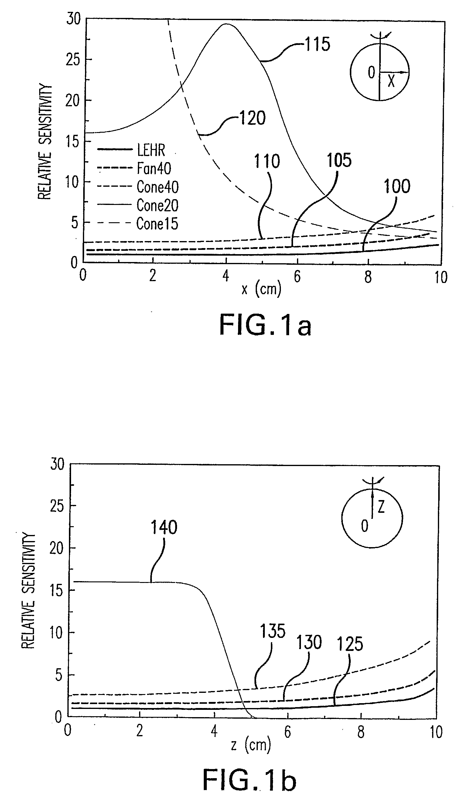
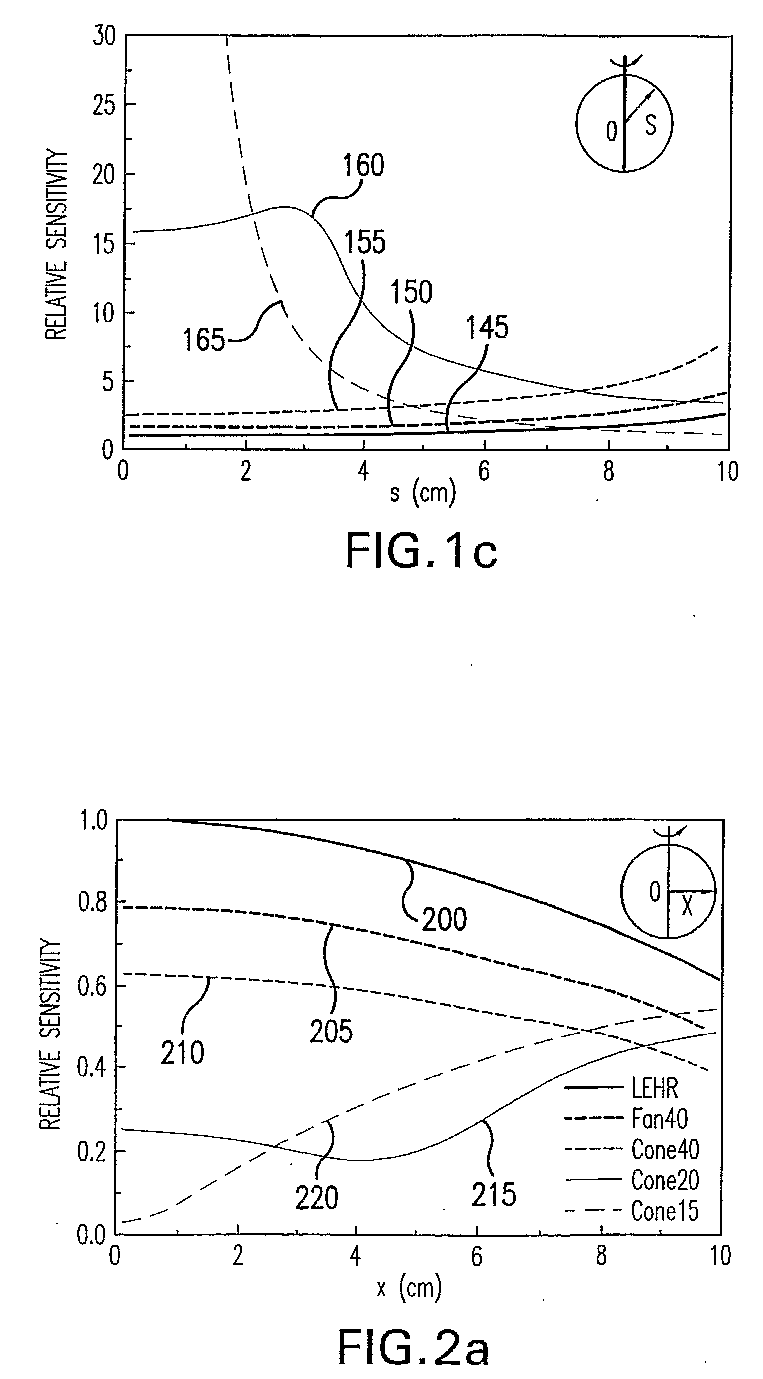
[0050]A geometric sensitivity can represent a ratio of the number of photons detected to the number emitted from a source. When the attenuation is negligible, the sensitivity of a parallel-hole collimating arrangement may be independent of a source position, while that of a converging collimating arrangement can be position-dependent. A sensitivity of converging collimating arrangements may be greater than that of parallel collimating arrangements, and cone-beam collimating arrangements can provide greater sensitivity than fan-beam collimating arrangements having the same focal length. The sensitivity of both fan- and cone-beam collimating arrangements can depend on their focal lengths.
[0051]The sensitivity of several collimating arrangements can be determined based on analytic collimating arrangement aperture functions such as those described in S. Genna et al., “Annular single-crystal emission tomography systems” in The fundamentals of PET and SPECT (Wernick M N and Asrsvold J N),...
PUM
 Abstract
Description
Claims
Application Information