Such a suturing method, however, is very
time consuming, even for an experienced surgeon.
In some cases the
blood flow in the newly joined vessels may be poor, and the surgeon must remove the stitches and repeat the suturing procedure.
In some
surgical procedures, the total time required for suturing is very substantial, increasing
ischemia time and / or resulting in prolonged anaesthesia time and increasing the cost of the surgical procedure.
In such procedures, the surgical opening and
visibility are significantly reduced, and hand suturing is more difficult.
Other new developments in
surgical procedures have made conventional suturing even more difficult.
However, the main
disadvantage of using sutures remains that the success and thus patency rate of the procedure is directly related to the skills and dexterity of the surgeon.
Also, the smaller the vessels are that need to be connected the more
time consuming the procedure is.
However such stapling devices are not generally suitable for use with much smaller and / or much more sensitive vessels such as blood vessels.
Disadvantages of such systems include the fact that the interior
diameter available for flow of body fluids (e.g. blood) is reduced by the tubular element.
Also, in such prior art systems, the tubular element is within the flow-path of the vessel and must therefore have suitable, very-hard-to-achieve
biocompatibility material properties to prevent its causing further medical problems for the patient (e.g. for blood vessels, such effects might include creating emboli that could migrate into the
systemic circulation.
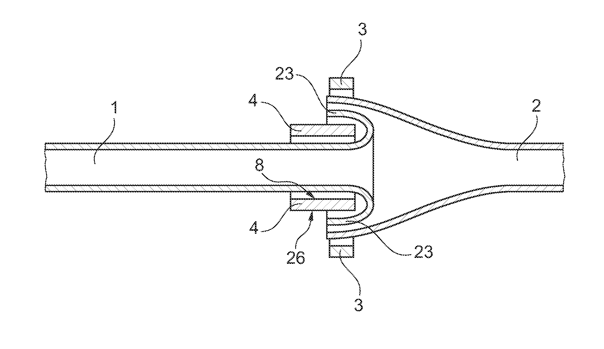
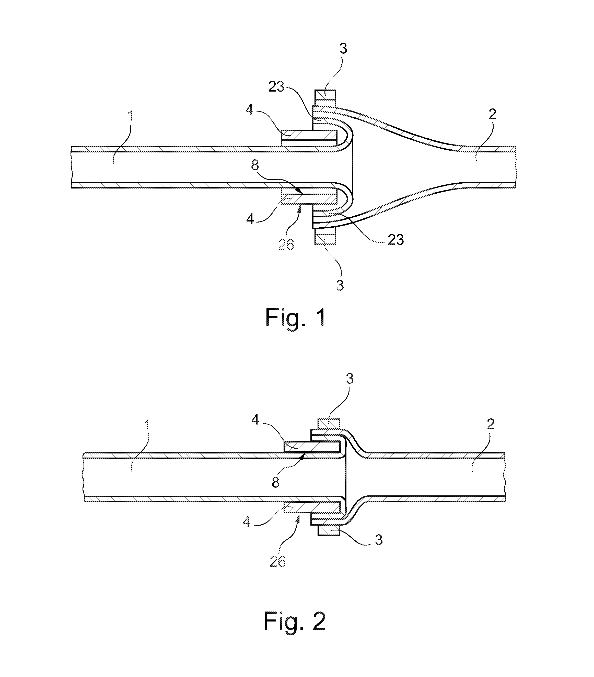
The
ring element also acts as an external tubular
scaffold—holding the vessels open at the joint, though remaining outside of the vessels at all times. However, this
system has a number of disadvantages including that, over time, the spikes may cause undesirable internal damage to other organs of the patient near to the anastomosis joint—particularly in bodily locations where many small delicate organs are very close to each other and often in motion (e.g. blood vessels in a finger).
Also, for vessels that may be in motion when in use, there is a risk that a vessel may become unhooked and, at best, cause a need to repeat the surgical procedure.
Another
disadvantage of the ‘hooked on’
system is that unintended leakage may occur at the joint prior to full healing and, depending on the particular type and location of the particular vessels, this may cause considerable undesirable patient complications.
Although this
system has enjoyed a certain amount of clinical success, it has several disadvantages including the continued use of spikes as outlined above and the fact that the form and size of the applicator device can make it difficult to make the joint in applications providing tight environments.
Also, the relatively large dimensions of the rings makes it prone to tip over and thereby constrict or compress the vessel.

