Medical device

a medical device and a technology for preventing leakage, which is applied in the field of medical devices, can solve the problems of poor blood flow in the newly joined vessels, high time requirements for suturing, and high cost, and achieves the effects of reducing the risk of leakage, and reducing the risk of infection

Active Publication Date: 2016-06-14
METESO
View PDF17 Cites 0 Cited by
  • Summary
  • Abstract
  • Description
  • Claims
  • Application Information

AI Technical Summary

Benefits of technology

[0020]The intima-to-intima anastomosis kit of the present invention has the advantage that it provides a system that is simple and relatively fast to apply and a substantially fluid-tight, secure joint may be formed without using spikes that might damage neighbouring organs. Thus, the main advantages of the intima-to-intima anastomosis kit of the present invention are providing better, substantially leak-free anastomosis; saving time during surgery; and making the task of anastomosis more reliably and safely reproducible even at levels of operator dexterity which are less than the highest achievable level.
[0021]The intima-to-intima anastomosis kit of the present invention also has the advantage that all elements of the kit remain, at all times, outside of the flow-path of the tubular organs being anastomosed. This both reduces danger to a patient (in particular reducing blood-clotting risks) and lowers biocompatibility requirements of the material used for the parts of the anastomosis kit which are to remain in a patient's body.
[0022]The intima-to-intima anastomosis kit of the present invention also has the advantage that it brings the interior surfaces (i.e. ‘intima’) of the tubular organs which are to be joined into close contact to maximise the chances of effective and rapid healing and patient recovery.
[0023]The intima-to-intima anastomosis kit of the present invention also has the advantage that the parts of the kit which are to remain in a patient's body may preferably be made as longitudinally short as possible to allow for use in as many different procedures as possible and to minimise the amount of foreign material placed inside a patient.

Problems solved by technology

Such a suturing method, however, is very time consuming, even for an experienced surgeon.
In some cases the blood flow in the newly joined vessels may be poor, and the surgeon must remove the stitches and repeat the suturing procedure.
In some surgical procedures, the total time required for suturing is very substantial, increasing ischemia time and / or resulting in prolonged anaesthesia time and increasing the cost of the surgical procedure.
In such procedures, the surgical opening and visibility are significantly reduced, and hand suturing is more difficult.
Other new developments in surgical procedures have made conventional suturing even more difficult.
However, the main disadvantage of using sutures remains that the success and thus patency rate of the procedure is directly related to the skills and dexterity of the surgeon.
Also, the smaller the vessels are that need to be connected the more time consuming the procedure is.
As long as microsurgery and free flap surgery in reconstructive procedures are time-consuming and dependent on technical skills, they will never be very popular.
However such stapling devices are not generally suitable for use with much smaller and / or much more sensitive vessels such as blood vessels.
Disadvantages of such systems include the fact that the interior diameter available for flow of body fluids (e.g. blood) is reduced by the tubular element.
Also, in such prior art systems, the tubular element is within the flow-path of the vessel and must therefore have suitable, very-hard-to-achieve biocompatibility material properties to prevent its causing further medical problems for the patient (e.g. for blood vessels, such effects might include creating emboli that could migrate into the systemic circulation.
The ring element also acts as an external tubular scaffold—holding the vessels open at the joint, though remaining outside of the vessels at all times. However, this system has a number of disadvantages including that, over time, the spikes may cause undesirable internal damage to other organs of the patient near to the anastomosis joint—particularly in bodily locations where many small delicate organs are very close to each other and often in motion (e.g. blood vessels in a finger).
Also, for vessels that may be in motion when in use, there is a risk that a vessel may become unhooked and, at best, cause a need to repeat the surgical procedure.
Another disadvantage of the ‘hooked on’ system is that unintended leakage may occur at the joint prior to full healing and, depending on the particular type and location of the particular vessels, this may cause considerable undesirable patient complications.
Although this system has enjoyed a certain amount of clinical success, it has several disadvantages including the continued use of spikes as outlined above and the fact that the form and size of the applicator device can make it difficult to make the joint in applications providing tight environments.
Also, the relatively large dimensions of the rings makes it prone to tip over and thereby constrict or compress the vessel.

Method used


Image

  • Medical device
  • Medical device
  • Medical device

Examples


Embodiment Construction

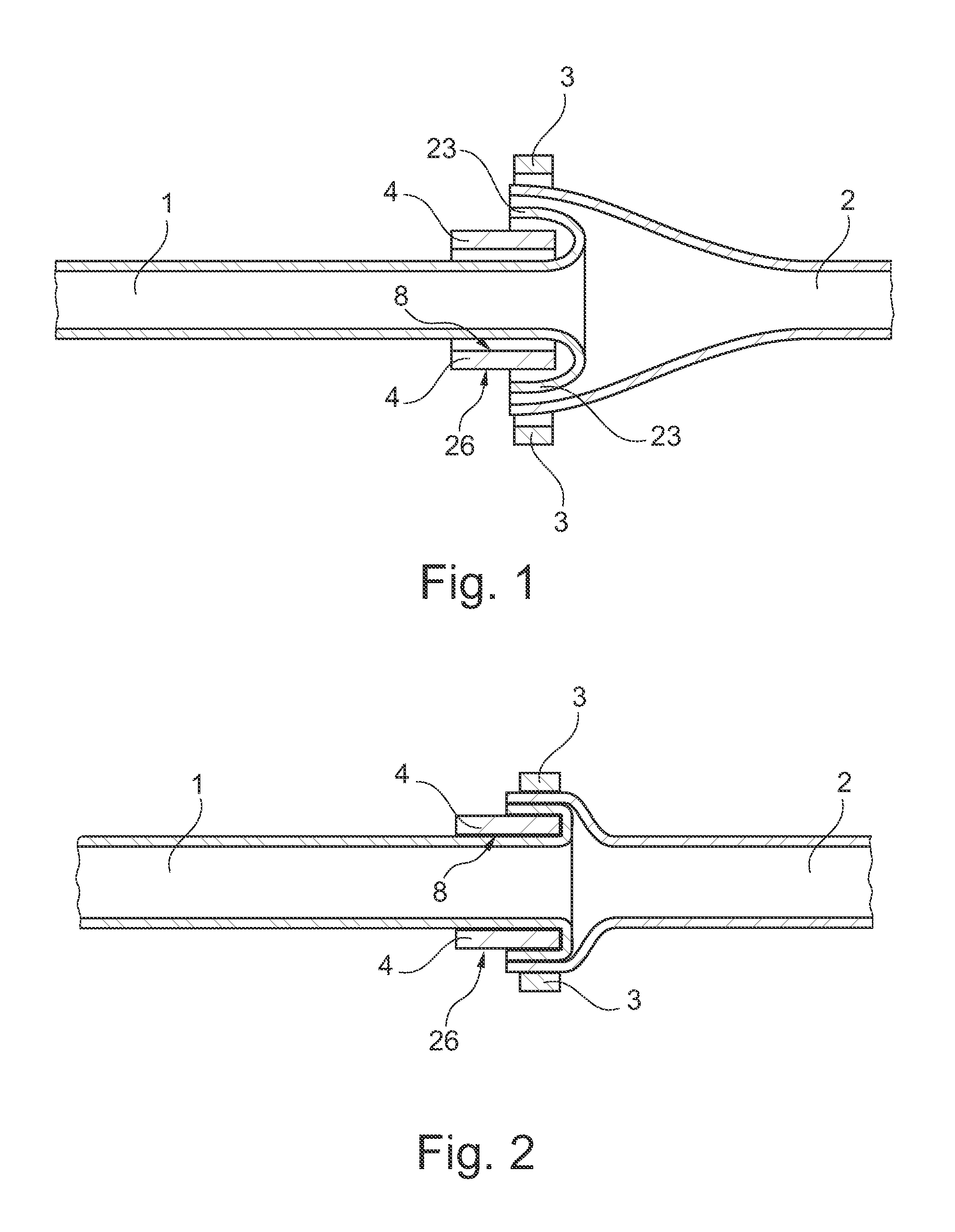
[0049]FIGS. 1 and 2 show in schematic form, the interoperational functionality of an external tubular scaffold 4 and a clip 3 when used to anastomose a first tubular organ 1, such as a blood vessel, in end-to-end fashion with a second tubular organ 2, such as another blood vessel.

[0050]In FIG. 1, the first tubular organ 1 has been threaded through the external tubular scaffold 4, such that an inner tubular surface 8 of the external tubular scaffold 4 is adjacent to the exterior surface of the wall of the first tubular organ 1. A cuff portion 23 of the first tubular organ 1, formed by folding the first tubular organ 1 back over itself, has been placed concentrically around an exterior surface 26 of the external scaffold structure 4 and a second tubular organ 2 has been placed concentrically around the cuff portion 23. An open clip 3 has been placed concentrically around the second tubular organ 2 and the cuff portion 23 and the exterior surface 26 of the external tubular scaffold 4.

[...


PUM


Abstract

A medical device is disclosed for joining a first tubular organ (1), such as a blood vessel, to a second tubular organ (2), such as a blood vessel. The kit includes an external tubular scaffold (4) with a removable scaffold handle (7, 18, 19) and a clip (3). In use, the clip (3) and the external tubular scaffold (4) remain outside of the tubular organs (1, 2) being joined. The clip (3) embraces a first longitudinal tubular section (6) of the external tubular scaffold (4) thereby holding together a longitudinal segment of an intima of the first tubular organ (1) and a longitudinal segment of an intima of the second tubular organ (2).

Description

CROSS-REFERENCE TO RELATED APPLICATIONS[0001]This application is a National Phase Application of PCT International Application No. PCT / EP2011 / 072886, International Filing Date Dec. 15, 2011, claiming priority of European Patent Application No. 10195212.5, filed Dec. 15, 2010, which is hereby incorporated by reference.DESCRIPTION[0002]The present invention relates generally to the area of sutureless anastomosis of tubular organs, such as blood vessels or intestines or tubular structures of the urogenital tract.[0003]More particularly, the present invention relates to intima-to-intima anastomosis kits for effecting intima-to-intima anastomosis as set out in the pre-characterising portion of claim 1.BACKGROUND ART[0004]Anastomosis is the connection of two tubular structures or organs. It refers to connections between blood vessels or between other tubular organs such as loops of intestine. Circulatory anastomosis is further divided into arterial and venous anastomosis. An example of su...

Claims


Application Information

Patent Timeline
Patent Type & Authority Patents(United States)
IPC IPC(8): A61B17/08A61B17/11A61B17/10
CPCA61B17/11A61B17/08A61B17/10A61B2017/1121A61B2017/1132
Inventor SGARZANI, ROSSELLASPINGLER, ROLF
Owner METESO
Why Eureka
Try Eureka