Cell analysis device

a cell analysis and cell technology, applied in the field of cell analysis devices, can solve the problems of high cost, cell damage on stage, blurred image due to chromatic aberration,

Inactive Publication Date: 2013-11-28
KINKI UNIVERSITY +4
View PDF1 Cites 20 Cited by
  • Summary
  • Abstract
  • Description
  • Claims
  • Application Information

AI Technical Summary

Benefits of technology

[0066]According to the present invention, it can be estimated what visual color is possessed by trace amounts of cells in a subject by absorption, absorption spectral characteristics can be analyzed, cells can be identified by the color or the intracellular absorption characteristic distribution, and target cells can be selectively recovered and purified, and thus correct genetic information and expression information of the target cells can be analyzed.

Problems solved by technology

When light components of a plurality of wavelengths are used at the same time with these microscopes, an image is blurred due to chromatic aberration.
However, this method has the problems that it costs high; the device is large; a high electric field of several thousand volts is necessary; a large amount of samples concentrated to a certain concentration are necessary; there is a risk that the cells may be damaged on the stage of creating the droplets; samples cannot be directly observed; and the like.
However, with this cell sorter formed by use of the microprocessing technology, the response speed of sample separation to the observation means is low.
In the case where trace amounts of samples are concentrated in another device, it is difficult to recover the concentrated solutions with no loss, and problems undesirable for reusing the recovered cells occur, for example, the problem that the cells are contaminated on a complicated pre-processing stage.
A known problem of a malignant tumor is that the tumor is spread to another organ by an ability of infiltrating from the tissue of the cancer cell itself into the blood vessel or lymphatic vessel.
However, this technology is not used for absorption spectrum measurement or the like.

Method used


Image

  • Cell analysis device
  • Cell analysis device
  • Cell analysis device

Examples


Embodiment Construction

[0072]Hereinafter, embodiments of the present invention will be described in more detail with reference to the drawings. The following embodiments are merely illustrative, and the present invention is not limited to these embodiments.

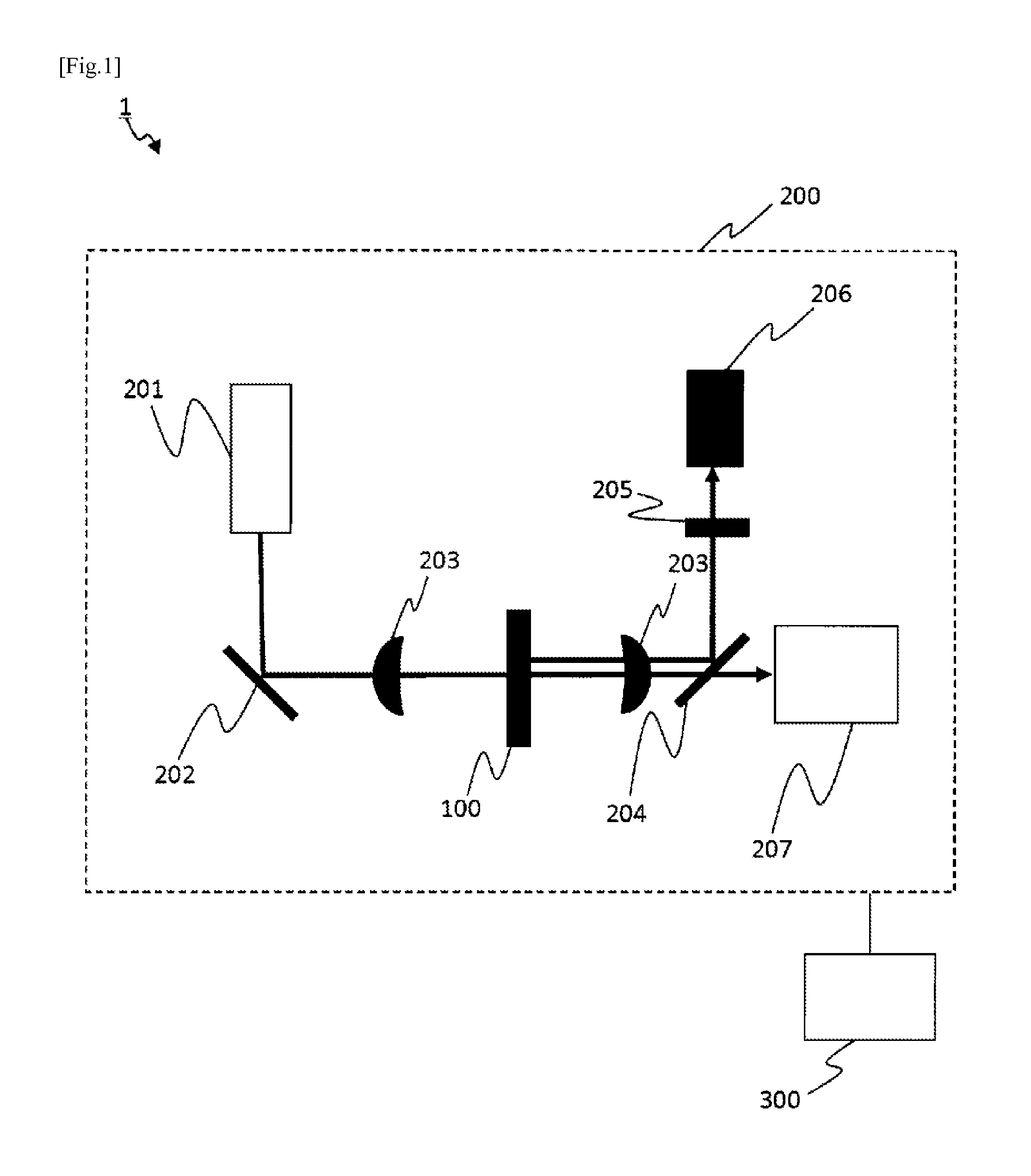
[0073]FIG. 1 is a conceptual view of a cell analysis device 1 in an embodiment according to the present invention.

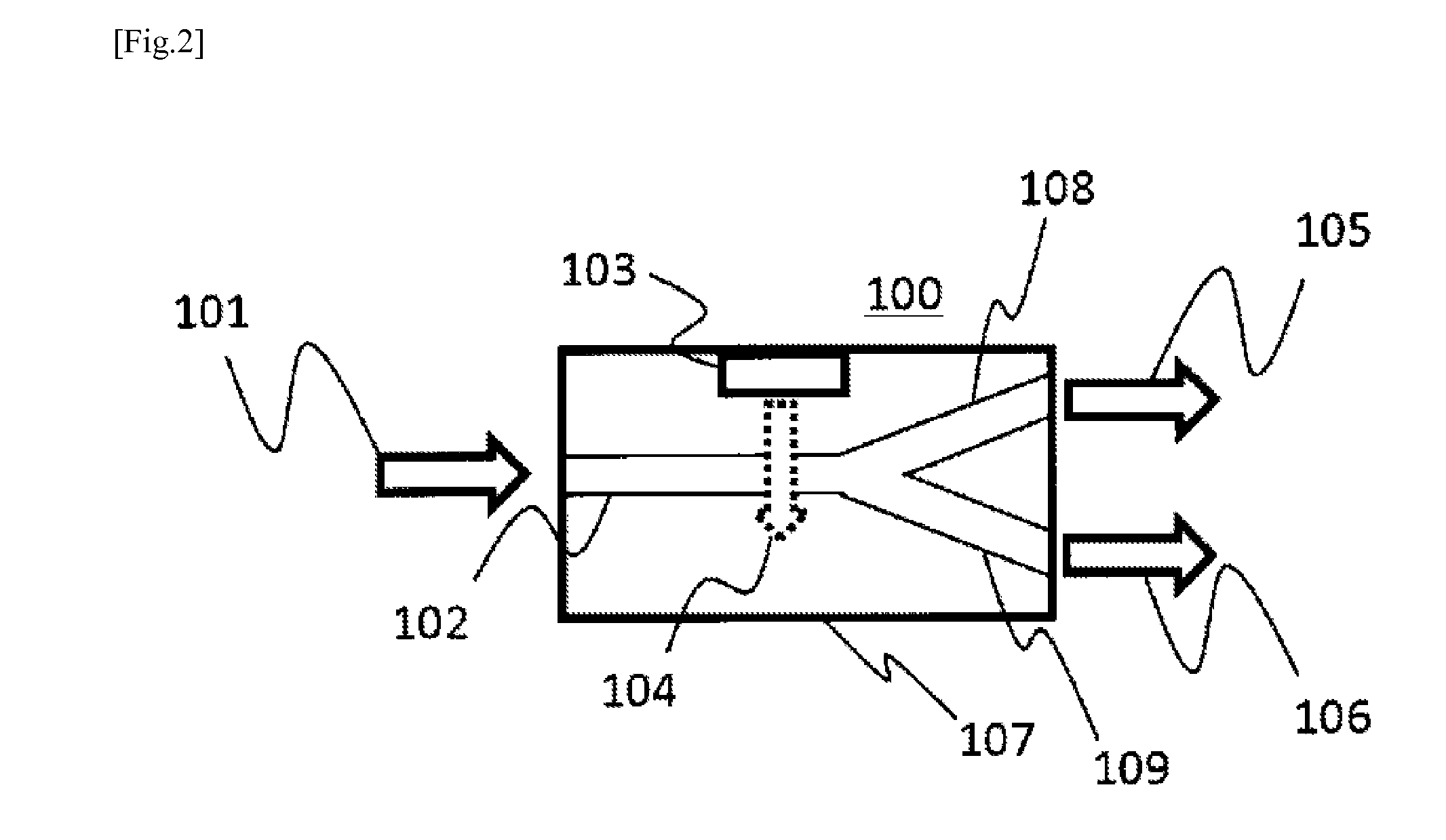
[0074]As shown in FIG. 1, the cell analysis device 1 according to the present invention typically includes an image detection type single-cell separation / purification module 200 for acquiring image data of about 10,000 cell images per second of cells flowing in a microchannel which is formed in a substrate of a cell sorter chip 100, and purifying and recovering cell populations or cell clusters by a processing capability of 10,000 cells per second at the maximum in real time based on the result of analyzing the image information; and a recording / control section 300 for recording / analyzing various data acquired by the module 200 to control va...


PUM


Abstract

Provided is a cell concentration/purification device having a function of successively locating cells in a specific area of a microchannel, and a function of sequentially capturing cell images by use of light from a plurality of monochromatic light sources on an image basis, and performing comparative analysis of the cell images to recognize individual cells based on information on the shape of the cells and an absorption spectral distribution of the cells or inside of the cells, thereby selectively separating/purifying the cells.

Description

TECHNICAL FIELD[0001]The present invention relates to a cell analysis device.BACKGROUND ART[0002]In biological tissues in multi-cell organisms, various cells each play its own role to keep the function thereof in a coordinated manner. When a part of the cell becomes a cancer cell (herein, the term “cancer” encompasses cancers and tumors), the part becomes a neoplasm different from the area around the part. Such a cancer area and a normal tissue area far from the cancer area are not necessarily separated along a border, and the area around the cancer area is influenced in some way. Therefore, in order to analyze the function of an organ tissue, a small number of cells present in a narrow area needs to be separated in a short time, as simply as possible, and with minimum loss.[0003]In the field of regenerative medicine, there is an attempt to separate a stem cell of an organ from the tissue and re-culture the stem cell for differentiation-induction in order to regenerate a target tiss...

Claims


Application Information

Patent Timeline
Patent Type & Authority Applications(United States)
IPC IPC(8): G06T7/00
CPCG06T7/0016G01N15/1475G01N15/1484G01N27/447G01N33/487G01N2015/1006G01N2015/149G01N2015/1497G01N2015/1472G01N2015/1493
Inventor YASUDA, KENJITERAZONO, HIDEYUKIKIM, HYONCHOLHAYASHI, MASAHITOHATTORI, AKIHIRONISHIO, KAZUTOARAO, TOKUZOMIYAGI, YOHEIOHTSU, TAKASHI
Owner KINKI UNIVERSITY
Why Eureka
Try Eureka