
Method and system for assessing visual disorder

a visual system and disorder technology, applied in the field of medical techniques, can solve the problems of affecting the normal performance of visual tasks, affecting the response of subjects, and affecting the quality of life of subjects,

Inactive Publication Date: 2013-11-28
HADASIT MEDICAL RES SERVICES & DEVMENT
View PDF3 Cites 12 Cited by
  • Summary
  • Abstract
  • Description
  • Claims
  • Application Information

AI Technical Summary

Benefits of technology

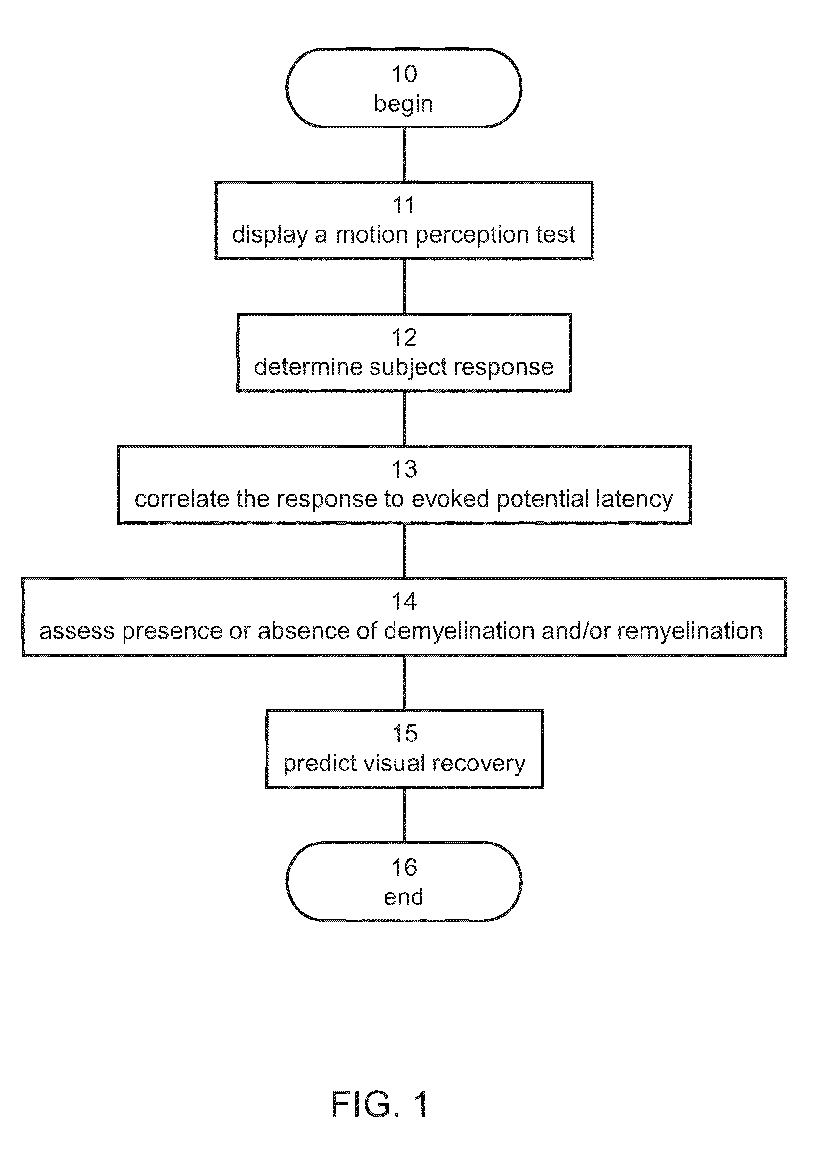
The present invention provides a method and system for diagnosing demyelination and remyelination using a motion perception test. The method involves presenting a motion perception test to a subject and determining the subject's response to the test. The response is then correlated to visual evoked potentials latency to assess the presence or absence of demyelination and remyelination. The method can be performed using a display device and can be automated or semi-automated. The invention can help in diagnosing optic neuritis and predicting the outcome of the disease. The method can also be used to assess the effect of treatment. The technical effects of the invention include providing a more accurate and reliable tool for diagnosing demyelination and remyelination, and aiding in the diagnosis of optic neuritis.

Problems solved by technology

It is one of the causes of acute loss of vision associated with pain.
However, subjects typically continue to perceive difficulties in performing everyday visual tasks following an ON attack.
In subjects with ON, the response may be slower because signal conduction along demyelinated nerve fibers is impaired.

Method used


Image

  • Method and system for assessing visual disorder
  • Method and system for assessing visual disorder
  • Method and system for assessing visual disorder

Examples


example 1

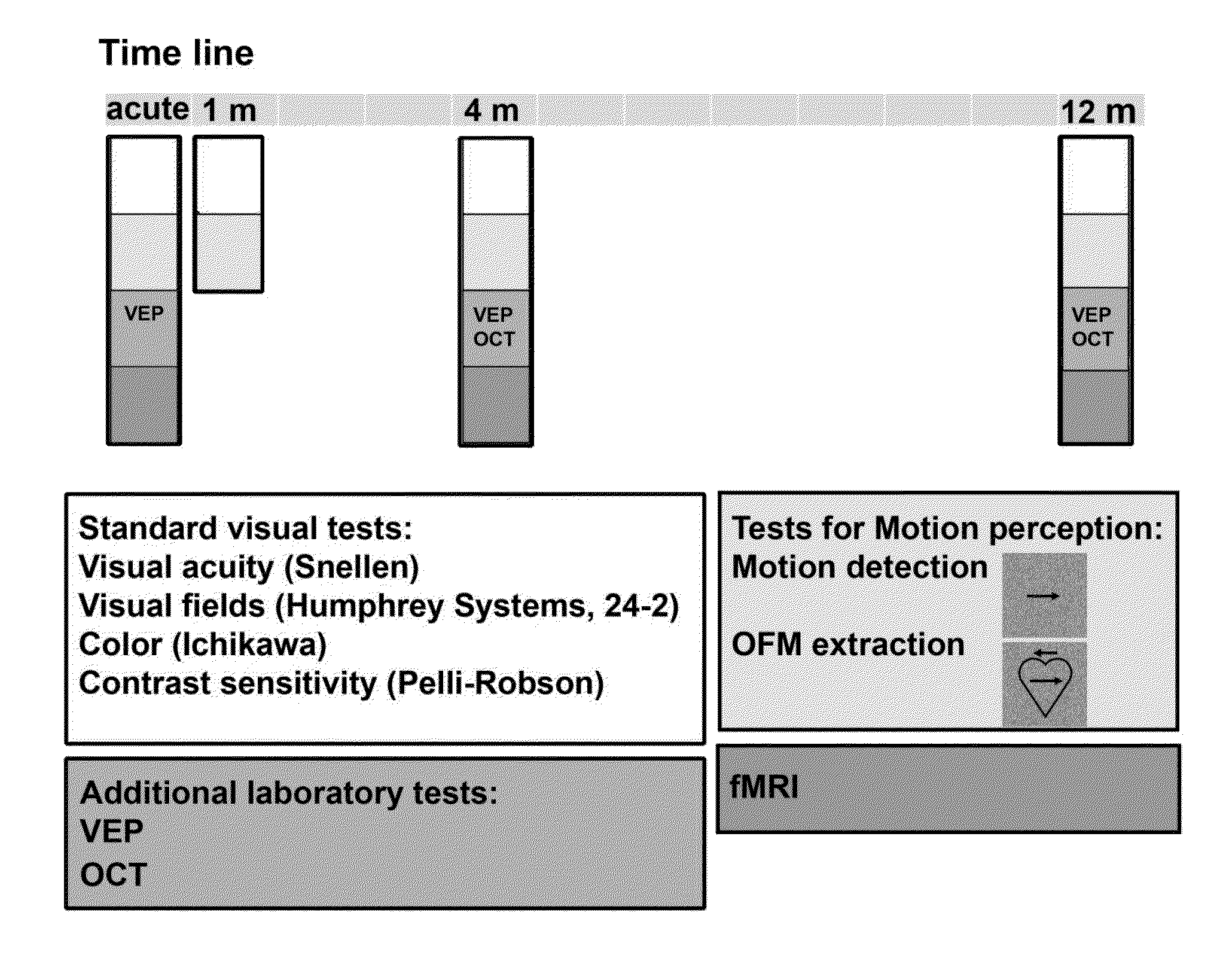
[0103]The present Example describes experiments performed according to some embodiments of the present invention to assess the recovery process in patients after an acute ON attack, and to compare static and dynamic visual functions.

[0104]Motion perception begins in the retina, mediated through the magnocellular pathway, containing cells with transient responses and fast-conductive axons. Cortically, the visual area MT (middle temporal), likely plays a major role in the integration of local motion signals into global percepts [7].

[0105]Recently, fMRI was used to evaluate the cortical response following an ON attack [8-12], suggesting that changes in cortical organization may have an adaptive role in visual recovery after ON, in addition to the remyelinating process in the nerve itself.

[0106]The present study assesses motion perception longitudinally following an ON attack, and to document its associated cortical response.

Methods

[0107]The Hadassah Hebrew University Medical Center Eth...

example 2

[0198]Example 1 above demonstrated a specific sustained deficit in dynamic visual functions following ON. The present Example describes a study directed to identify the mechanism of this deficit. In the current study, patients were followed-up longitudinally after an ON attack providing the timeline for recovery and visual outcome predictability.

Methods

[0199]Twenty-one patients aged 18-59 (mean±STDEV 29±9.5) years presenting with a first-ever episode of acute ON participated in the study. Patients were enrolled during hospitalization. All patients presented with unilateral visual loss, a relative afferent pupillary defect, and an otherwise normal neuro-ophthalmological examination. Two patients had a recurrent attack during study follow-up, and their data were therefore excluded from subsequent analyses. Twenty-one control subjects who were matched to the patients for age, gender and dominant eye on a subject-by-subject-basis were included in the study. The Hadassah Hebrew Universit...


PUM


Abstract

A method of diagnosis is disclosed. The method comprises using a display device for presenting a motion perception test to a subject and determining a subject response to the motion perception test. The response can be used for assessing presence or absence of demyelination and / or remyelination.

Description

RELATED APPLICATION[0001]This application claims the benefit of priority under 35 USC 119(e) of U.S. Provisional Patent Application No. 61 / 651,012 filed May 24, 2012, the contents of which are incorporated herein by reference in their entirety.FIELD AND BACKGROUND OF THE INVENTION[0002]The present invention, in some embodiments thereof, relates to medical techniques and, more particularly, but not exclusively, to a method and system for assessing a disorder in the visual system.[0003]The retinal ganglion cell is a retinal output cell, and its-axon is also called an optic nerve fibers, runs in the retinal inner layer and the nerve fibers layer (nearest side to the vitreous body), gathers at the optic disc, leaves the eye ball, forms an optic nerve and undertakes a role of transmitting visual information to the cerebral cortex.[0004]The retinal ganglion cell is distributed over the entire area of the retina. A retinal damage due to, e.g., inflammation and the like can be caused by var...

Claims


Application Information

Patent Timeline
IPC IPC(8): A61B3/02
CPCA61B3/022A61B3/032A61B5/40A61B5/4848G01R33/4806A61B5/055A61B5/377
Inventor LEVIN, NETTARAZ, NOABEN-HUR, TAMIR
Owner HADASIT MEDICAL RES SERVICES & DEVMENT
Learn More