Multi-phase computed tomography image reconstruction

a computed tomography and image reconstruction technology, applied in the field of multi-phase computed tomography image reconstruction, can solve the problems of difficult to provide useful phase selection (such as cardiac phase of interest) to a reviewer of image data, artifacts or other imperfections in the reconstructed image,

Inactive Publication Date: 2014-01-16
GENERAL ELECTRIC CO
View PDF11 Cites 15 Cited by
  • Summary
  • Abstract
  • Description
  • Claims
  • Application Information

AI Technical Summary

Benefits of technology

[0004]In a further embodiment, one or more non-transitory computer-readable media are provided. The computer-readable media encode one or more routines which, when executed by a processor, cause the processor to perform acts comprising: accessing a set of calibrated X-ray projection data acquired over at least a partial rotation; reconstructing a plurality of initial images at different phases based on the set of calibrated X-ray projection data; performing a frequency transform on each initial image to generate a frequency domain representation of the respective initial image; for each frequency element of the frequency domain represen...

Problems solved by technology

However, various physical limitations or constraints on acquisition may result in artifacts or other imperfections in the reconstructed imag...

Method used


Image

  • Multi-phase computed tomography image reconstruction
  • Multi-phase computed tomography image reconstruction
  • Multi-phase computed tomography image reconstruction

Examples


Embodiment Construction

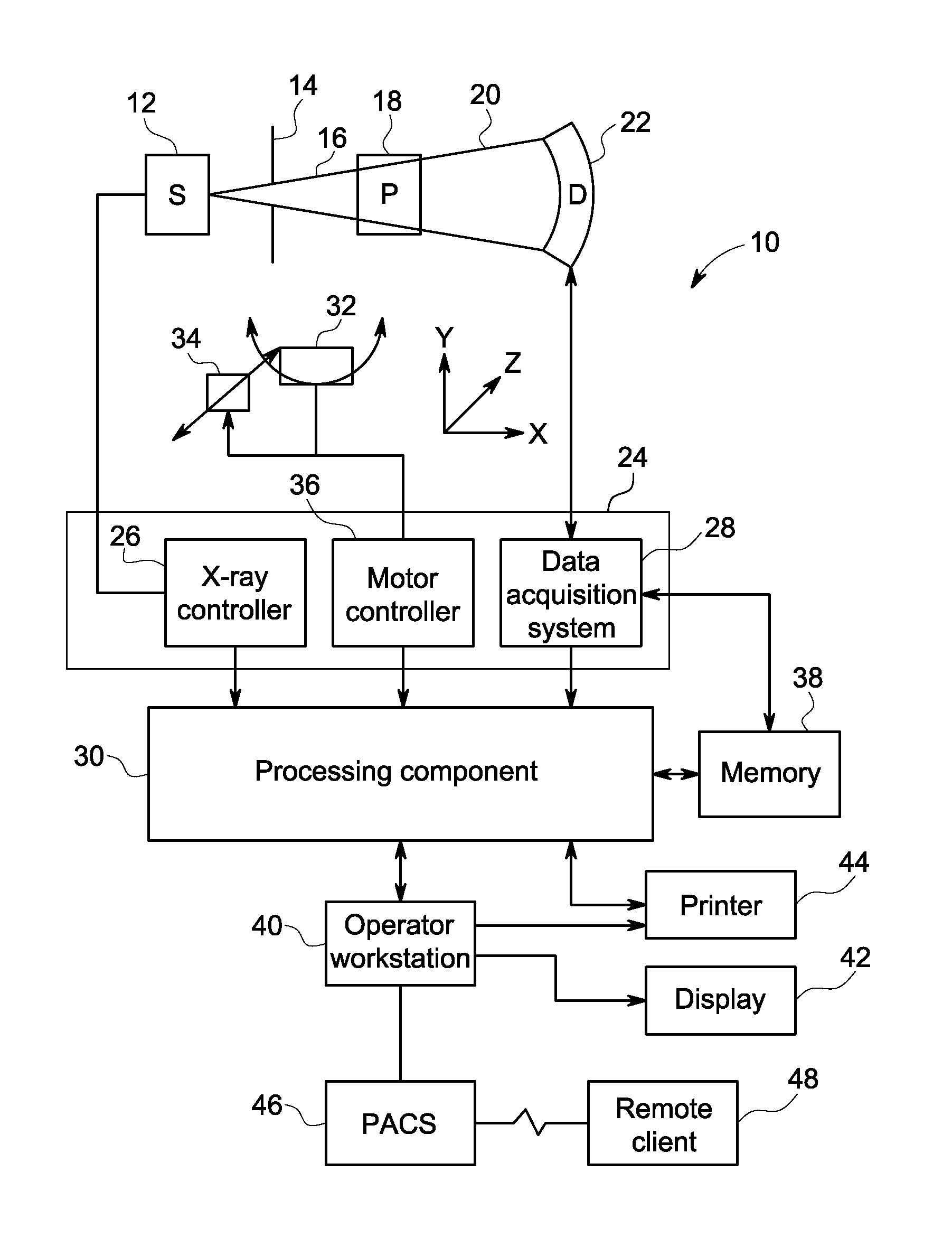
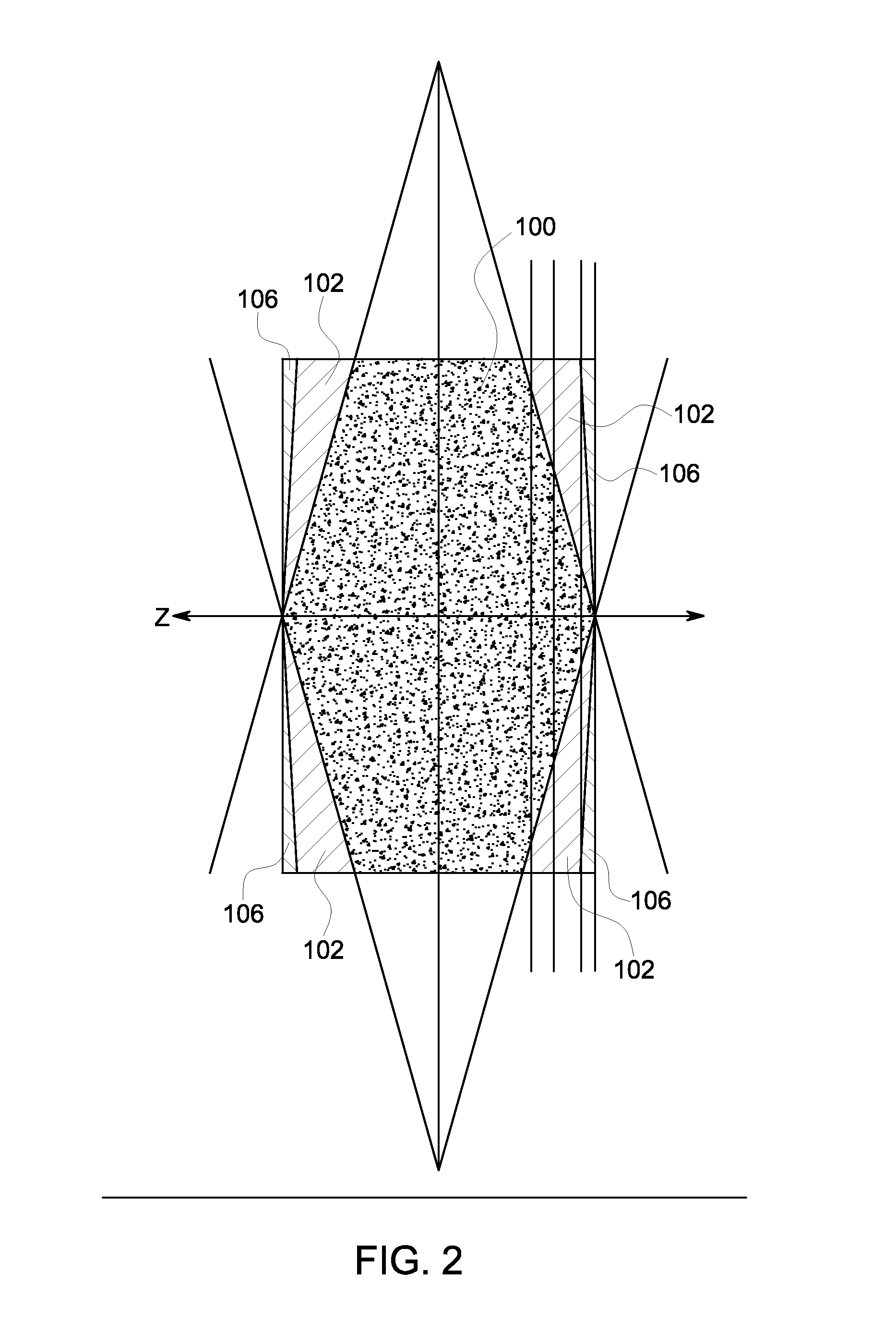
[0015]In the context of dynamic image acquisition / reconstruction it may be useful to reconstruct images with different gating windows. For instance in the case of cardiac computed tomography (CT) these different gating windows correspond to different phases in the cardiac cycle. In cardiac CT imaging there is no a priori knowledge of the best phase for imaging each coronary artery. Therefore, clinicians often specify data to be collected over a range of cardiac phases. Typically, each image volume is reconstructed completely independently. For example, in clinical workflow phases are typically reconstructed at a limited number of discrete cardiac phases, such as every 5% or 10% in the R-R interval. In addition to diagnostic visualization, there may be other reasons to reconstruct multiple phases, such as Fourier Image Deblurring (FID) or Motion Evoked Artifact Deconvolution (MEAD). As discussed herein, an approach for reconstructing multiple image volumes with a significant time sav...


PUM


Abstract

Approaches for reconstructing multi-phase images are disclosed. In certain embodiments, calibrated X-ray projection data acquired over at least a partial axial or low-pitch helical rotation is accessed and used to reconstruct one or more initial images. A frequency transform is performed on the images to generate respective frequency domain representations. Elements of the frequency domain representations are weighted based on at least the difference between the phase associated with the elements and a specified phase of interest. The weighted frequency domain representations are combined to generate a frequency domain representation at the phase of interest, which can be used to generate an image at the phase of interest.

Description

BACKGROUND[0001]Non-invasive imaging technologies allow images of the internal structures of a patient or object to be obtained without performing an invasive procedure on the patient or object. In particular, technologies such as computed tomography (CT) use various physical principles, such as the differential transmission of X-rays through the target volume, to acquire image data and to construct tomographic images (e.g., three-dimensional representations of the interior of the human body or of other imaged structures). However, various physical limitations or constraints on acquisition may result in artifacts or other imperfections in the reconstructed image.[0002]For example, in certain dynamic imaging contexts, such as cardiac imaging, it may be desirable to generate images corresponding to different phases of motion for the object or organ of interest (such as corresponding to different phases of the cardiac cycle). However, in general, it is difficult to provide useful phase...

Claims


Application Information

Patent Timeline
IPC IPC(8): G06K9/00
CPCG06T11/008G06T2211/412
Inventor NETT, BRIAN EDWARDPACK, JED DOUGLASZENG, KAI
Owner GENERAL ELECTRIC CO
Why Eureka
Try Eureka