Method and apparatus for forming x-ray mammogram

a mammogram and x-ray technology, applied in the field of x-ray imaging methods, can solve problems such as difficulty in distinguishing normal tissues from lumps, and achieve the effect of reducing nois

Inactive Publication Date: 2014-03-13
SAMSUNG ELECTRONICS CO LTD
View PDF3 Cites 20 Cited by
  • Summary
  • Abstract
  • Description
  • Claims
  • Application Information

AI Technical Summary

Benefits of technology

[0009]One or more exemplary embodiments provide an X-ray imaging method and apparatus by which an X-ray image having reduced noise and showing a clear boundary of a lesion region may be obtained.

Problems solved by technology

In particular, inner tissues of dense breasts overlap each other to a large extent, and thus it is difficult to distinguish normal tissues from lumps.

Method used


Image

  • Method and apparatus for forming x-ray mammogram
  • Method and apparatus for forming x-ray mammogram
  • Method and apparatus for forming x-ray mammogram

Examples


Embodiment Construction

[0022]Reference will now be made in detail to exemplary embodiments, examples of which are illustrated in the accompanying drawings. However, the present inventive concept may be embodied in many different forms and should not be construed as being limited to the exemplary embodiments set forth herein. Rather, these exemplary embodiments are provided so that the present disclosure will be thorough and complete, and will fully convey the scope of the present inventive concept to those skilled in the art, and the spirit and scope of the present inventive concept should be defined by the appended claims.

[0023]Hereinafter, exemplary embodiments of an X-ray imaging method and apparatus will be described in detail with reference to the accompanying drawings. In the drawings, like reference numerals refer to like elements.

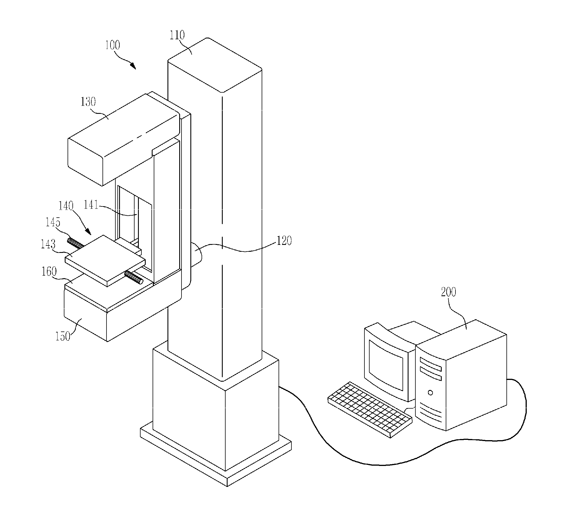
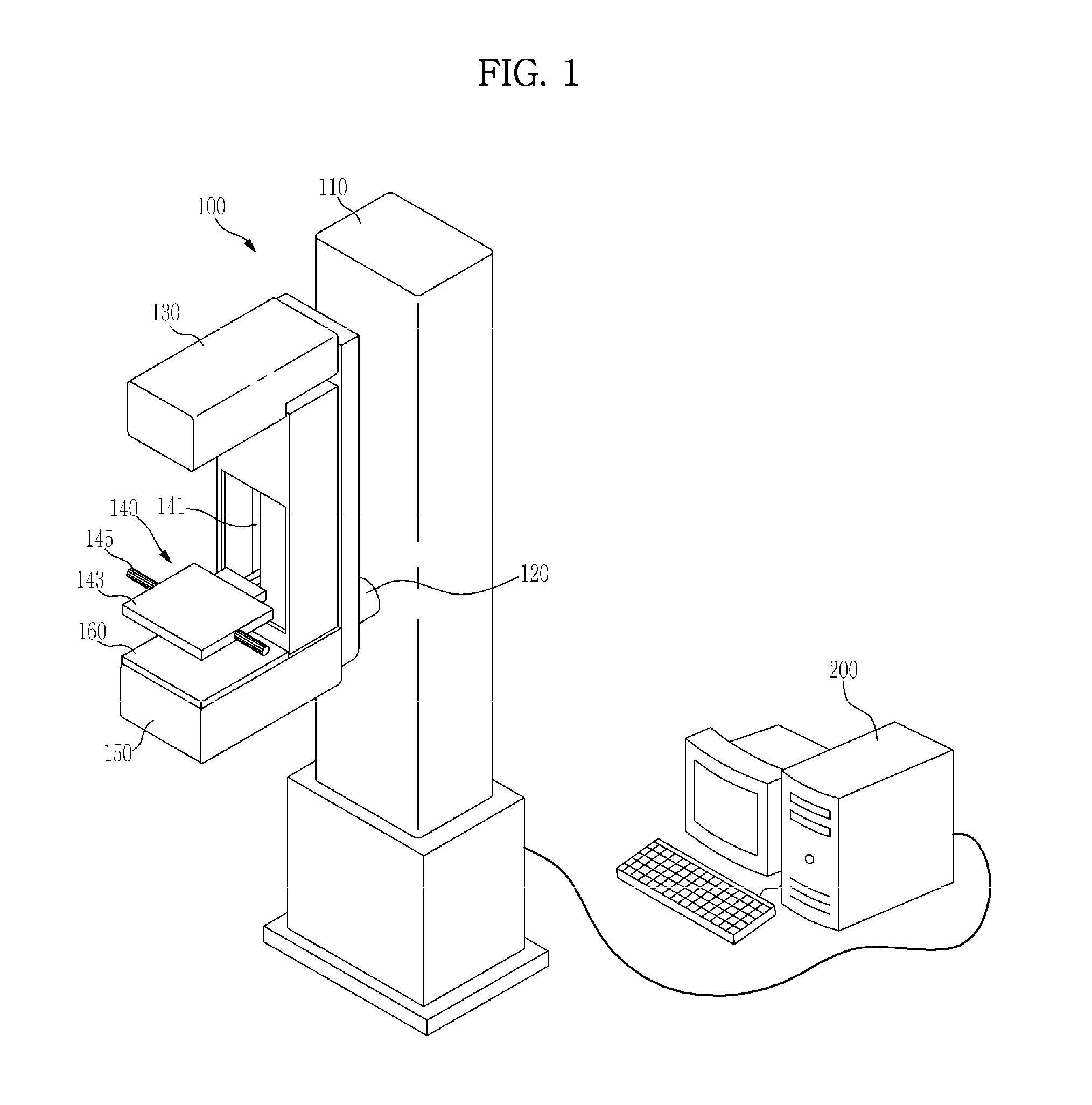
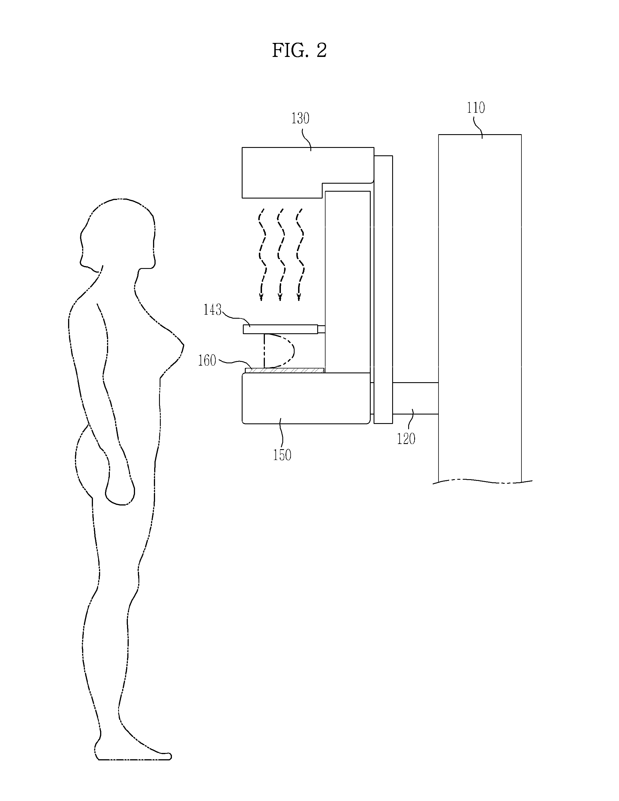
[0024]First, an X-ray imaging apparatus according to an exemplary embodiment will be described with reference to FIGS. 1 through 5.

[0025]FIG. 1 is a perspective view of a...


PUM


Abstract

An X-ray imaging method and apparatus are provided for forming an X-ray image having reduced noise and showing a clear boundary of a lesion region. The X-ray imaging method includes performing a first main shot which irradiates an object in a compressed state by using X-rays at least once to obtain a single two-dimensional image, performing a second main shot which irradiates the object by using X-rays at different positions to obtain a plurality of two-dimensional images, and forming a two-dimensional final image by removing a lesion region having a unclear boundary from each of the plurality of two-dimensional images and substituting a lesion region of the single two-dimensional image into an area which corresponds to the removed lesion region.

Description

CROSS-REFERENCE TO RELATED APPLICATION[0001]This application claims priority from Korean Patent Application No. 10-2012-0099442, filed on Sep. 7, 2012 in the Korean Intellectual Property Office, the disclosure of which is incorporated herein by reference in its entirety.BACKGROUND[0002]1. Field[0003]Exemplary embodiments relate to an X-ray imaging method and apparatus for performing X-ray imaging by transmitting X-rays toward an object, reducing noise in soft tissue images of the object, and forming a clear X-ray image of a lesion region inside the object.[0004]2. Description of the Related Art[0005]Mammography uses radiation, such as X-rays, to capture an X-ray image of breasts, which image can then be used for diagnosing breast cancer. An apparatus using mammography may be, for example, a mammography system. The mammography system irradiates a breast with X-rays in a state in which the breast is compressed in order to obtain a two-dimensional X-ray image of the breast.[0006]The ma...

Claims


Application Information

Patent Timeline
Patent Type & Authority Applications(United States)
IPC IPC(8): A61B6/02A61B6/00
CPCA61B6/502A61B6/022A61B6/025A61B6/0414A61B6/5217A61B6/5235G16H50/30A61B6/03A61B6/488A61B6/5252G06F18/21G06F18/253
Inventor JANG, KWANG EUNLEE, JONG HASUNG, YOUNG HUNLEE, KANG EUILEE, JAE HAK
Owner SAMSUNG ELECTRONICS CO LTD
Why Eureka
Try Eureka