Ultrasound diagnostic apparatus

a diagnostic apparatus and ultrasonic technology, applied in diagnostics, medical science, applications, etc., can solve problems such as operational errors and complicated operations of operators, and achieve the effects of reducing the number of steps in operations, accurately executing a series of examinations, and easy recognition of the flow of operations

Active Publication Date: 2014-03-27
FUJIFILM CORP
View PDF6 Cites 18 Cited by
  • Summary
  • Abstract
  • Description
  • Claims
  • Application Information

AI Technical Summary

Benefits of technology

[0019]According to the invention, in ultrasound diagnosis, it is possible to easily recognize the flow of operations and to reduce t...

Problems solved by technology

However, in ultrasound diagnosis, since in many cases, a series of examinations are performed while sequentially changing diagnostic portions, and since portions to be diagnosed and the number thereof in the series of examinations may be different depending on facilities such as hospitals or the like, an operation performed by an operator is complicated in the pr...

Method used


Image

  • Ultrasound diagnostic apparatus
  • Ultrasound diagnostic apparatus
  • Ultrasound diagnostic apparatus

Examples


first embodiment

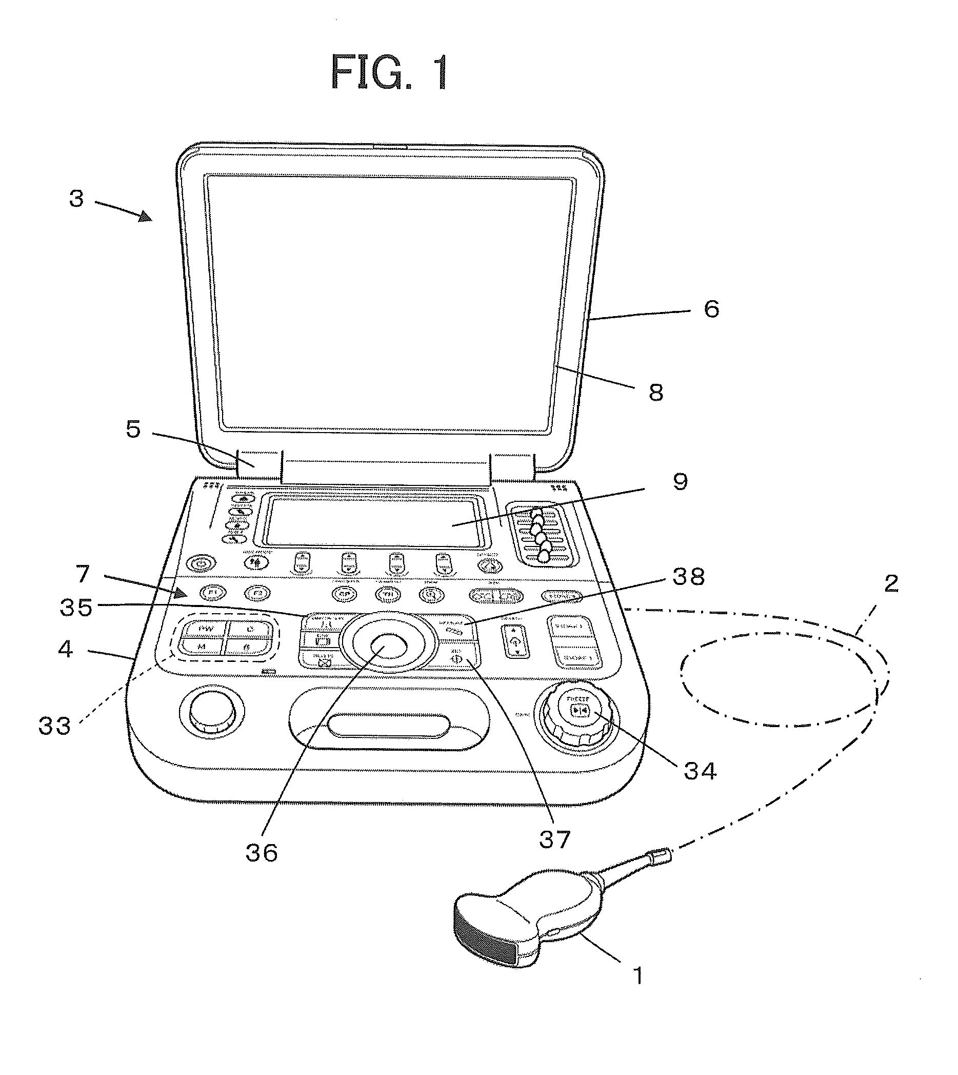
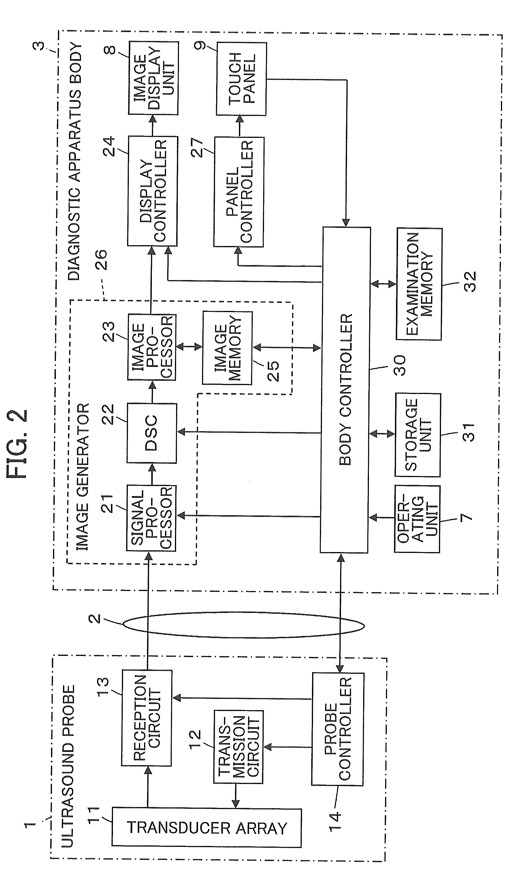
[0035]FIG. 1 shows the configuration of an ultrasound diagnostic apparatus according to a first embodiment of the invention. The ultrasound diagnostic apparatus includes an ultrasound probe 1 and a diagnostic apparatus body 3 connected to the ultrasound probe 1 through a communication cable 2.

[0036]The ultrasound diagnostic apparatus body 3 includes a housing 4 and a cover 6 that is rotatably installed at an end of the housing 4 through a hinge unit 5. The housing 4 has a substantially flat plate shape, and an operating unit 7 for various operations by an operator is formed on the surface thereof. A touch panel 9 is installed in the operating unit 7 on the side of the hinge unit 5. The cover 6 also has a substantially flat plate shape, and an image display unit 8 is formed on an inner surface thereof. The image display unit 8 faces the operating unit 7 of the housing 4 with the rotational moving by the hinge unit 5.

[0037]The operating unit 7 includes an imaging mode selecting button...

second embodiment

[0079]Next, a second embodiment of the invention will be described.

[0080]The configuration of an ultrasound diagnostic apparatus according to the second embodiment is the same as that of the first embodiment illustrated in FIGS. 1 and 2.

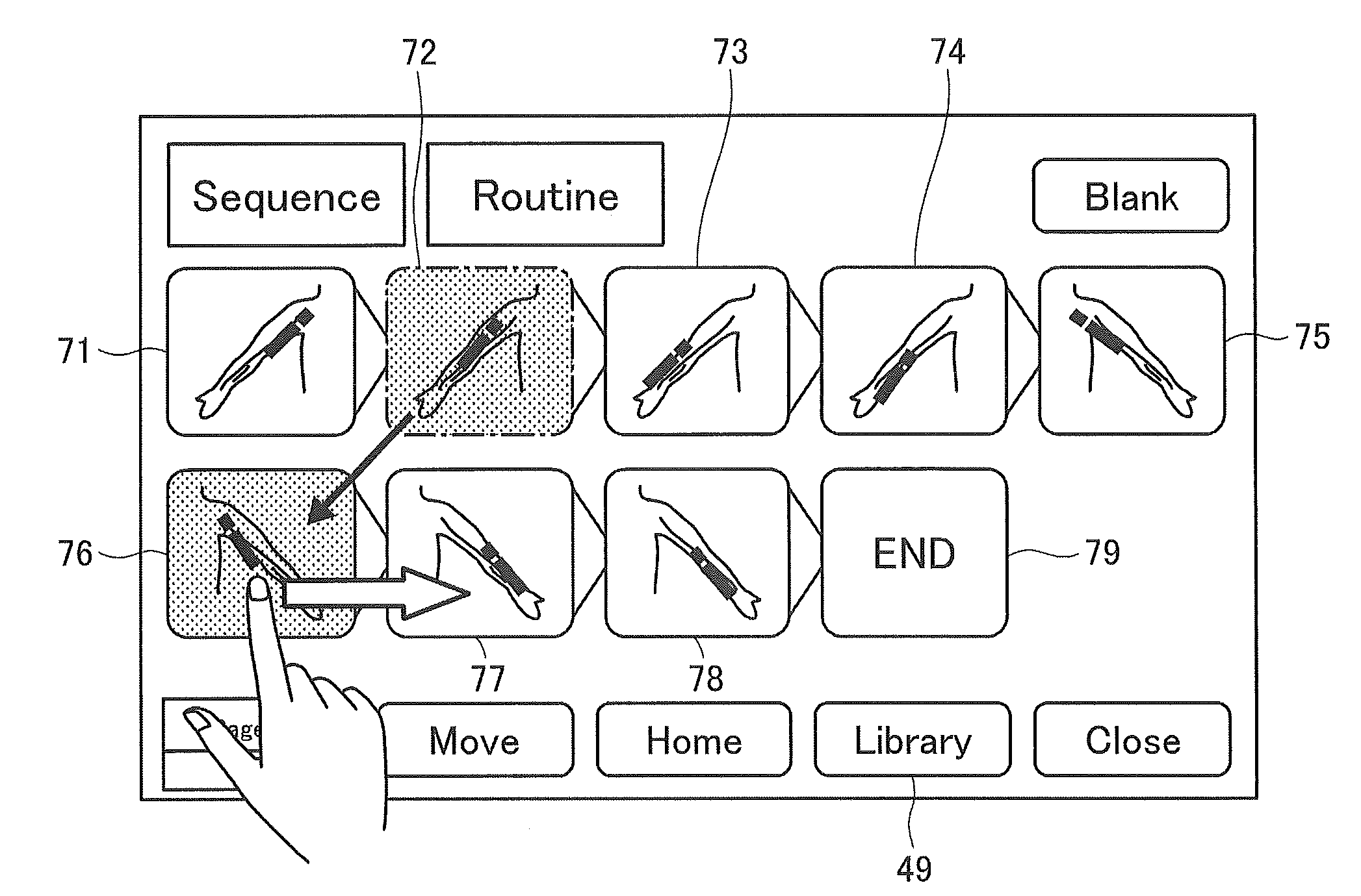
[0081]At the time of ultrasound diagnosis, if the sequence button 48 that is displayed on the touch panel 9 illustrated in FIG. 3 is selected by the operator, the body controller 30 detects that the sequence button 48 is selected, calls up a series of routine examinations relating to the artery of upper extremity that is stored in advance in the storage unit 31, for example, and sequence-displays body marks 71 to 78 and an end button 79 on the touch panel 9 through the panel controller 27, as illustrated in FIG. 7.

[0082]Further, before the ultrasound diagnosis is started, for example, if the body mark 73 which corresponds to an examination in the middle of the series of examinations is selected by the operator from among the body marks 71 to 78 seque...

third embodiment

[0085]Next, a third embodiment of the invention will be described. A configuration of an ultrasound diagnostic apparatus according to the third embodiment is the same as those of the first and second embodiments.

[0086]In the series of examinations in the first and second embodiments, the body controller 30 detects that the freeze button 34 is pushed again and performs switching of the highlighting of the body mark in a predetermined timing when the freezing of the ultrasonic image is released, but the invention is not limited thereto. The body controller 30 may detect that the imaging mode selecting button 33 is pushed and may perform switching of the highlighting of the body mark in a predetermined timing when the imaging mode is switched, instead of performing the switching in a predetermined timing when the freezing of the ultrasonic image is released.

[0087]For example, in ultrasound diagnosis, in a case where a B mode image is obtained and then a PW mode image is obtained at a p...


PUM


Abstract

An ultrasound diagnostic apparatus in which an ultrasonic wave is transmitted toward a subject by an ultrasound probe, an ultrasound image is generated by a diagnostic apparatus body based on reception data thus obtained and an examination is performed based on the ultrasound image, including a display unit on which a plurality of examination portions corresponding to a series of examinations relating to ultrasound diagnosis are displayed in an order of time series in a single screen at a time, and a controller which highlights an examination portion corresponding to an examination being executed among the plurality of examination portions in the display unit, in which each of the plurality of examination portions is displayed as a body mark which indicates an examination position by the ultrasound probe.

Description

BACKGROUND OF THE INVENTION[0001]The present invention relates to an ultrasound diagnostic apparatus and in particular, to an ultrasound diagnostic apparatus to generate an ultrasound image based on reception signal obtained by transmission and reception of the ultrasonic waves using an ultrasound probe and to display the ultrasound image on a display unit.[0002]In the medical field, an ultrasound diagnostic apparatus using an ultrasound image has been put into practical use. Generally, in this kind of ultrasound diagnostic apparatus, the ultrasound image is generated by transmitting an ultrasonic wave toward a subject from a transducer array of an ultrasound probe, receiving an ultrasonic echo from the subject by the transducer array, and processing the reception signal electrically in a diagnostic apparatus body.[0003]In recent years, an ultrasound diagnostic apparatus has been developed in which various examinations are performed on the basis of ultrasound images and an assist of...

Claims


Application Information

Patent Timeline
IPC IPC(8): A61B8/00
CPCA61B8/465A61B8/463A61B8/08A61B8/4427A61B8/462A61B8/469A61B8/54A61B8/585
Inventor TASHIRO, RIKA
Owner FUJIFILM CORP
Why Eureka
Try Eureka