Assessment of iron deposition post myocardial infarction as a marker of myocardial hemorrhage

a myocardial infarction and marker technology, applied in the field of diagnosis of reperfusion and non-reperfusion hemorrhage, can solve the problems of ami, limited value of individual patient differentiation strategies, and the inability to distinguish patients with acute myocardial infarctions, so as to prevent iron deposition

Active Publication Date: 2014-04-24
CEDARS SINAI MEDICAL CENT
View PDF5 Cites 6 Cited by
  • Summary
  • Abstract
  • Description
  • Claims
  • Application Information

AI Technical Summary

Benefits of technology

[0064]This invention provides a method for classifying an infarction (both reperfused and non-reperfused types) to be hemorrhagic or non-hemorrhagic based on MRI in order to provide therapeutic interventions to prevent iron deposition in the chronic peri

Problems solved by technology

Significant narrowing of epicardial coronary arteries due to atherosclerotic disease or acute embolic obstruction can impede blood flow and oxygen to the myocardium resulting in acute myocardial infarction (AMI).
However, reperfusion therapies are also associated with post-infarct complications that are often associated with fatal heart failure.
If undetected and untreated, this can degenerate into asystole leading to hemodynamic impairment causing death.
Although there are diagnostic and therapeutic strategies for managing and treating ischemic heart disease, these strategies have limited value for distinguishing individual patients at risk for arrhythmias and SCD.
However, there are no diagnostic strategies for predict

Method used


Image

  • Assessment of iron deposition post myocardial infarction as a marker of myocardial hemorrhage
  • Assessment of iron deposition post myocardial infarction as a marker of myocardial hemorrhage
  • Assessment of iron deposition post myocardial infarction as a marker of myocardial hemorrhage

Examples


example 1

Experimental Methods

Surgical Procedure

[0065]Canines (n=23, 20-25 kg) were enrolled and studied according to the protocols approved by the Institutional Animal Care and Use Committee. Each dog was given an intramuscular injection of the pre-anesthetic tranquilizer Innovar (0.4 mg / ml of fentanyl and 20 mg / ml of droperidol) at a dose of 1 ml / 25-50 kg of body weight. Subsequently, the dog was anesthetized with an intravenous injection of Propofol (5.0-7.5 mg / kg), endotracheally intubated and maintained on gas anesthesia (2.0-2.5% isoflurane with 100% oxygen). Animals were artificially ventilated at 1-2 L / min with the respiration rate being continuously adjusted to maintain partial pressure of CO2 in blood (PaCO2) between 30 and 35 mmHg. Left lateral thoracotomy was performed at the fourth intercostal space, and the exposed heart was suspended in a pericardial cradle (FIG. 10). Aortic and left atrial catheters were inserted and secured for invasive blood pressure monitoring and drug deli...

example 2

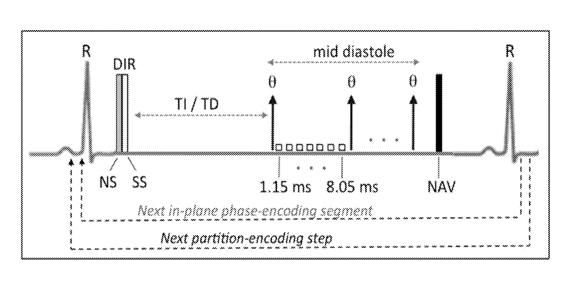
Free-Breathing, ECG-Triggered, Dark-Blood Prepared 3D T2* MRI

[0094]Breath-held, ECG-triggered, 2D T2* mapping at 1.5T is the current standard for identifying iron overload in the heart. However, this approach has a number of limitations for our application: (i) Our early studies and the literature suggest that, in the setting of large infarcts, breath holding may trigger arrhythmias. In our experience, repetitive breath-held image acquisitions have led to fatal arrhythmias in canines with hemorrhage; and non-fatal arrhythmias demand undesirably long breath holding times; (ii) Partial volume effects in the through-plane direction can significantly reduce the conspicuity of the regions with an iron overload; (iii) Bright blood T2* maps are prone to significant image artifacts (ghosts and smears), particularly when TEs are long. At 1.5T, the sensitivity for visualizing smaller iron depositions can be limited and require the use of longer TEs in spite first-order flow compensation at ev...

example 3

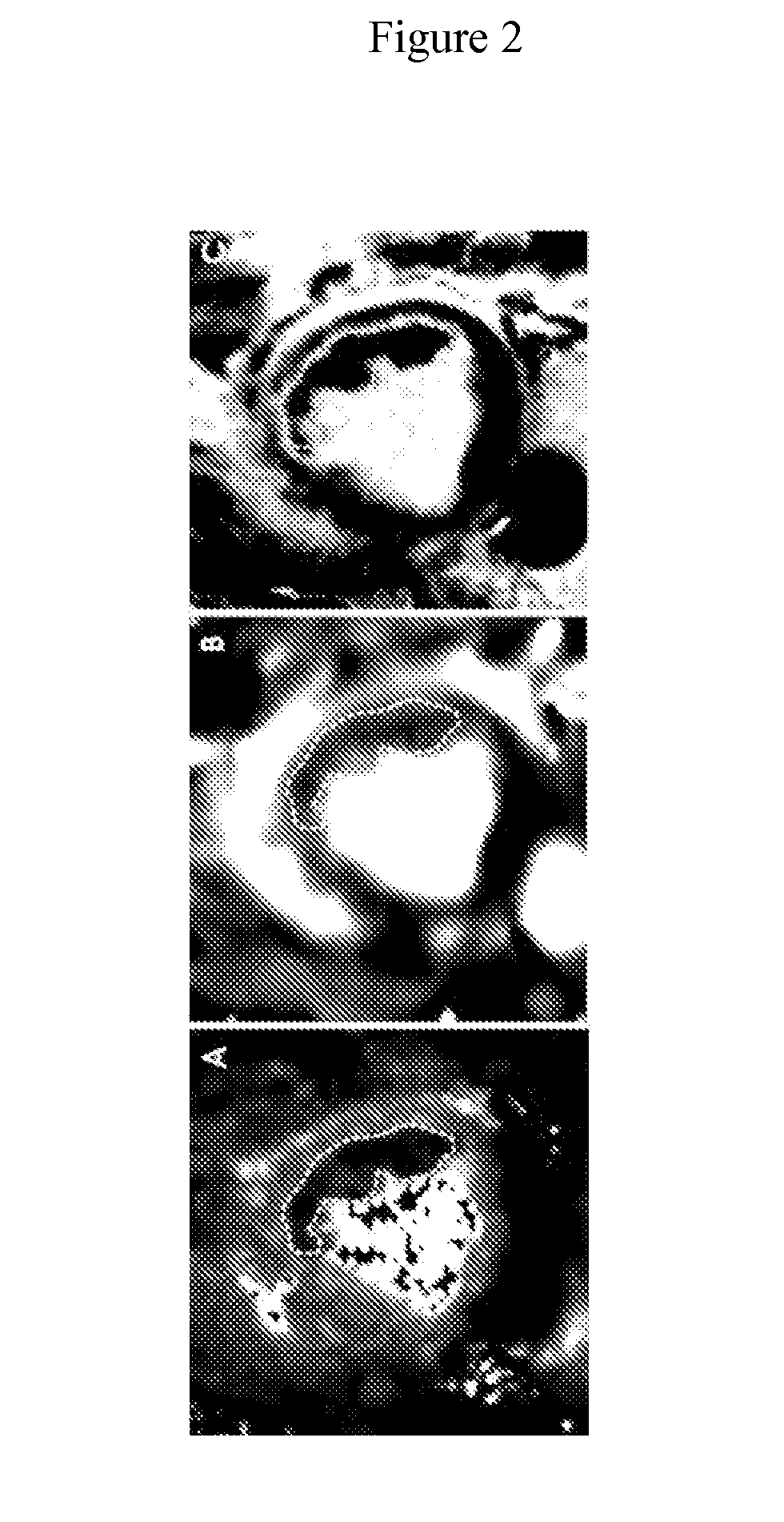
Detecting Acute Myocardial Reperfusion Hemorrhage (aMRH) with MRI

[0096]T2 and T2* MRI have both been shown to be sensitive for detecting aMRH. However, there is (i) no consensus on which of the two methods yield the most desirable means for detecting aMRH, and (ii) no histology studies that confirm T2 or T2* MRI can and do identify myocardial hemorrhage (O'Regan D P, Ahmed R, Karunanithy N, et al. Reperfusion hemorrhage following acute myocardial infarction: assessment with T2* mapping and effect on measuring the area at risk. Radiology 2009; 250:916-922. Ganame J, Messalli G, Dymarkowski S, et al. Impact of myocardial haemorrhage on left ventricular function and remodelling in patients with reperfused acute myocardial infarction. Eur Heart J 2009; 30:1440-1449). The inventors determined the optimal quantitative MRI approach for detecting a MRH and to validate that iron composites are found within hemorrhagic infarctions on the basis tissue histology.

[0097]Ischemia reperfusion injur...


PUM

PropertyMeasurementUnit
Timeaaaaaaaaaa
Mechanical propertiesaaaaaaaaaa
Conductionaaaaaaaaaa

Abstract

The invention is directed to methods for diagnosing reperfusion/non-reperfusion hemorrhage and predicting cardiac arrhythmias and sudden cardiac death in subjects comprising using imaging techniques to detect regional iron oxide deposition. The invention also provides treatment methods for subject at increased risk of sudden cardiac death.

Description

GOVERNMENT RIGHTS[0001]The invention was made with government support under Grant No. HL091989 awarded by the National Institutes of Health. The government has certain rights to the invention.FIELD OF INVENTION[0002]The invention is directed to methods for diagnosing reperfusion and non-reperfusion hemorrhage, predicting cardiac arrhythmias, sudden cardiac death, and adverse remodeling in subjects post myocardial infarction. The invention also provides treatment methods for subjects at increased risk of sudden cardiac death and heart failure.BACKGROUND[0003]All publications herein are incorporated by reference to the same extent as if each individual publication or patent application was specifically and individually indicated to be incorporated by reference. The following description includes information that may be useful in understanding the present invention. It is not an admission that any of the information provided herein is prior art or relevant to the presently claimed inve...

Claims


Application Information

Patent Timeline
IPC IPC(8): A61B5/026A61B5/02A61N1/362A61B5/0472A61B5/00A61N1/39A61K45/06A61B5/024A61B5/366
CPCA61B5/0263A61B5/0044A61B5/02042A61N1/3956A61B5/0472A61K45/06A61N1/362A61B5/02405A61B5/055A61B5/14542A61B2576/023A61N1/3629G16H30/40A61K49/06A61B5/366
Inventor DHARMAKUMAR, ROHANCOKIC, IVAN
Owner CEDARS SINAI MEDICAL CENT
Why Eureka
Try Eureka