Apparatus and method for tissue biopsy

a tissue biopsy and apparatus technology, applied in the field of apparatus and method for tissue biopsy, can solve the problems of extended healing time and leave marks, and achieve the effects of facilitating access to tissue samples, facilitating grasping and manipulating needles, and facilitating identification of distances along the tissue sampl

Active Publication Date: 2014-07-17
THE GENERAL HOSPITAL CORP
View PDF15 Cites 27 Cited by
  • Summary
  • Abstract
  • Description
  • Claims
  • Application Information

AI Technical Summary

Benefits of technology

[0009]The exemplary hollow needle can optionally be provided with one or more openings or windows provided on the wall thereof. Such openings can facilitate access to the tissue sample without removing the sample from the needle. A plurality of markings can optionally be provided along at least a portion of the length of the needle, e.g., proximal to the one or more openings, to facilitate an identification of distances along the tissue sample in the needle.
[0010]The one or more exemplary hollow needles can be affixed or coupled to a handle or substrate to facilitate grasping and manipulation of the needle and / or to provide mechanical stability to the needle. The handle or substrate can be adjustably coupled to the one or more hollow needles, e.g., using a friction fitting, a clamp, a threaded arrangement, or the like. Such adjustable coupling can be adapted to vary the protrusion length of one or more of the hollow needles beyond a lower surface of the handle or substrate, e.g., to vary the effective needle length or maximum penetration distance when the apparatus is pressed into a tissue region. The coupling of the needles to the substrate can be adapted such that lengths of the needles can be adjusted either individually or as a group. Such adjustment can be performed using one or more threaded or screw arrangements, clamps or friction fittings, or the like.

Problems solved by technology

However, conventionally, the removal of such large tissue samples may require an extended healing time and / or stitches, and can further leave markings after healing has occurred.

Method used


Image

  • Apparatus and method for tissue biopsy
  • Apparatus and method for tissue biopsy
  • Apparatus and method for tissue biopsy

Examples


Embodiment Construction

[0006]The herein described exemplary embodiments of the present invention pertain to a method and apparatus. Synergetic effects may arise from different combinations of the features and embodiments described herein, although all such combinations might not be described in detail. Further, it shall be noted that embodiments of the present invention concerning a method might be carried out with the order of the steps as described, nevertheless this need not be the only or essential order of the steps of the method unless otherwise specified.

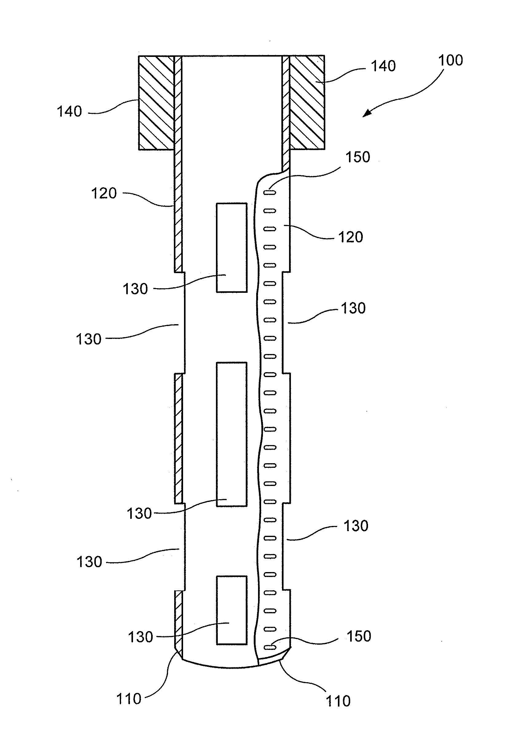
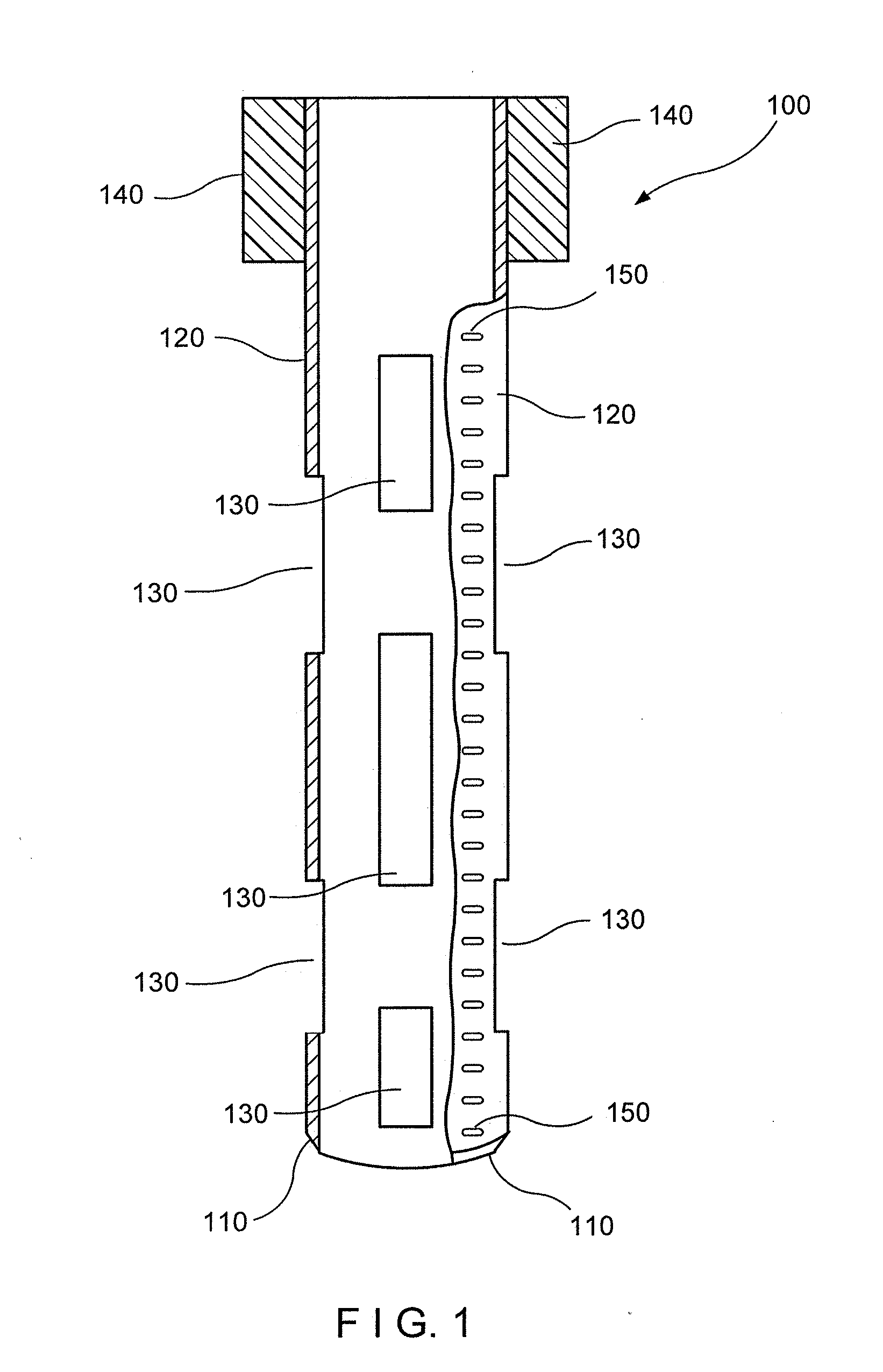
[0007]Exemplary embodiments of the present invention relate to simple, inexpensive, and safe methods, apparatus and devices for a removal of one or more tissue samples that can be smaller than certain samples that can be removed in conventional punch biopsy procedures, such that the removal of such small tissue samples is well-tolerated, for example, the removal sites may heal quickly without significant bleeding or risk of infection, and that may ...


PUM


Abstract

Exemplary embodiments of an apparatus can be provided for obtaining portions or samples of tissue from a target region of a biological tissue. One or more needles can be provided that have a small internal diameter, e.g., about 1 mm or less, and the needles can be configured to extract the tissue portions when the needles are inserted into and withdrawn from the tissue. Windows and/or markings can be provided on the wall of the needles to facilitate access to the sample. The needles can be provided in an enclosure, and an actuator can be provided to direct the needles into the tissue and/or withdraw them. A plurality of tissue portions having known relative locations in the target region can be obtained, and extraction of the tissue portions can be well-tolerated by the tissue as compared with conventional punch biopsies or the like.

Description

CROSS REFERENCE TO RELATED APPLICATION[0001]The present application relates to and claims priority from U.S. Provisional Patent Application Ser. No. 61 / 437,493 filed Jan. 28, 2011, the disclosure of which is incorporated herein by reference in its entirety.TECHNICAL FIELD[0002]The present invention relates to methods and apparatus for obtaining biological tissue samples, e.g., for biopsies or the like.BACKGROUND INFORMATION[0003]A punch biopsy is a procedure that removes a sample of skin or other tissue, which can be used for external analysis and / or detection of any of a variety of conditions within the tissue. A punch biopsy can be used, e.g., for the diagnosis of squamous cell carcinoma and melanomas. Punch biopsies can generally include a removal of a relatively large tissue sample using, for example, a round shaped knife or tube typically having a diameter of about 2 mm or greater, up to about 4-6 mm. The underlying portion of the sample may be cut using a scalpel or the like, ...

Claims


Application Information

Patent Timeline
Patent Type & Authority Applications(United States)
IPC IPC(8): A61B10/02A61B19/00
CPCA61B19/5202A61B10/0266A61B10/0233A61B2010/0208A61B17/32053A61B10/0275A61B2010/0225A61B90/30A61B90/92A61B90/94A61B10/0096A61B10/025
Inventor AUSTEN, WILLIAMMANSTEIN, DIETER
Owner THE GENERAL HOSPITAL CORP
Why Eureka
Try Eureka