Image processing apparatus and image processing method

a technology of image processing and image processing equipment, which is applied in the field of image processing equipment and image processing methods, can solve the problems of requiring concentration and a long time to observe all of these images

Active Publication Date: 2014-10-09
OLYMPUS CORP
View PDF8 Cites 21 Cited by
  • Summary
  • Abstract
  • Description
  • Claims
  • Application Information

AI Technical Summary

Benefits of technology

[0101]As described above, in the first embodiment, between the present image group and the past image group, the images in the region greatly different in the imaging time periods between the feature images are emphasized, or displayed in a format distinguishable by the user. Accordingly, the user is able to make observation intensively and perform diagnosis on an image of a site in a subject having a possibility of having a certain change caused between the past examination and the present examination. Therefore, it becomes possible to increase a discovery efficiency of an abnormal site and to shorten an observation time period as a whole, and thus to improve an observation efficiency.

Problems solved by technology

Therefore, to observe all of these images takes a very long time and requires an ability to concentrate.

Method used


Image

  • Image processing apparatus and image processing method
  • Image processing apparatus and image processing method
  • Image processing apparatus and image processing method

Examples


first embodiment

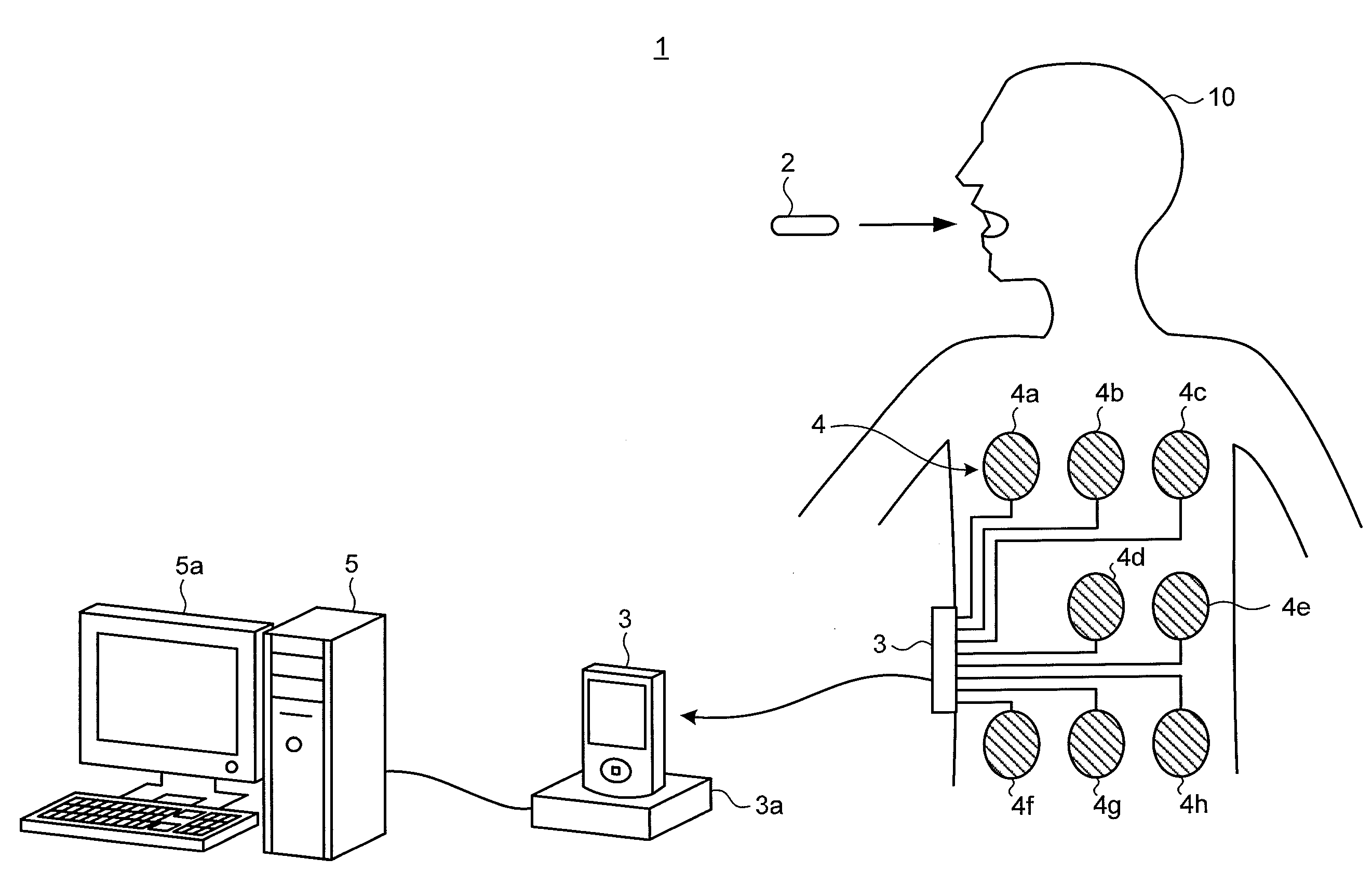
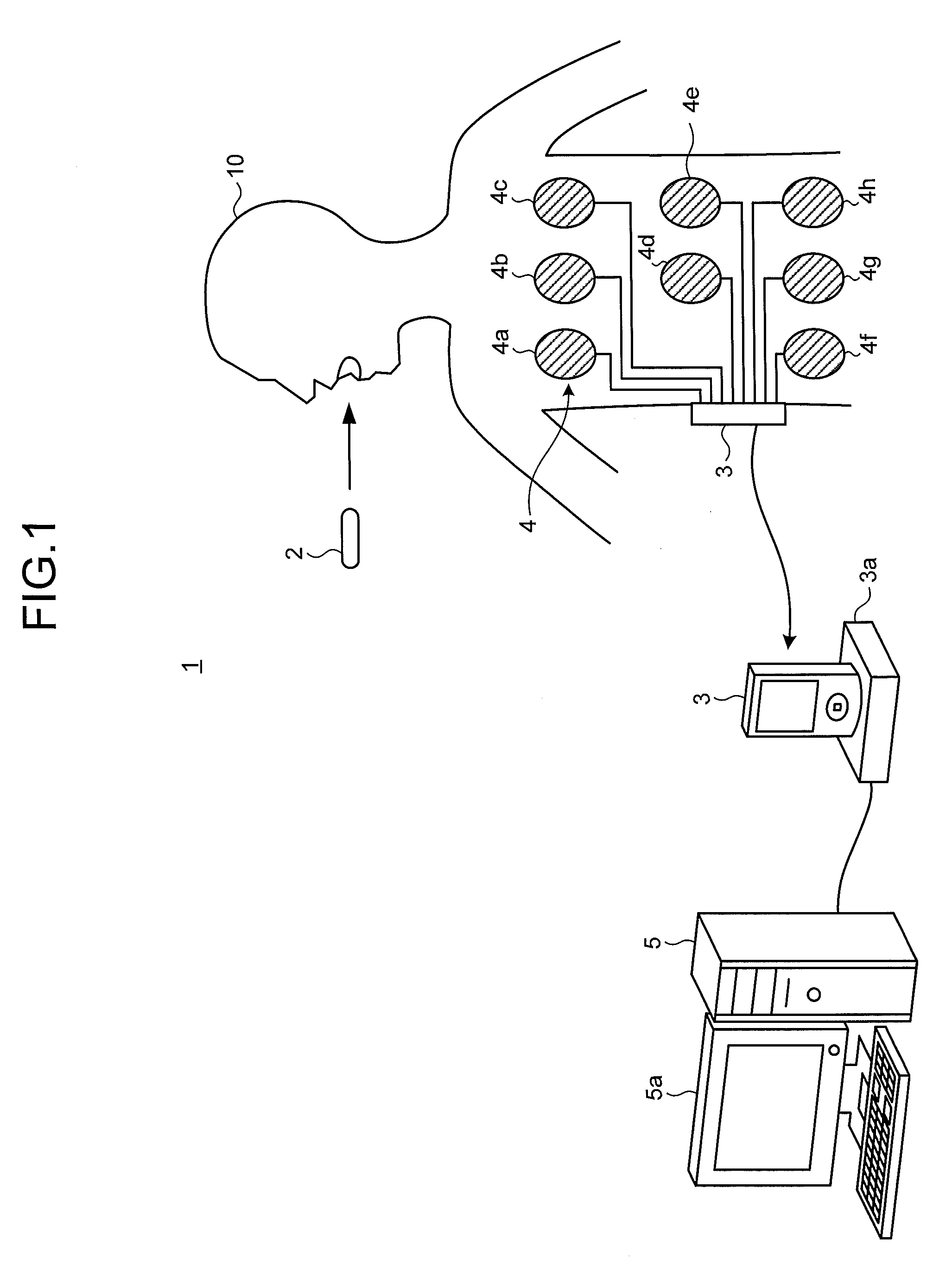
[0029]FIG. 1 is a schematic diagram illustrating a schematic configuration of a capsule endoscopic system including an image processing apparatus according to a first embodiment of the present invention. A capsule endoscopic system 1 illustrated in FIG. 1 includes: a capsule endoscope 2, which generates image data by being introduced into a subject 10 and capturing an image of inside of the subject 10, and superimposes and transmits the image data on a wireless signal; a receiving device 3, which receives the wireless signal transmitted from the capsule endoscope 2 via a receiving antenna unit 4 that is attached to the subject 10; and an image processing apparatus 5, which acquires the image data from the receiving device 3 and performs predetermined image processing on the image data.

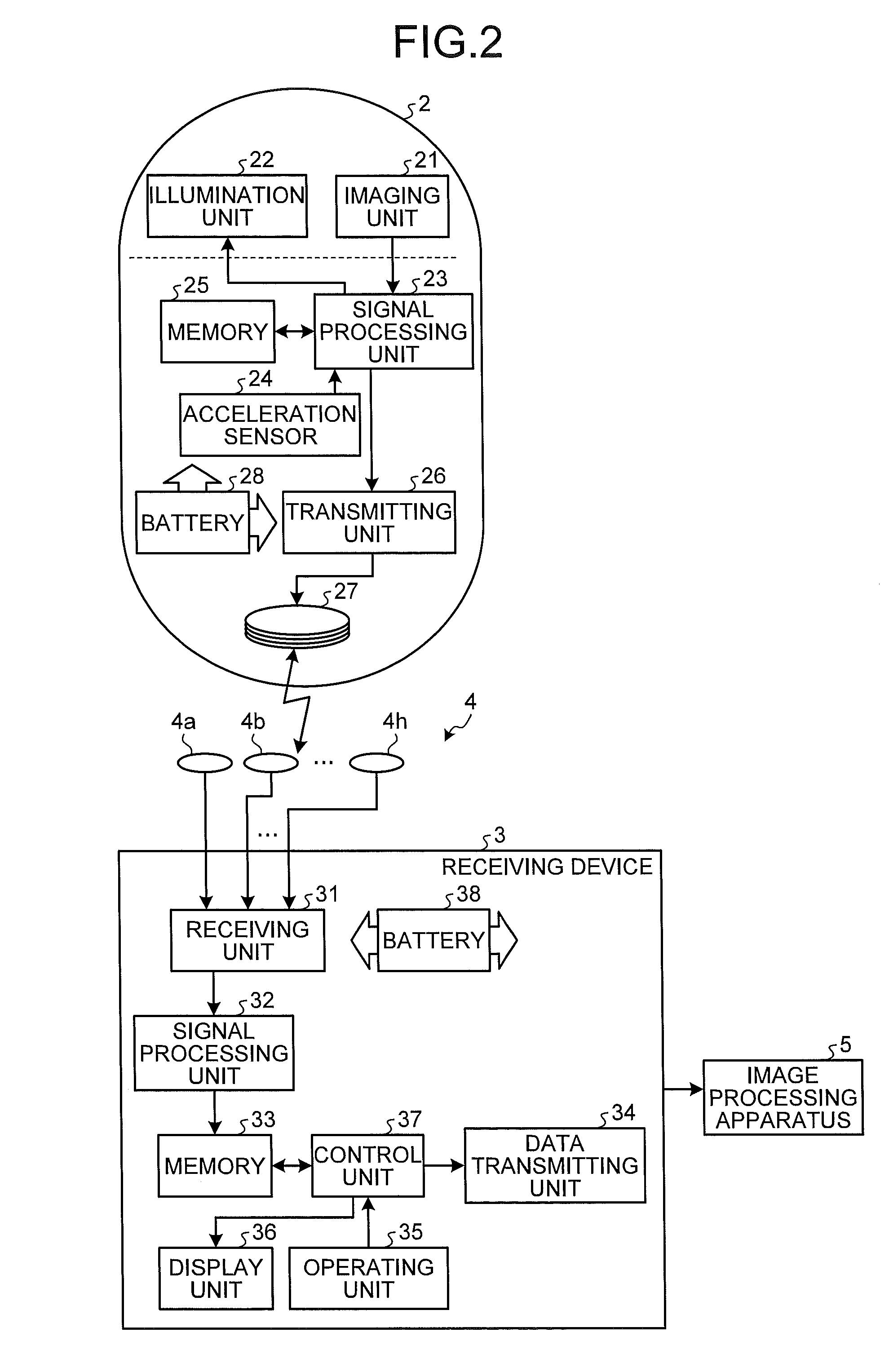
[0030]FIG. 2 is a block diagram illustrating a schematic configuration of the capsule endoscope 2 and the receiving device 3.

[0031]The capsule endoscope 2 is a device, which has various built-in parts ...

modified example 1-1

[0102]Next, a modified example 1-1 of the first embodiment of the present invention will be described.

[0103]In the first embodiment, as a quantity corresponding to a time period between the imaging times of the plurality of feature images extracted respectively from the present image group and the past image group, the imaging time period is acquired (see step S13 and S16). However, instead of the imaging time period, the number the series of images captured between a certain feature image and another feature image may be acquired. Since image capturing is performed at a constant imaging frame rate normally in the capsule endoscope 2, the imaging time period and the number of images are corresponding quantities. In this case, at step S17 of FIG. 4, whether or not a difference between the number of images of an interval acquired from the present image group and the number of images of a corresponding interval acquired from the past image group is equal to or greater than a predetermi...

modified example 1-2

[0104]Next, a modified example 1-2 of the first embodiment of the present invention will be described.

[0105]When the images added with the careful observation flag are displayed on the observation image D1, the display may be performed after performing further predetermined image processing on these images. For example, on the images added with the careful observation flag, an image analysis process of extracting a predetermined lesion region may be performed, and when these images are displayed in the main display area d3, a result of that analysis may be displayed therewith.

[0106]In addition, the careful observation flag may be used as a parameter of various aiding functions for generating and displaying the observation screen.


PUM


Abstract

A image processing apparatus includes an image extraction unit that extracts first and second feature images representing first and second feature, respectively from a first image group acquired by sequentially capturing images of inside of a subject, and that further extracts third and fourth feature images representing first and second feature, respectively from a second image group acquired before the first image group, a feature data acquiring unit that acquires first and second feature data characterizing a movement of the capsule endoscope between the first and second feature images and between the third and the fourth feature images, respectively, a comparing unit that compares the first feature data with the second feature data, and a display control unit that performs, with respect to the first image group, display control based on a result of the comparison by the comparing unit.

Description

CROSS REFERENCES TO RELATED APPLICATIONS[0001]This application is a continuation of PCT international application Ser. No. PCT / JP2013 / 077612 filed on Oct. 10, 2013 which designates the United States, incorporated herein by reference, and which claims the benefit of priority from Japanese Patent Applications No. 2012-231182, filed on Oct. 18, 2012, incorporated herein by reference.BACKGROUND OF THE INVENTION[0002]1. Field of the Invention[0003]The present invention relates to an image processing apparatus and an image processing method, which process images acquired by a capsule endoscope that is introduced into a subject and captures images of inside of the subject.[0004]2. Description of the Related Art[0005]In recent years, examinations using capsule endoscopes that are introduced into subjects such as patients and capture images of inside of the subjects are known in the field of endoscopes. A capsule endoscope is an apparatus that has built-in imaging function, wireless communic...

Claims


Application Information

Patent Timeline
Patent Type & Authority Applications(United States)
IPC IPC(8): A61B1/00A61B1/04
CPCA61B1/00009A61B1/00045A61B1/00016A61B1/041A61B1/0002A61B1/00006A61B1/0005G06T7/0016G06T2207/10068G06T2207/30028A61B1/000094
Inventor TANIGUCHI, KATSUYOSHI
Owner OLYMPUS CORP
Why Eureka
Try Eureka