Breast biopsy system using mr and gamma imaging

a breast biopsy and gamma imaging technology, applied in the field of breast biopsy systems using can solve the problems of not having a real-time mri biopsy system available, proving difficult to achieve both from the patient comfort perspective as well as engineering and product design perspectives, and no one has discussed equipment configurations for optimizing breast biopsy using combined mr and gamma imaging

Inactive Publication Date: 2014-10-09
CUBRESA
View PDF4 Cites 7 Cited by
  • Summary
  • Abstract
  • Description
  • Claims
  • Application Information

AI Technical Summary

Benefits of technology

[0022]There may be two

Problems solved by technology

This is available in ultrasound biopsy, in which the ultrasound transducer is used at the same time as the needle sampling occurs, however there are no real-time MRI biopsy systems currently available.
This is because to make real-time MRI biopsy guidance available requires the patient to be in the m

Method used


Image

  • Breast biopsy system using mr and gamma imaging
  • Breast biopsy system using mr and gamma imaging
  • Breast biopsy system using mr and gamma imaging

Examples


Embodiment Construction

[0034]Unless defined otherwise, all technical and scientific terms used herein have the same meaning as commonly understood by one of ordinary skill in the art to which the invention belongs. Although any methods and materials similar or equivalent to those described herein can be used in the practice or testing of the present invention, the preferred methods and materials are now described. All publications mentioned hereunder are incorporated herein by reference.

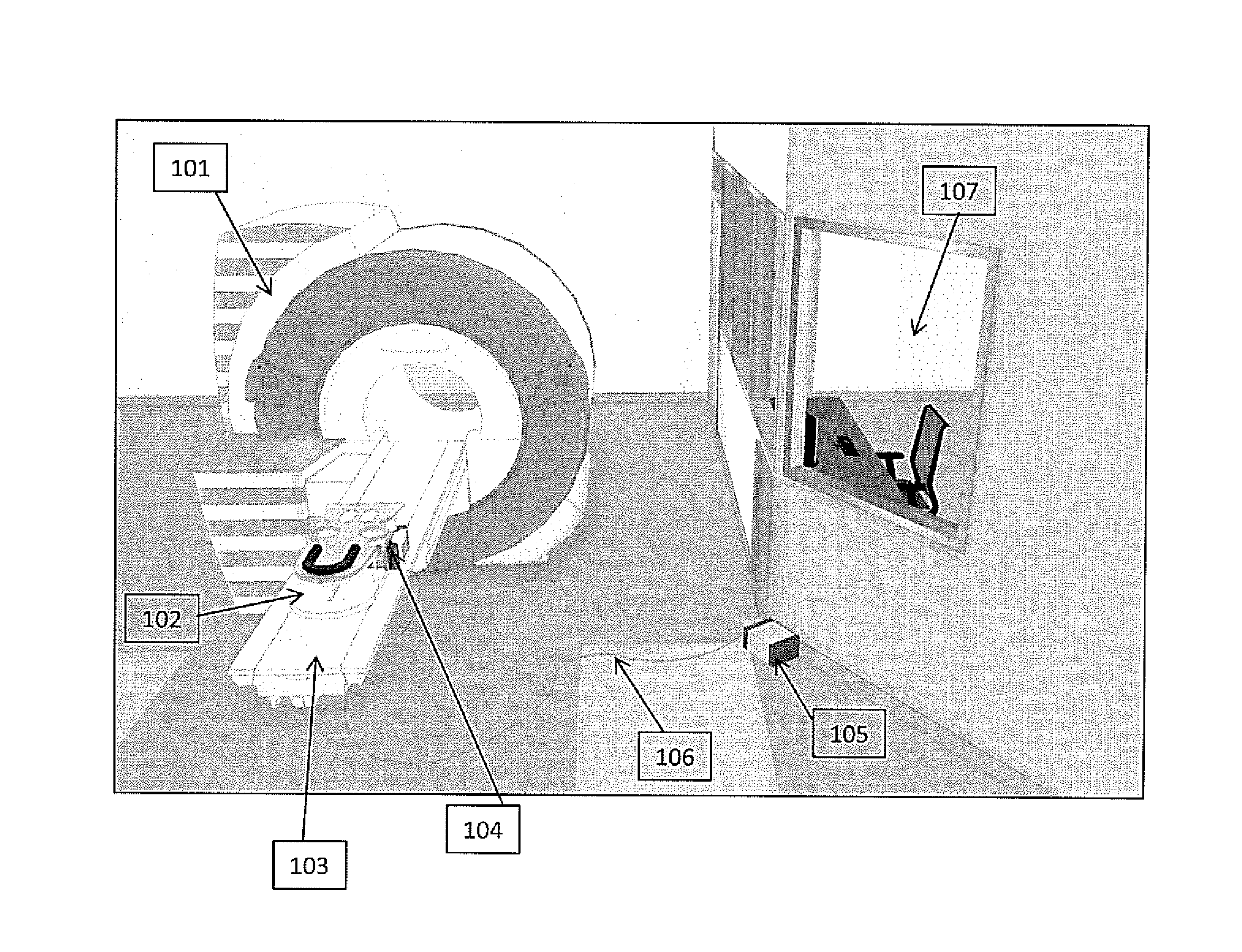
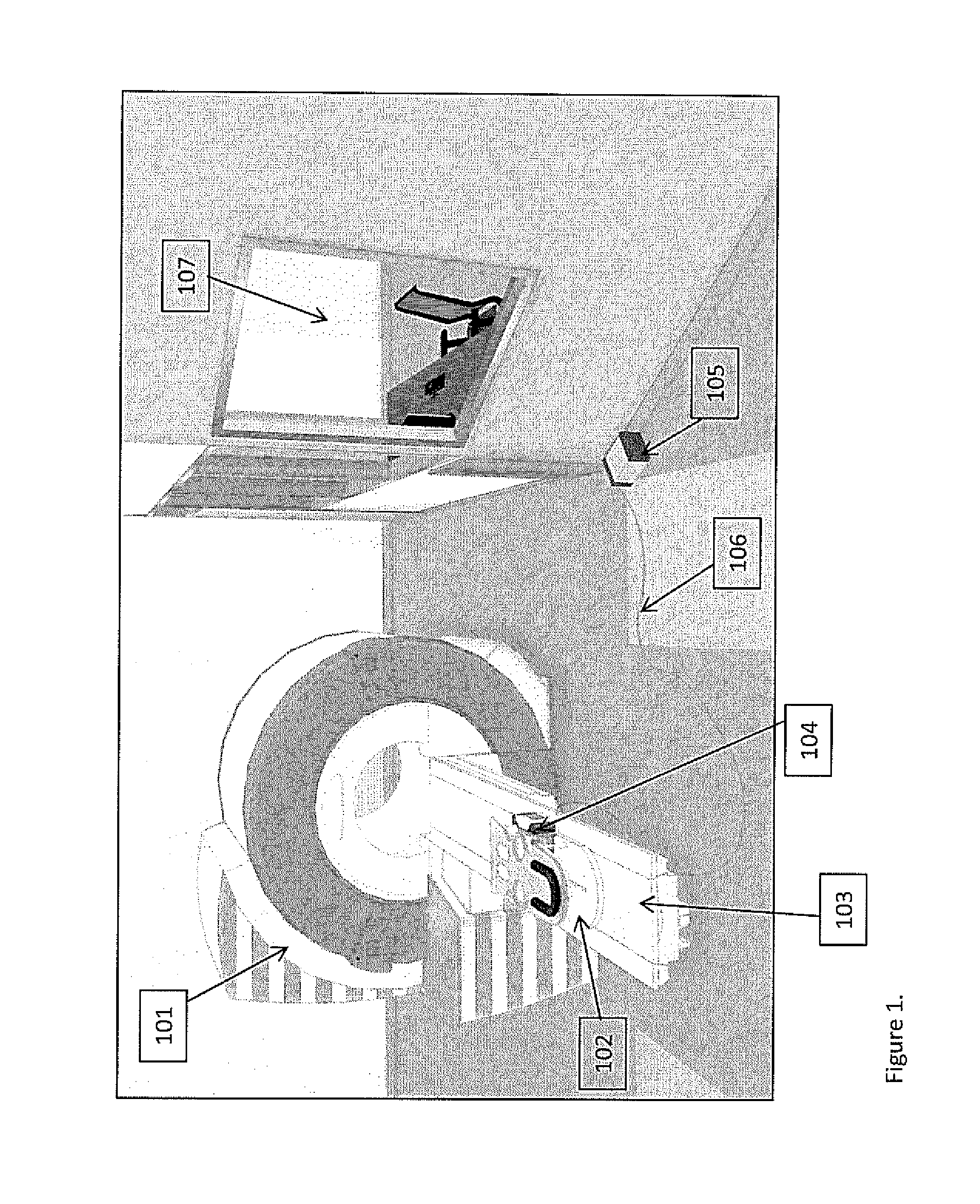
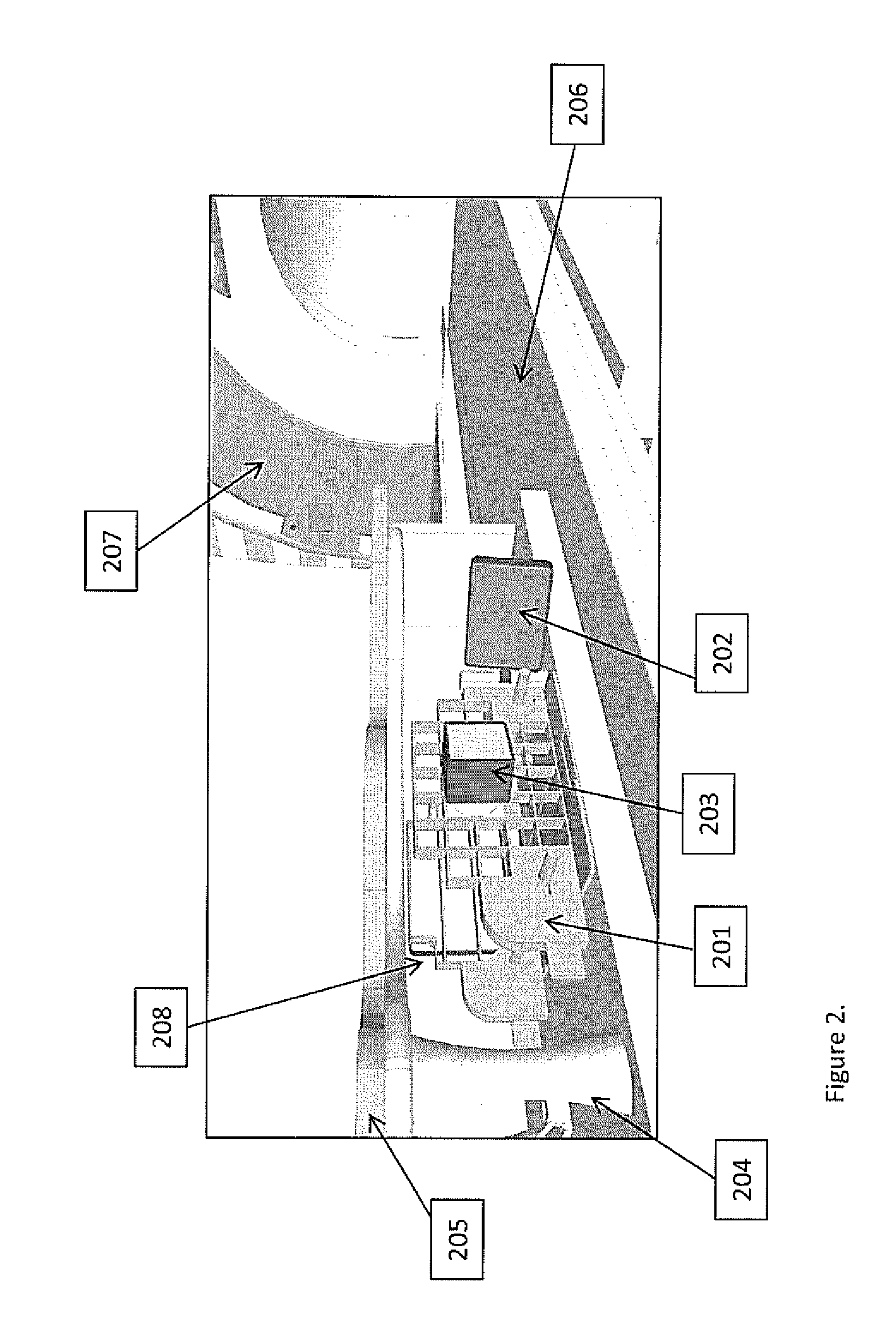
[0035]For MRI Breast Biopsy using a MR Safe Gamma Camera, the equipment requirements include:[0036]an MRI system with software and hardware[0037]a patient rest and breast immobilization system[0038]a biopsy needle system[0039]a software capability for needle positioning and guidance[0040]a method of gadolinium and radioisotope injection into the patient, and[0041]an MR Safe gamma camera system consisting of:[0042]one or more gamma camera heads which are MR safe[0043]an interface module which allows connection between the g...


PUM


Abstract

Described herein is the use of gamma cameras in the fringe field of the MRI system. Specifically, an MR image of the breast with lesion identification is first produced. Then, a gamma camera is attached to the existing breast immobilization system for generating one or more gamma images of the breast. The gamma camera is then removed from the breast immobilization system, and a breast biopsy is performed. The gamma camera can then be used to image the biopsy cores that have been removed from the patient in order to verify that the biopsy cores are radioactive, that the biopsy cores extend from one end of the lesion to the other and have a radioactive profile in which the tip is not as radioactive as the middle, and in which the ratio of amount of radioactivity in the middle of the core to the amount of radioactivity that is present in the tip of the core can be expressed as a ratio.

Description

PRIOR APPLICATION INFORMATION[0001]This application claims the benefit of U.S. Provisional Patent Application 61 / 500,421, filed Jun. 23, 2011.BACKGROUND OF THE INVENTION[0002]MR Breast Biopsy can be characterized as a 1 to 2 hour procedure, in which typically 1, 2, or 3 lesions may be biopsied [1,2,3]. Each biopsy procedure requires approximately 4-5 insertions of the patient into the MRI system. The first insertion is to obtain the starting image; the second insertion is to confirm the guide position; the third insertion is to confirm the needle position prior to insertion into the lesion; and the fourth insertion is often required as a result of patient movement or other reasons. For example, 30% of the time, the guide is placed in the wrong position, and the guide must be moved and the image repeated. If a second and / or third lesion is / are sampled, additional MRI insertions are required. For each lesion site, there may be a need for multiple needle insertions, with between 6 and ...

Claims


Application Information

Patent Timeline
IPC IPC(8): A61B6/12A61B10/02A61B5/00A61B6/00A61B5/055G01R33/48
CPCA61B6/12A61B5/0555G01R33/4808A61B5/708A61B6/4266A61B10/0233A61B6/481A61B6/502A61B6/4258A61B10/0041A61B6/584A61B90/37A61B2090/371A61B2090/397A61B5/055
Inventor SCHELLENBERG, JAMESDIKA, CHERYLRIEDIGER, ERIN
Owner CUBRESA
Why Eureka
Try Eureka