Methods and Systems for Automatically Determining Magnetic Field Inversion Time of a Tissue Species

a tissue species and automatic determination technology, applied in the field of automatic determination of magnetic inversion time of tissue species, can solve problems such as difficult automaticization of systems

Inactive Publication Date: 2014-10-23
THE UNITED STATES OF AMERICA AS REPRESENTED BY THE SEC DEPT OF HUMAN HEALTH & SERVICES +1
View PDF16 Cites 8 Cited by
  • Summary
  • Abstract
  • Description
  • Claims
  • Application Information

AI Technical Summary

Benefits of technology

[0005]Embodiments of the present invention address and overcome one or more of the above shortcomings and drawbacks, by providing methods, systems, and apparatuses for automatically determining magnetic field inversion time of a tissue species based on a combination of a T1 parametric map, a-priori knowledge about the whereabouts of the tissue, and a T1-based healthy tissue classification algorithm. This technology is particularly well-suited for, but by no means limited to, cardiac-focused clinical applications.

Problems solved by technology

In conventional systems, T1 and the optimal TI are estimated using a segmented cine inversion recovery sequence, but these systems are difficult to automate as they are based on a series of images which differ both in contrast and cardiac phase.

Method used


Image

  • Methods and Systems for Automatically Determining Magnetic Field Inversion Time of a Tissue Species
  • Methods and Systems for Automatically Determining Magnetic Field Inversion Time of a Tissue Species
  • Methods and Systems for Automatically Determining Magnetic Field Inversion Time of a Tissue Species

Examples


Embodiment Construction

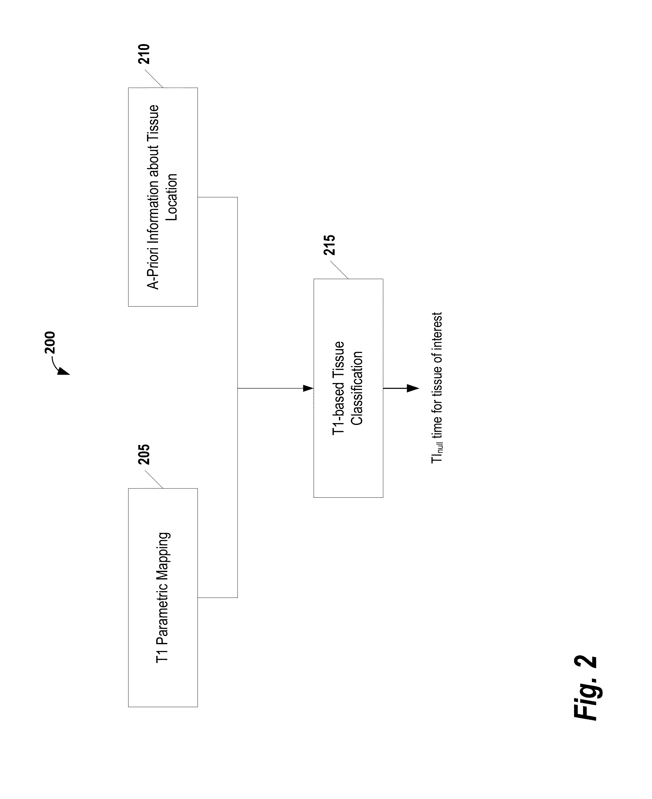
[0025]The following disclosure describes the present invention according to several embodiments directed at methods, systems, and apparatuses for automatically determining a null inversion time of a tissue species. In some embodiments, a system automatically determines the null inversion time using a T1 parametric map, a-priori anatomical information, and T1-based tissue classification. The system may be used, for example, to improve image contrast in inversion recovery MR imaging and workflow for late enhancement myocardial viability imaging. Additionally, the system may be used to improve inter-scan reliability in MRI myocardial viability imaging by automatically identifying an optimal inversion time to null MR signal in healthy myocardium.

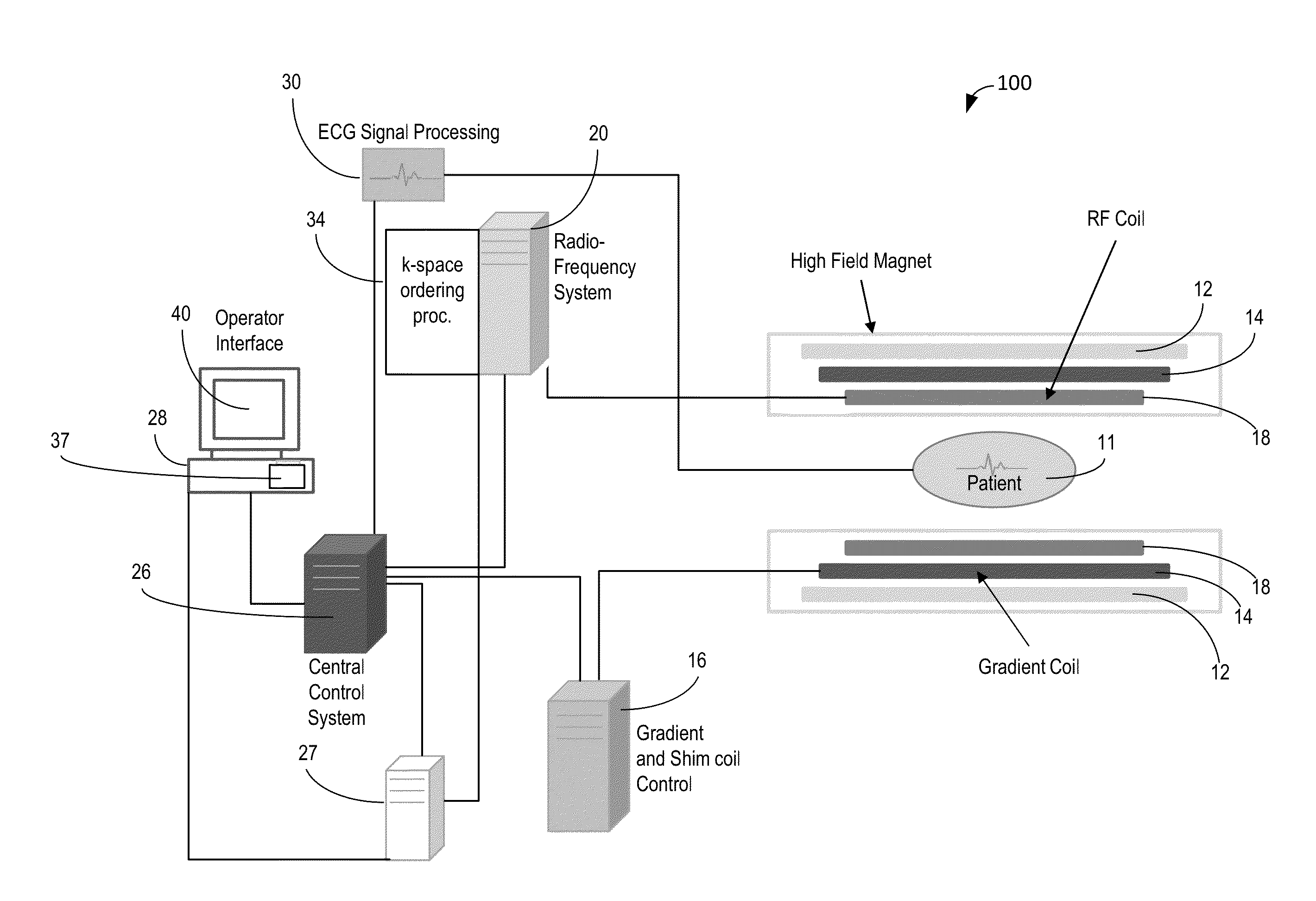
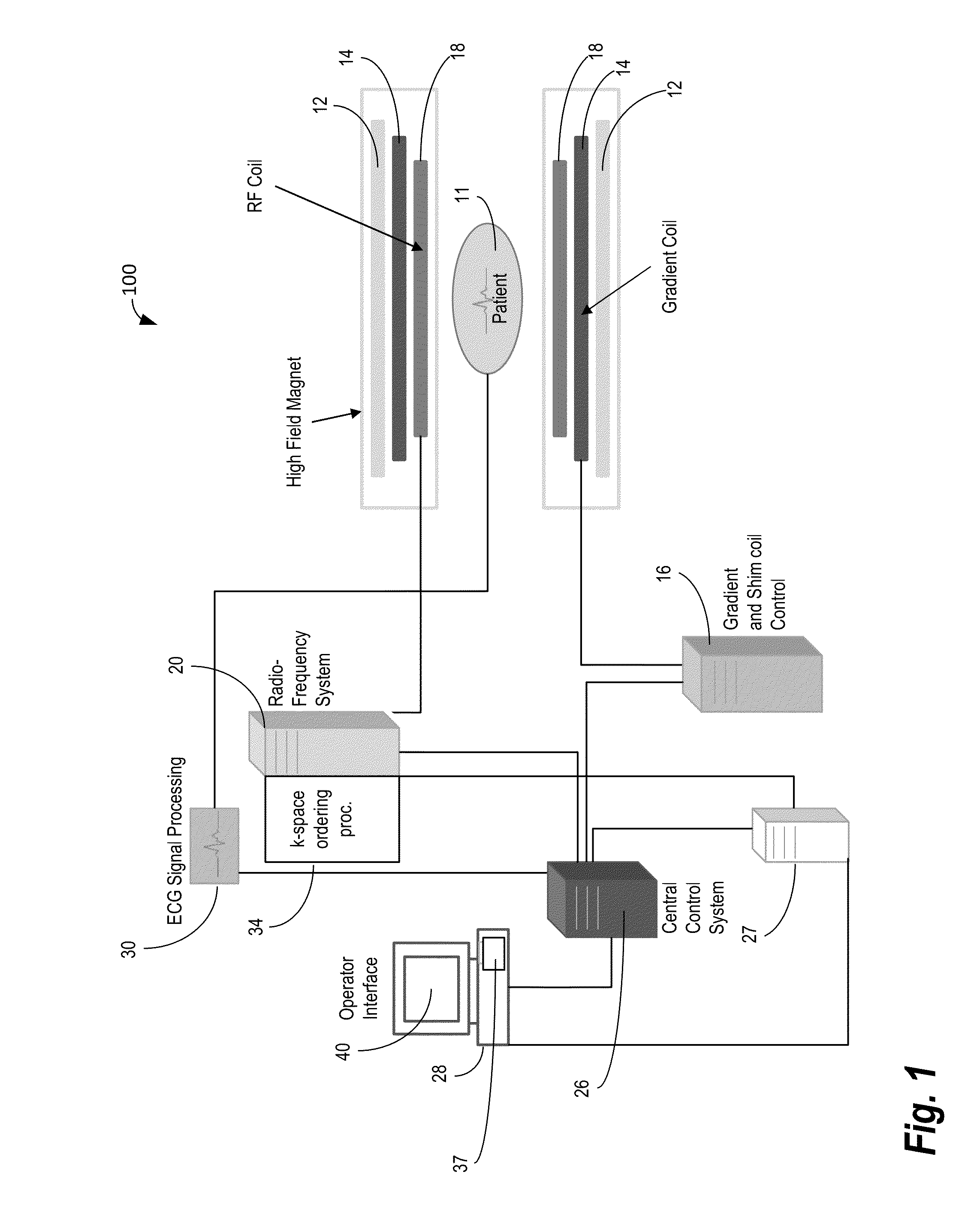
[0026]FIG. 1 shows system 100 for ordering acquisition of frequency domain components representing MR image data for storage in a k-space storage array. In system 100, magnet 12 creates a static base magnetic field in the body of patient 11 to b...


PUM


Abstract

A computer-implemented method for determining magnetic field inversion time of a tissue species includes generating a T1-mapping image of a tissue of interest, the T1-mapping image comprising a plurality of T1 values within an expected range of T1 values for the tissue of interest. An image mask is created based on predetermined identification information about the tissue of interest. Next, an updated image mask is created based on a largest connected region in the image mask. The updated image mask is applied to the T1-mapping image to yield a masked image. Then, a mean relaxation time value is determined for the largest connected region. The mean relaxation time value is then used to determine a time point for nulling longitudinal magnetization.

Description

CROSS-REFERENCE TO RELATED APPLICATIONS[0001]This application claims priority to U.S. provisional application Ser. No. 61 / 812,743 filed Apr. 17, 2013 which is incorporated herein by reference in its entirety.TECHNICAL FIELD[0002]The present invention relates generally to methods, systems, and apparatuses for automatically determining magnetic inversion time of a tissue species using a T1-mapping image, a-priori information about the region of interest, and a tissue classification algorithm. The disclosed methods, systems, and apparatuses may be applied to, for example, to identify areas of healthy tissue in cardiac applications.BACKGROUND[0003]Conventional inversion recovery (IR) methods in magnetic resonance imaging (MRI) can be used to suppress a signal from a specific tissue and, as such, leverage the image contrast in adjacent tissues. For example late enhancement IR imaging may be used to diagnose a wide range of ischemic and non-ischemic cardiomyopathies. In this application, ...

Claims


Application Information

Patent Timeline
Patent Type & Authority Applications(United States)
IPC IPC(8): G06T7/00
CPCG06T7/0012G06T2207/30048G06T2207/10088
Inventor SPOTTISWOODE, BRUCE S.LU, XIAOGUANGBI, XIAOMINGXUE, HUIGLIELMI, CHRISTOPHERKELLMAN, PETERGREISER, ANDREAS
Owner THE UNITED STATES OF AMERICA AS REPRESENTED BY THE SEC DEPT OF HUMAN HEALTH & SERVICES
Why Eureka
Try Eureka