Detection of tooth fractures in CBCT image

a technology of cbct image and tooth fracture, applied in the field of dental imaging, can solve the problems of difficult detection of vertical root fractures in periapical images, and difficulty in detecting some types of conditions

Inactive Publication Date: 2014-10-23
CARESTREAM DENTAL TECH TOPCO LTD
View PDF2 Cites 16 Cited by
  • Summary
  • Abstract
  • Description
  • Claims
  • Application Information

AI Technical Summary

Benefits of technology

[0009]Features offered by the present invention promote the art of dental imaging, particularly for v

Problems solved by technology

However, detection of some types of conditions remains a challenge with conventional 2-D images.
For example, some types of vertical root fracture (VRF) can be difficult to detect in the periapical image.
Vertical root fracture is a type of tooth fracture that affects the root, causing pain due to infection and inflammation and often necessitating tooth extraction.
While CBCT imaging can be used to improve detection of VRF and other conditions requiring endodontic treatment or other types of treatment, however, difficulties remain.
Manipulating the CBCT image can be challenging, particularly for practitioners/technicians who are new to volume

Method used


Image

  • Detection of tooth fractures in CBCT image
  • Detection of tooth fractures in CBCT image
  • Detection of tooth fractures in CBCT image

Examples


Embodiment Construction

[0044]The following is a description of exemplary embodiments of the invention, reference being made to the drawings in which the same reference numerals identify the same elements of structure in each of the several figures. Where they are used, the terms “first”, “second”, and so on, do not necessarily denote any ordinal or priority relation, but may be used for more clearly distinguishing one element or time interval from another.

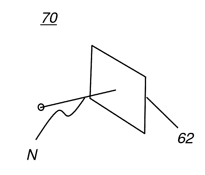
[0045]In the context of the present disclosure, the term “image” refers to multi-dimensional image data that is composed of discrete image elements. For 2-D images, the discrete image elements are picture elements, or pixels. For 3-D images, also termed volume images, the discrete image elements are volume image elements, or voxels. In the context of the present disclosure, the term “spline” is equivalent to a curve, free-form curve, or line.

[0046]In the context of the present disclosure, the term “index” refers to an alphanumeric character or string tha...


PUM


Abstract

A method for analyzing a subject tooth. The method includes obtaining volume image data including at least the subject tooth and segments the subject tooth from within the volume data according to one or more operator instructions. An index is generated that is indicative of a suspected fracture or lesion identified for the segmented subject tooth. The subject tooth is displayed with the suspected fracture or lesion highlighted. The generated index also displays.

Description

CROSS REFERENCE TO RELATED APPLICATIONS[0001]This application claims priority to U.S. Ser. No. 61 / 814,470, provisionally filed on Apr. 22, 2013, entitled “DETECTION OF TOOTH FRACTURES IN CBCT IMAGE”, in the name of Souza et al., incorporated herein in its entirety.FIELD OF THE INVENTION[0002]The invention relates generally to dental imaging, and in particular to a radiographic imaging apparatus for viewing volume images having highlighted fracture information for teeth.BACKGROUND[0003]Cone beam computed tomography (CBCT) is an X-ray imaging modality capable of acquiring three-dimensional information from the human anatomy with a lower radiation dose to the patient as compared to conventional medical computed tomography (CT) systems. Cone beam CT systems capture volumetric data sets by using a high frame rate digital radiography (DR) detector and an x-ray source. The detector and source are typically affixed to a gantry that rotates about the subject to be imaged, with the source dir...

Claims


Application Information

Patent Timeline
IPC IPC(8): G06T7/00
CPCG06T2207/30036G06T7/0012G06T2207/10081G06T2207/20096
Inventor SOUZA, ANDRERAY, LAWRENCE A.FALCAO, ALEXANDRE X.
Owner CARESTREAM DENTAL TECH TOPCO LTD
Why Eureka
Try Eureka