
Method and system for integrated radiological and pathological information for diagnosis, therapy selection, and monitoring

a integrated technology, applied in the field of integrating radiological and pathological information, can solve the problems of inability to effectively communicate, perform correlation and gather consensus of findings' concordance, and lack of access to larger sets of data with the known outcom

Active Publication Date: 2014-10-23
SIEMENS MEDICAL SOLUTIONS USA INC
View PDF3 Cites 39 Cited by
  • Summary
  • Abstract
  • Description
  • Claims
  • Application Information

AI Technical Summary

Benefits of technology

The present invention is a method and system that combines information from different medical tests such as MRIs, CT scans, PET scans, and ultrasounds to help diagnose and treat diseases. This is especially useful when a biopsy is the main way of analyzing a patient's condition. The invention takes into account the locations where the biopsy was taken and the results from the tests. It uses advanced technology and analysis to automatically read and evaluate the information from the tests.

Problems solved by technology

In the exemplary conventional workflow above, there it can be difficult to effectively communicate, perform correlation, and gather consensus of findings' concordance.
These difficulties are primarily due to physical separation, geographic constraints, time, resources, and labor.
Also, there is no access to larger sets of data with the known outcome, from which a clinician can draw resemblances and similarities for better final diagnosis and clinical decisions.
While the recently proposed system facilitates much need communication, it falls short in many aspects.
For example, it is nearly impossible to establish spatial correlation between biopsy samples and groups of pixels within radiological images, meaning that one cannot tell where the biopsies were taken relative to the anatomical information provided by the radiological images.
This can be a cause of discordance, for example, for micro-calcifications in breast cancer cases.
Another cause of discordance is inter and intra operator variability, which can cause the need for repeat biopsies.
When discordance between the radiological imaging and the histology cannot be resolve, an expensive surgical procedure may be required.

Method used


Image

  • Method and system for integrated radiological and pathological information for diagnosis, therapy selection, and monitoring
  • Method and system for integrated radiological and pathological information for diagnosis, therapy selection, and monitoring
  • Method and system for integrated radiological and pathological information for diagnosis, therapy selection, and monitoring

Examples


Embodiment Construction

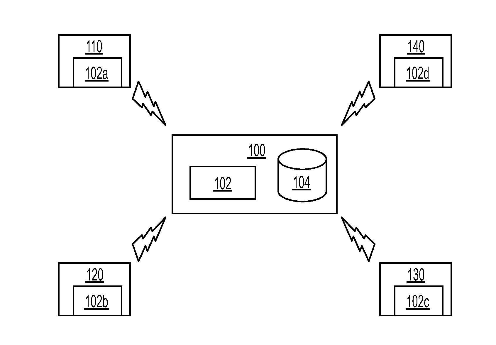
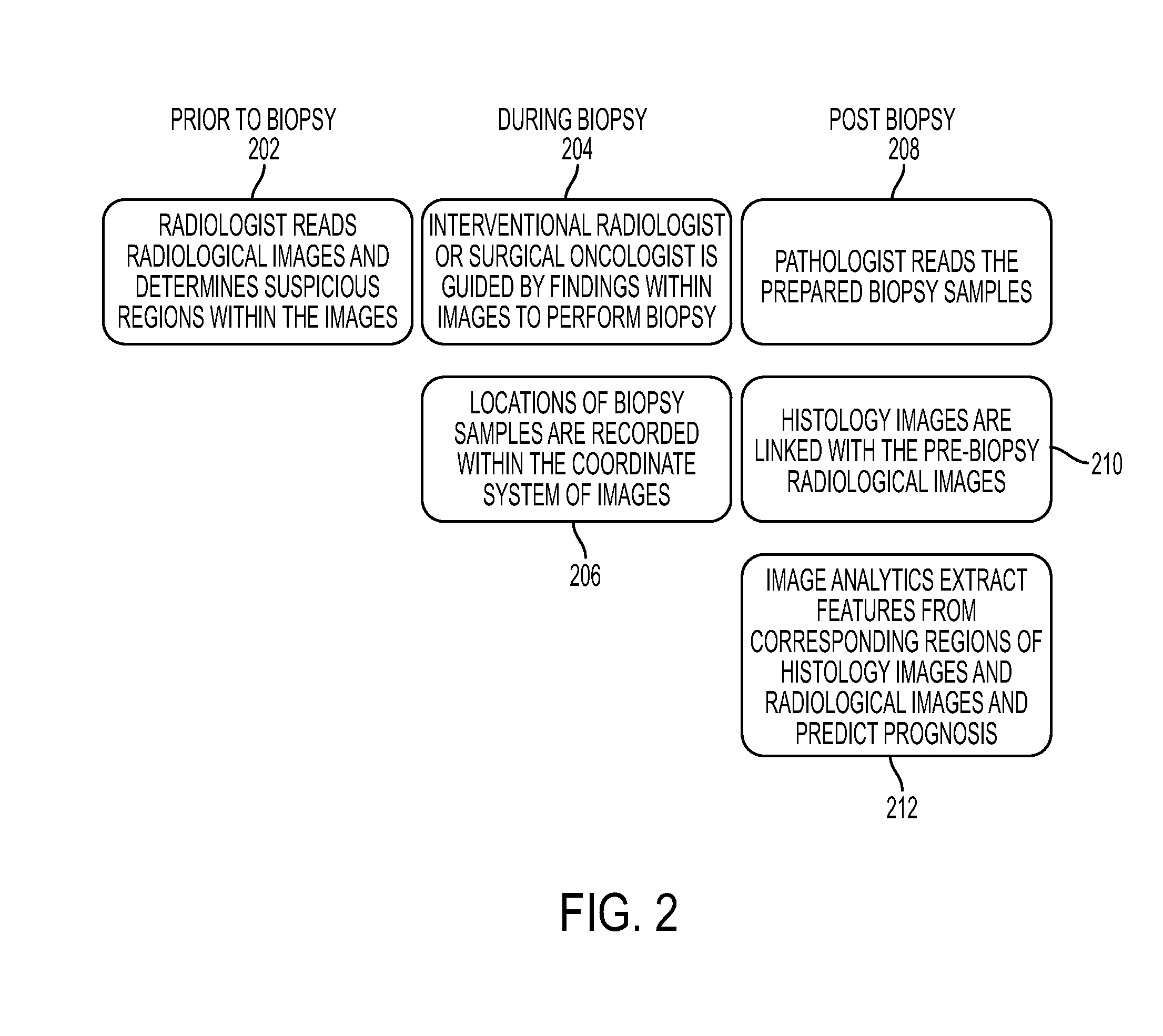
[0016]The present invention is directed to a method and system for integrating radiological and pathological information. Embodiments of the present invention are described herein to give a visual understanding of radiological and pathological information integration method. A digital image is often composed of digital representations of one or more objects (or shapes). The digital representation of an object is often described herein in terms of identifying and manipulating the objects. Such manipulations are virtual manipulations accomplished in the memory or other circuitry / hardware of a computer system. Accordingly, it is to be understood that embodiments of the present invention may be performed within a computer system using data stored within the computer system.

[0017]Accurate cancer diagnosis, therapy selection, and treatment and disease monitoring heavily depends on both information from radiological scans (e.g., magnetic resonance (MR), computed tomography (CT), positron e...


PUM


Abstract

A method and system for integrating radiological and pathological information for cancer diagnosis, therapy selection, and monitoring is disclosed. A radiological image of a patient, such as a magnetic resonance (MR), computed tomography (CT), positron emission tomography (PET), or ultrasound image, is received. A location corresponding to each of one or more biopsy samples is determined in the at least one radiological image. An integrated display is used to display a histological image corresponding to the each biopsy samples, the radiological image, and the location corresponding to each biopsy samples in the radiological image. Pathological information and radiological information are integrated by combining features extracted from the histological images and the features extracted from the corresponding locations in the radiological image for cancer grading, prognosis prediction, and therapy selection.

Description

[0001]This application claims the benefit of U.S. Provisional Application No. 61 / 525,929, filed Aug. 22, 2011, the disclosure of which is herein incorporated by reference.BACKGROUND OF THE INVENTION[0002]The present invention relates to integrating radiological and pathological information, and more particularly to integrating information from radiological scans, such as but not limited to magnetic resonance (MR), computed tomography (CT), positron emitted tomography (PET), and ultrasound (US) scans, and pathological information derived from biopsy samples for diagnosis, therapy selection, and treatment or disease monitoring.[0003]In conventional clinical workflows, physicians perform biopsies of suspicious lesions or masses. For example, a urologist may perform a transrectal random-core biopsy of the prostate gland in order to search for cancerous tissue samples in patients with high prostate-specific antigen (PSA) blood test results. Specimens from the biopsy are sent to a patholo...

Claims


Application Information

Patent Timeline
IPC IPC(8): G06T7/00
CPCG06T2207/30004G06T7/0012A61B6/12A61B6/463A61B6/467A61B8/0841A61B8/463A61B8/465A61B8/467A61B10/02G16H20/40G16H30/20G16H50/20
Inventor KAMEN, ALIGRADY, LEOPALADINI, GIANLUCAKHURD, PARMESHWARKUTTER, OLIVERCOMANICIU, DORIN
Owner SIEMENS MEDICAL SOLUTIONS USA INC
Learn More