

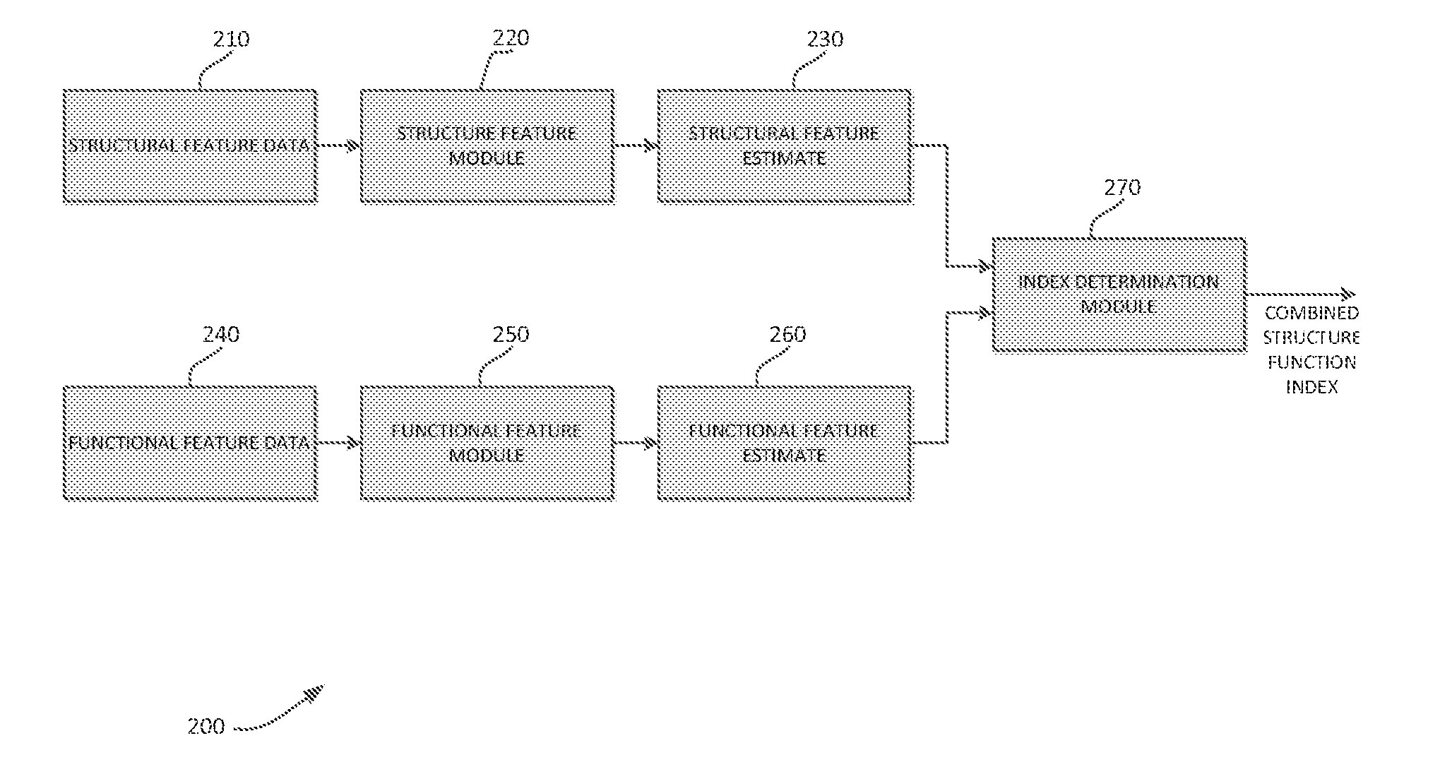
Systems and methods for determining retinal ganglion cell populations and associated treatments
a retinal ganglion cell and retinal ganglion cell technology, applied in the field of optic neuropathy, can solve the problems of increasing the risk of functional impairment of patients with severe damage, increasing the risk of vision-related quality of life, and more aggressive treatmen
- Summary
- Abstract
- Description
- Claims
- Application Information
AI Technical Summary
Benefits of technology
Problems solved by technology
Method used
Image

Examples
case examples
[0123]FIG. 18 illustrates an eye that had an estimated RGC count of 520 950 cells at the time of development of the initial visual field defect on SAP, corresponding to a 43% RGC loss compared with the healthy group. The defect was confirmed on subsequent tests based on the criterion of 3 consecutive abnormal fields with PSD, with P<5%. The optic disc photograph shows extensive neuroretinal rim loss in agreement with the RNFL loss assessed by SD-OCT, which showed an average RNFL thickness of 58 μm. Despite the extensive RGC loss, the visual field defect on the pattern deviation plot was apparently small with only an inferior cluster of abnormal points, although there was evidence of diffuse loss of sensitivity as indicated by the MD.
[0124]FIG. 19 shows an eye with an estimated RGC count of 800 369 at the time of development of the initial visual field defect, which corresponded to a 12% RGC loss compared with the healthy group. The optic disc photograph shows inferior neuroretinal r...
PUM
 Abstract
Description
Claims
Application Information