Diagnosis assistance apparatus, method and program

a technology of diagnostic assistance and equipment, applied in the field of diagnostic assistance equipment, method and program, can solve the problems of erroneous extraction of a small intestine as a part of the large intestine, high risk of erroneous extraction caused by organ deformation, and high risk of all plural three-dimensional images, so as to reduce erroneous extraction, easy and accurate organ extraction, the effect of reducing the risk of erroneous extraction

Active Publication Date: 2015-01-29
FUJIFILM CORP
View PDF6 Cites 7 Cited by
  • Summary
  • Abstract
  • Description
  • Claims
  • Application Information

AI Technical Summary

Benefits of technology

[0009]It is an object of the present invention to reduce a burden of manually correcting an organ region for plural three-dimensional images by a user when doctors or the like extracts the organ region from the plural three-dimensional images that have been obtained by imaging the same tubular structure organ of the same patient at the same time. The present invention aims at reducing such burden on the user by providing an improved diagnosis assistance apparatus, method and program. It is also an object of the present invention to easily and accurately extract the organ region from plural three-dimensional images.
[0020]According to the diagnosis assistance apparatus, method and program of the present invention, each of a first image and a second image obtained by imaging a region including the same tubular structure organ of the same subject at the same time are obtained. An organ region of the organ is extracted from the first image. The extracted organ region is displayed on a display screen. An input of an air region included in the first image and an input of exceeding or lacking portion information representing that the air region is a lacking portion or an exceeding portion of the organ are received. The received air region and the received exceeding or lacking portion information corresponding to the air region are obtained, as correction information. Positions are matched with each other by calculating position-matching information that correlates corresponding positions to each other between the first image and the second image. Further, the organ region extracted from the first image is corrected, based on the correction information, by adding a portion corresponding to the lacking portion or by deleting the exceeding portion. Further, at least an air region in the second image located at a position corresponding to the air region corresponding to the correction information is extracted, based on the position-matching information, as a part of the organ region in the second image. Therefore, erroneous extraction occurred at a position in the first image does not occur at a corresponding position in the second image. Hence, a user only needs to perform manual correction on the organ only in the first image, and does not need to perform manual correction on the same position of the organ region in the second image. In this way, it is possible to easily and accurately extract the organ from plural three-dimensional images.
[0021]Further, when an air region in the second image located at a position corresponding to the corrected organ region is extracted, based on the position-matching information, as the organ region in the second image, a user can extract an organ region in the second image corresponding to the organ region in the first image that reflects correction performed on a region having a risk of erroneous extraction. Therefore, it is possible to reduce erroneous extraction caused by deformation of the organ in the second image, and to easily and accurately extract the organ from the second image.

Problems solved by technology

Further, as an opposite case, a part of the small intestine is erroneously extracted as a part of the large intestine in some cases, because a boundary between the large intestine and an adjacent organ, such as the small intestine, is not sufficiently identifiable depending on the shape of the connection part between the large intestine and the adjacent organ.
When an organ is automatically extracted from plural three-dimensional images obtained by imaging the same tubular organ of the same patient at the same time, as in the supine position image and the prone position image, a risk of erroneous extraction caused by deformation of the organ, as described above, is high for all of the plural three-dimensional images.

Method used


Image

  • Diagnosis assistance apparatus, method and program
  • Diagnosis assistance apparatus, method and program
  • Diagnosis assistance apparatus, method and program

Examples


second embodiment

[0059]In a second embodiment, an example in which organ region Q in second image V2 located at a position corresponding to organ region P in first image V1 is extracted by using a different position matching method will be described. FIG. 5A is a diagram explaining processing for representing organ region P in first image V1 by a graph and processing for representing air regions B1 through B8 in second image V2 by a graph. FIG. 5B is a diagram explaining processing for extracting an organ region from second image V2.

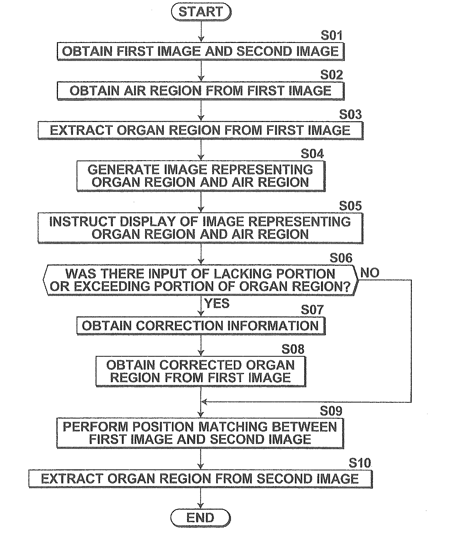
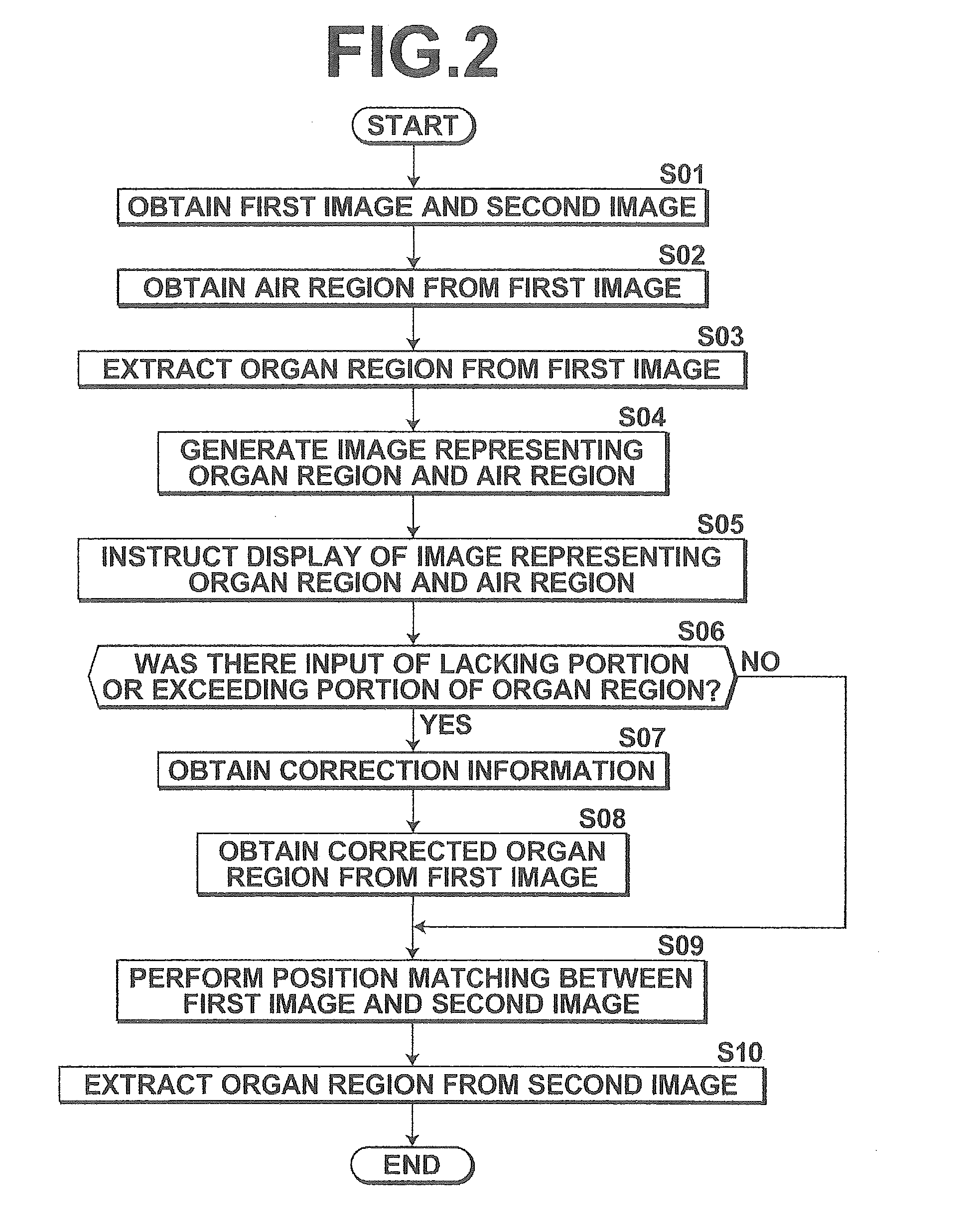
[0060]In the second embodiment, only position matching processing by the position matching unit 14 in S09 illustrated in FIG. 2 and processing for extracting an organ from second image V2 in S10 illustrated in FIG. 2 differ from the first embodiment. Regarding the other processing, the function of each unit and processing are same as those of the first embodiment. Therefore, in the second embodiment, the part different from the first embodiment will be mainly described. ...

first embodiment

[0062]Next, the organ region extraction unit 12 performs the following processing, as processing corresponding to processing for extracting an organ region from second image V2 (S10) in the First, the organ region extraction unit 12 creates a first graph structure (Gpa, Gpb, Gpc in FIG. 5A) representing corrected organ region P (Pa, Pb, Pc), and which is defined by plural nodes and at least one edge connecting the nodes. Here, thinning is performed on organ regions Pa, Pb, Pc, and a segment corresponding to a core line of the organ region is extracted. Further, ends of the segment are defined as nodes, and a node dividing the segment with predetermined intervals is defined. Further, the divided segments are defined as plural edges. Accordingly, first graph structures Gpa, Gpb, Gpc representing segments are generated.

[0063]Further, the organ region extraction unit 12 obtains plural air regions (B1 through B8 in FIG. 5A) included in second image V2, and creates plural second graph st...


PUM


Abstract

A first-image and a second-image representing the same organ of the same subject imaged at the same time are obtained, and an organ-region is extracted from the first-image. The extracted organ-region is displayed on a display screen. An input of an air-region included in the first-image and an input of exceeding or lacking portion information representing that the air-region is a lacking portion or an exceeding portion of the organ are received, and the received air-region and the received exceeding or lacking portion information corresponding to the air-region are obtained, as correction information. Corresponding positions are matched with other between the first-image and the second-image. The organ-region extracted from the first-image is corrected based on the correction information. At least an air-region in the second-image located at a position corresponding to the air-region corresponding to the correction information is extracted, as a part of the organ-region in the second-image.

Description

CROSS-REFERENCE TO RELATED APPLICATIONS[0001]The present application claims priority under 35 U.S.C. §119 to Japanese Patent Application No. 2013-155378, filed on Jul. 26, 2013. The above application is hereby expressly incorporated by reference, in its entirety, into the present application.BACKGROUND OF THE INVENTION[0002]1. Field of the Invention[0003]The present invention is appropriately applicable to medical fields, and relates to an apparatus, method and computer program for assisting a user in image-based diagnosis performed based on three-dimensional image data.[0004]2. Description of the Related Art[0005]In recent years, high quality three-dimensional image data became obtainable by an advance in modalities, such as a multi-slice CT (MDCT: Multi Detector Computer Tomography). In image-based diagnosis using such image data, not only a high definition tomographic image but also a virtual pseudo-three-dimensional image of a subject to be examined became used. Further, as disc...

Claims


Application Information

Patent Timeline
Patent Type & Authority Applications(United States)
IPC IPC(8): G06T5/50G06T7/00G06T3/00
CPCG06T5/50G06T3/0068G06T2207/30028G06T2207/10081G06T2207/20216G06T7/0012G06T2207/10088G06T2207/20036G06T2207/20072G06T2207/20101G06T2207/20128G06T7/11G06T7/162G06T7/187
Inventor IHARA, SATOSHI
Owner FUJIFILM CORP
Why Eureka
Try Eureka