

Diagnosis and treatment of traumatic brain injury
a brain injury and traumatic brain technology, applied in the field of diagnosis and treatment of traumatic brain injury, can solve the problems of toxic ccl20 to cultured oligodendrocytes
- Summary
- Abstract
- Description
- Claims
- Application Information
AI Technical Summary
Benefits of technology
Problems solved by technology
Method used
Image

Examples
example 1
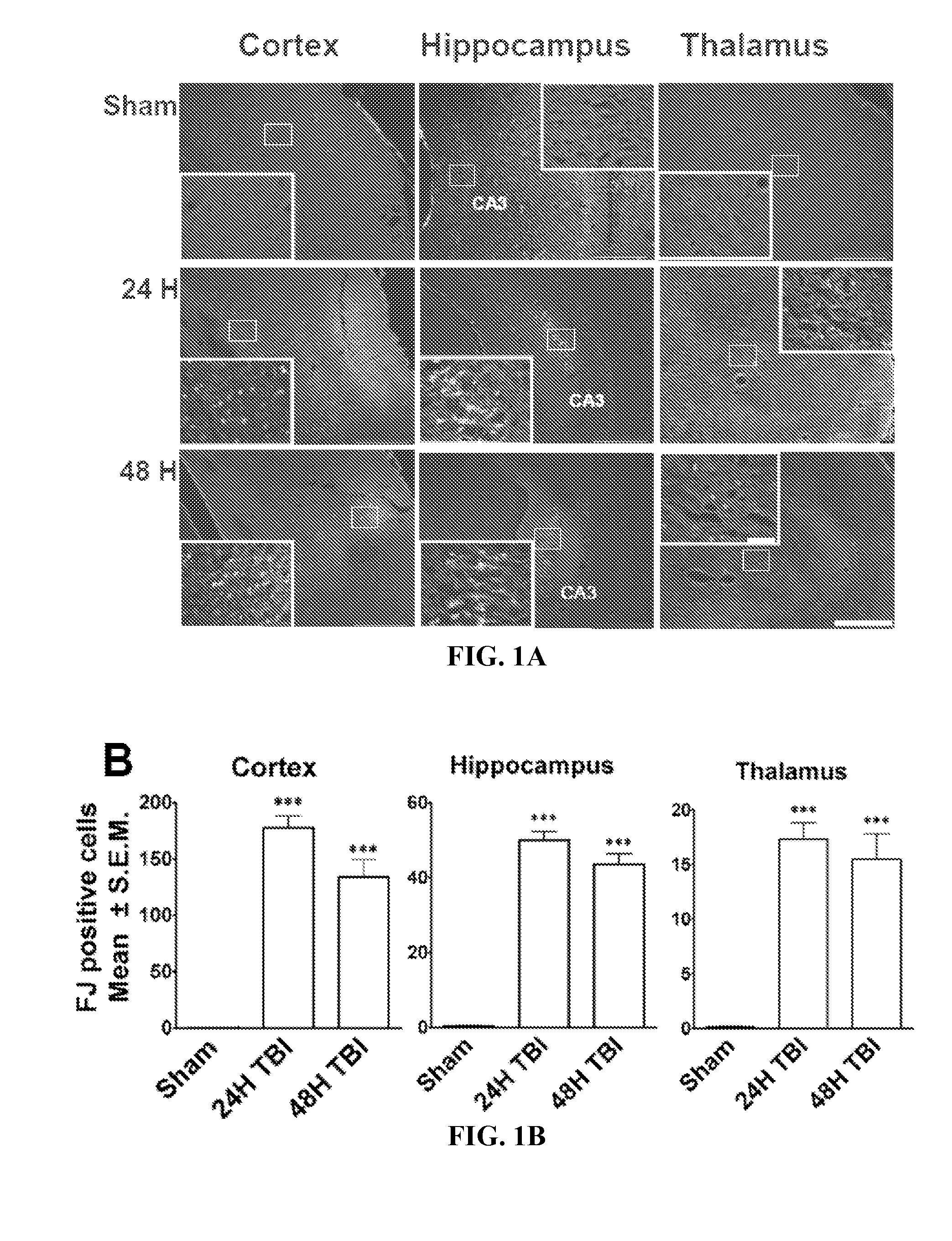
Regional Distribution of Neurodegeneration after TBI
[0193]To date, the assessment of TBI injury has been inconsistent across the laboratories. In addition, there is a lack of reliable, quantitative approaches for assessing neural injury. These have impeded efforts to develop novel treatments for TBI.
[0194]This Example conducts a detailed investigation throughout the brain to determine which regions of the brain exhibit consistent, prominent neurodegeneration. Briefly, rats were subjected to mild LFPI, and the brains were sectioned and stained with Fluoro-Jade. FIG. 1 shows a consistent profile, where the majority of Fluoro-Jade-positive cells were found within the cortex, hippocampus and thalamus. Cortical Fluoro-Jade was ubiquitous and was present at various levels throughout the brain. Hippocampal FJ staining was localized to the pyramidal cell layers (FIG. 1), while some diffuse labelling throughout the general structure was also evident. The thalamic staining was diffuse and spa...
example 2
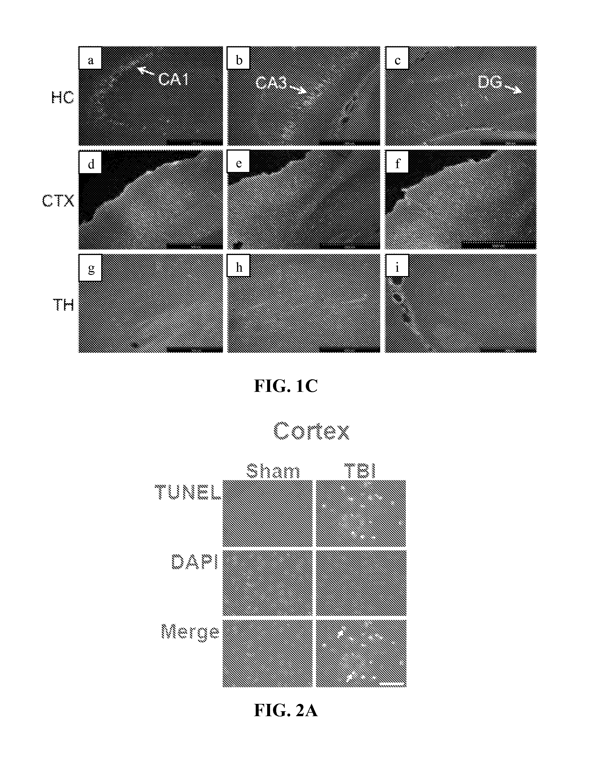
Mild TBI Induced Internucleosomal DNA Fragmentation in the Cortex and Hippocampus
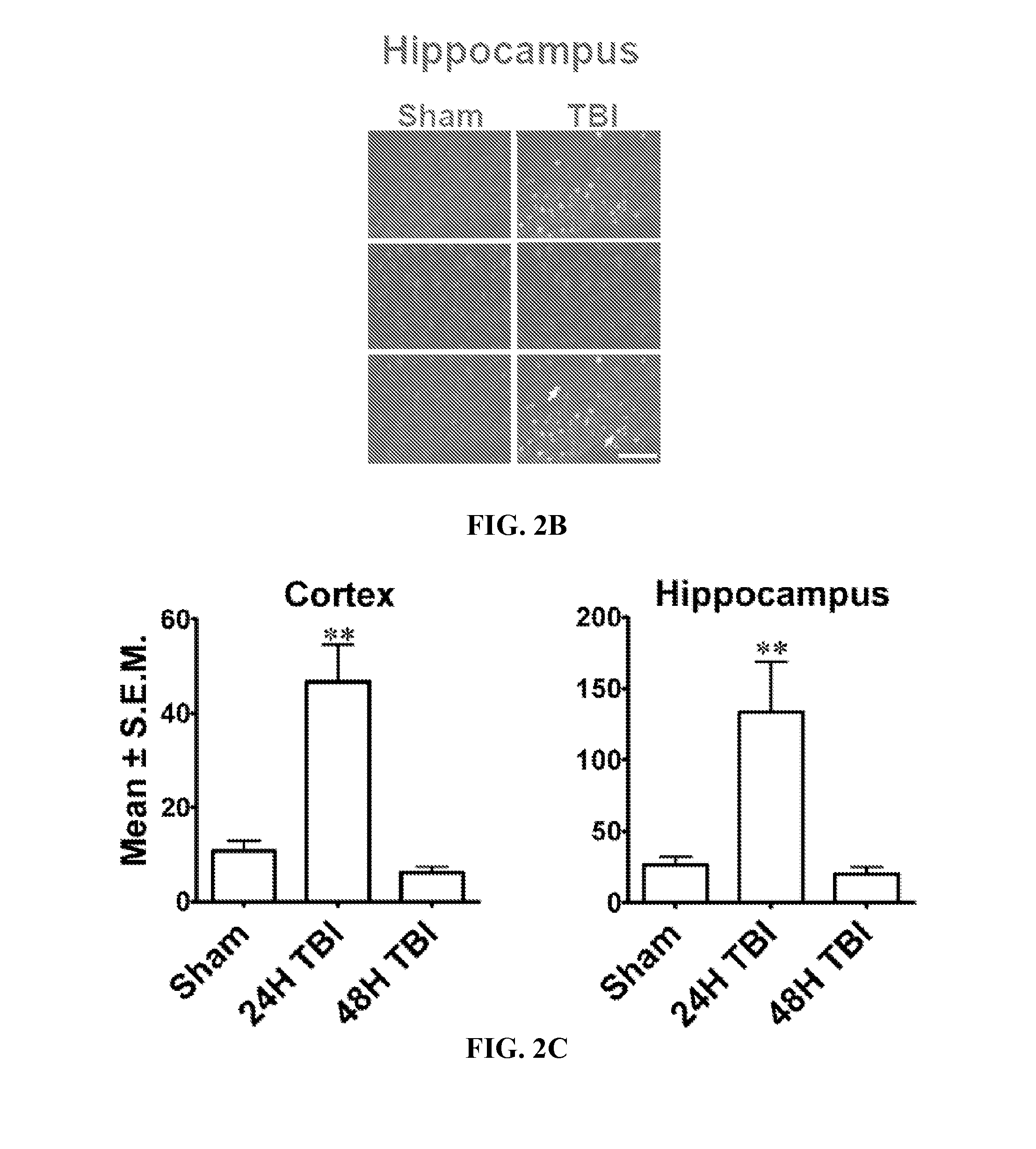
[0196]Internucleosomal DNA fragmentation, an important marker for apoptotic cells, was assessed by the Terminal Deoxynucleotidyl Transferase Biotin-dUTP Nick End Labeling (TUNEL) histochemistry. Few TUNEL-positive cells were detected in the contralateral hemisphere. While the ipsilateral thalamus showed sparse TUNEL staining in some sections, this was not a consistent finding throughout the experiment. The majority of TUNEL-stained nuclei were detected at 24 h post-TBI in the ipsilateral cortex (FIG. 2A) and hippocampus (FIG. 2B), while sections from sham-operated controls were predominantly devoid of TUNEL staining in these regions (FIG. 2A-2B) and showed only background levels of fluorescence. By 48 h after TBI, sections showed very few TUNEL-positive cells in the cortex and hippocampus and resembled sham-operated controls. Quantitation revealed a significant increase in TUNEL-positive cells in both c...
example 3
Activation of Microglia in the Brain Following Mild TBI
[0197]Isolectin-IB4, a 114 kD protein isolated from the seeds of the African legume—Griffonia simplicifolia, has been shown to have a strong affinity for brain microglial cells. To elucidate the local inflammatory response following mild TBI, Alexafluor 488 conjugated IB4 was used to label activated microglia in the rat brains (FIG. 3). While IB4 labelling was primarily restricted to the ipsilateral hemisphere, sparse labelling was detected within the contralateral hippocampus (data not shown). IB4-positive cells were abundant in the hippocampus, especially in the dentate gyrus (FIG. 3A). Microglia cells were also found in the cortex and thalamus following TBI.
[0198]CD11b, an activated microglial marker, was also found in the cells of the cortex and hippocampus (dentate gyrus, FIG. 3A) of the ipsilateral side. Confocal microscopy revealed that most but not all IB4+ cells in the cortex or hippocampus were also CD11b+ (FIG. 3A). Q...
PUM
| Property | Measurement | Unit |
|---|---|---|
| Time | aaaaa | aaaaa |
| Biological properties | aaaaa | aaaaa |
Abstract
Description
Claims
Application Information