Region extraction system

a region extraction and processing system technology, applied in image enhancement, diagnostic recording/measuring, instruments, etc., can solve the problems of increasing the labor burden of a doctor in tumor diagnosis, and achieve the effect of convenient region extraction, high precision, and easy extraction

Inactive Publication Date: 2015-02-19
HITACHI LTD
View PDF1 Cites 2 Cited by
  • Summary
  • Abstract
  • Description
  • Claims
  • Application Information

AI Technical Summary

Benefits of technology

[0014]According to the region extraction system of the present invention, when the luminance value distribution of a region concerned is to be estimated, it becomes possible to estimate the luminance value distribution of the region concerned with higher precision by using the luminance value distribution of a region which is similar to it in luminance value and is readily extracted and thereby the problem of false positives in region extraction can be overcome. Thereby, it be

Problems solved by technology

Since it becomes possible to acquire a large number of images, in particular, in an X-ray CT (Computed Tomography) device, an immense amount of labor has become necessary for a

Method used


Image

  • Region extraction system
  • Region extraction system
  • Region extraction system

Examples


embodiment 1

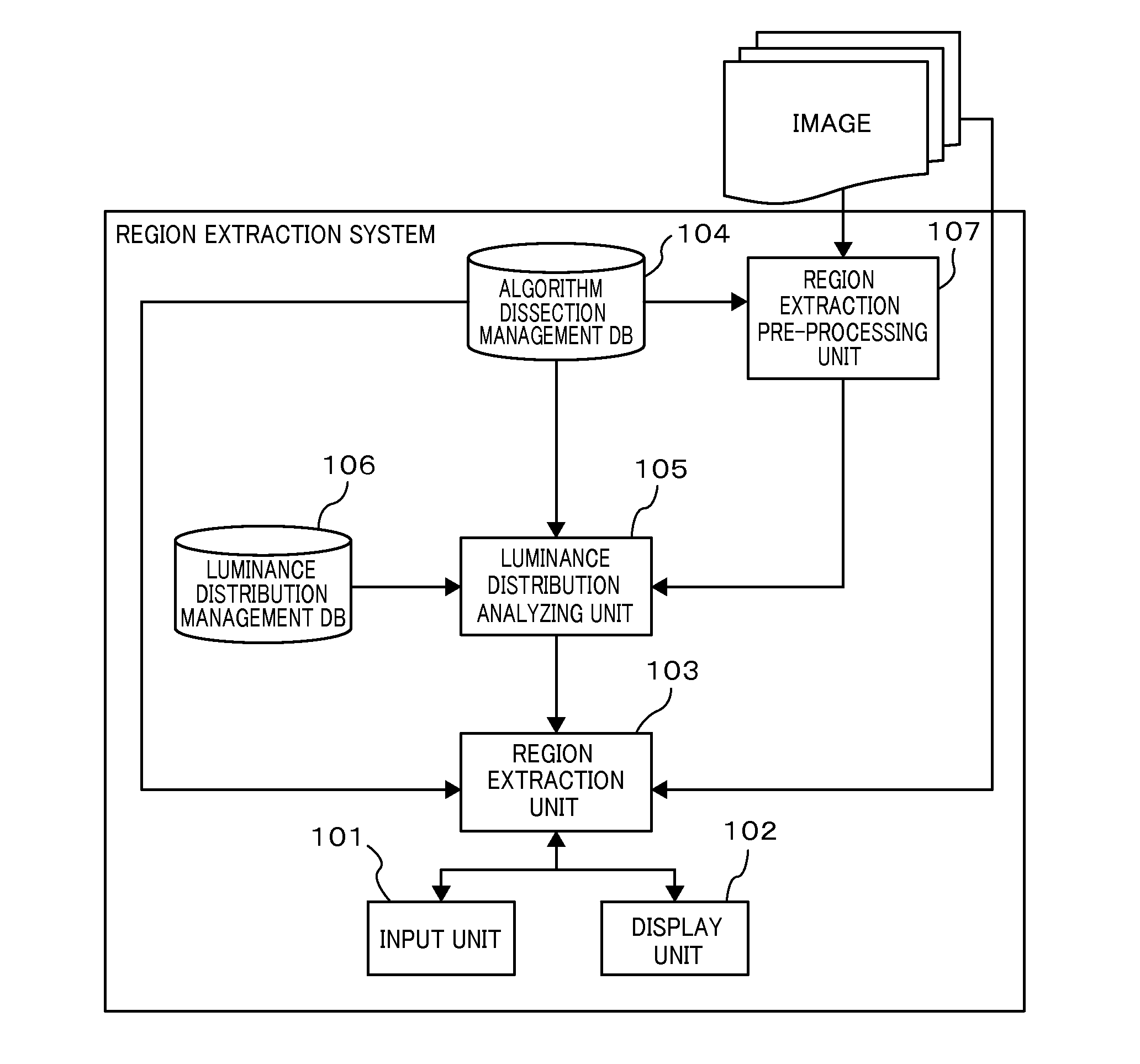
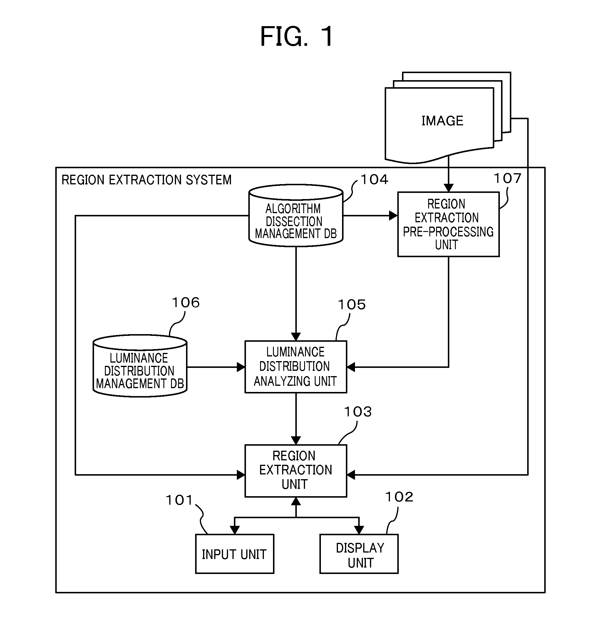
[0034]FIG. 1 is a configuration diagram of a region extraction system in the present invention. The region extraction system shown in FIG. 1 is configured by an input unit 101, a display unit 102, a region extraction unit 103, an algorithm dissection management database 104, a luminance distribution analyzing unit 105, a luminance distribution management database 106 and a region extraction pre-processing unit 107.

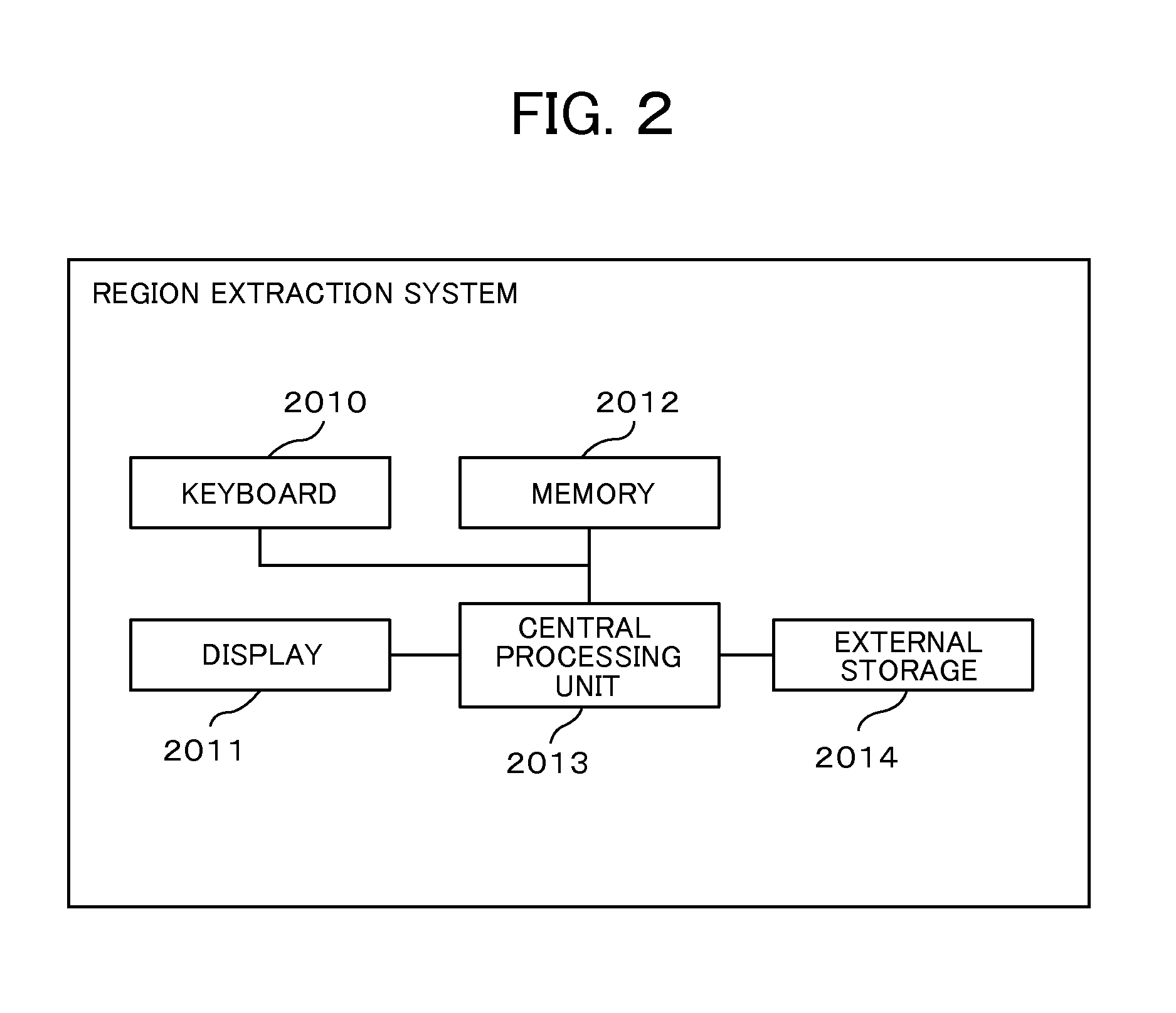
[0035]A hardware configuration of the present system will be described. A configuration diagram of hardware implementing the region extraction system in the present invention is shown in FIG. 2. The algorithm dissection management database 104 and the luminance distribution management database 106 are configured by an external storage 2014 and so forth represented by an HDD (Hard Disk Drive) device. The region extraction unit 103, the luminance distribution analyzing unit 105 and the region extraction pre-processing unit 107 can implement various kinds of processing by dev...

embodiment 2

[0050]FIG. 11 is a second configuration diagram of the region extraction system in the present invention. The processing method of the region extraction unit 103 is altered by newly adding an information system coordination unit 108 to the configuration diagram shown in FIG. 1 particularly for the purpose of acquiring the region of interest by coordinating with another information system such as the electronic medical record system and so forth. Although in the present embodiment, it coordinates with the electronic medical record system, it may coordinate with a system that manages the region of interest such as a radiological information system and so forth. The information system coordination unit 108 can implement various kinds of processing by developing / starting the predetermined program in the central processing unit 2013, the memory 2012 and so forth shown in FIG. 2. In addition, in FIG. 11, the electronic medical record system has at least an electronic medical record databa...

embodiment 3

[0055]FIG. 15 is a second flowchart showing detailed processing (S304) of the luminance distribution analyzing unit 105 in the present invention. Comparing it with the flowchart shown in FIG. 8, the processing of S3043 is replaced with that of S3045 in order to cope with a situation that there exist plural choices of algorithms and parameters to be acquired from the algorithm dissection management database 104. Thus, a flow of processing focusing on S3045 will be described by using a concrete example of the algorithm dissection management database 104 shown in FIG. 16. First, in S3042, in a case where the region of interest is the HCC, sets of the application algorithms and the parameter patterns for extracting the intrahepatic blood vessel, the portal vein and the HCC and sets of the application algorithms and the parameter patterns for extracting the intrahepatic blood vessel and the portal vein are acquired from the algorithm dissection management database 104 shown in FIG. 16.

[0...


PUM


Abstract

Provided is a region extraction system that uses an image data analysis process, which has overcome the problem of false positives in region extraction and assists in more accurate region extraction. A luminance distribution-analyzing unit (105) is used to determine a parameter of unknown quantity for extracting a region of interest. The luminance distribution-analyzing unit (105) searches a luminance distribution management database (106) for a region, which has a luminance distribution similar to the region of interest and is easily extracted, as a similar area. The region of the similar area is extracted using a region extraction pre-processing unit (107) and the value of the parameter of unknown quantity is determined on the basis of an image feature value calculated from the region extraction results.

Description

TECHNICAL FIELD[0001]The present invention relates to an analysis processing system for medical image data.BACKGROUND ART[0002]The number of images to be acquired in one checkup has been enormously increased and loads on diagnosticians and radiologists have been more and more increased with development of medical image diagnostic devices. Since it becomes possible to acquire a large number of images, in particular, in an X-ray CT (Computed Tomography) device, an immense amount of labor has become necessary for a doctor in tumor diagnosis. Since an increase in labor taken for such diagnosis may induce an oversight and an erroneous diagnosis, it is demanded to reduce it as much as possible. Therefore, a technology (CAD: Computer Aided Detection) that automatically extracts a region concerned such as a tumor and so forth and a device with that technology loaded are proposed.[0003]In Non-Patent Literature 1, for the purpose of extracting a liver tumor, a technique of applying a Region G...

Claims


Application Information

Patent Timeline
IPC IPC(8): G06K9/46G06T7/00
CPCG06K9/4661G06K2009/4666G06T7/0012A61B5/4244G06T2207/20076G06T2207/30056G06T7/11
Inventor YUI, SHUNTAROMATSUZAKI, KAZUKIMIYAKOSHI, JUNICHI
Owner HITACHI LTD
Why Eureka
Try Eureka