Quantitative in situ characterization of biological samples

a biological sample and quantitative technology, applied in the field of immuno-profiling of biological samples, can solve the problems of limited sample amount, loss of original location information, and limited ability to determine relative characteristics of targets

Active Publication Date: 2015-05-14
LEICA MICROSYSTEMS CMS GMBH
View PDF2 Cites 21 Cited by
  • Summary
  • Abstract
  • Description
  • Claims
  • Application Information

AI Technical Summary

Benefits of technology

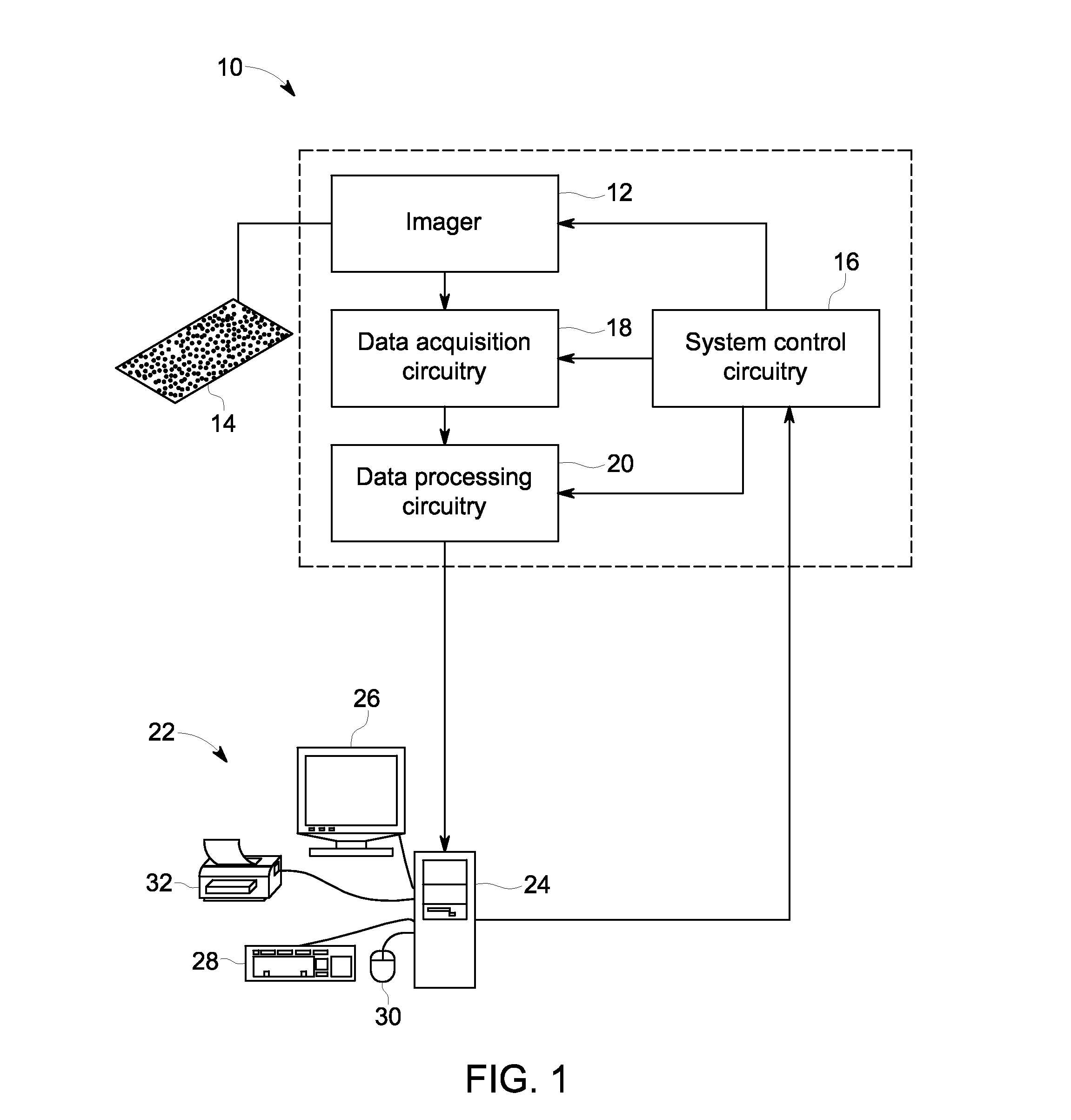
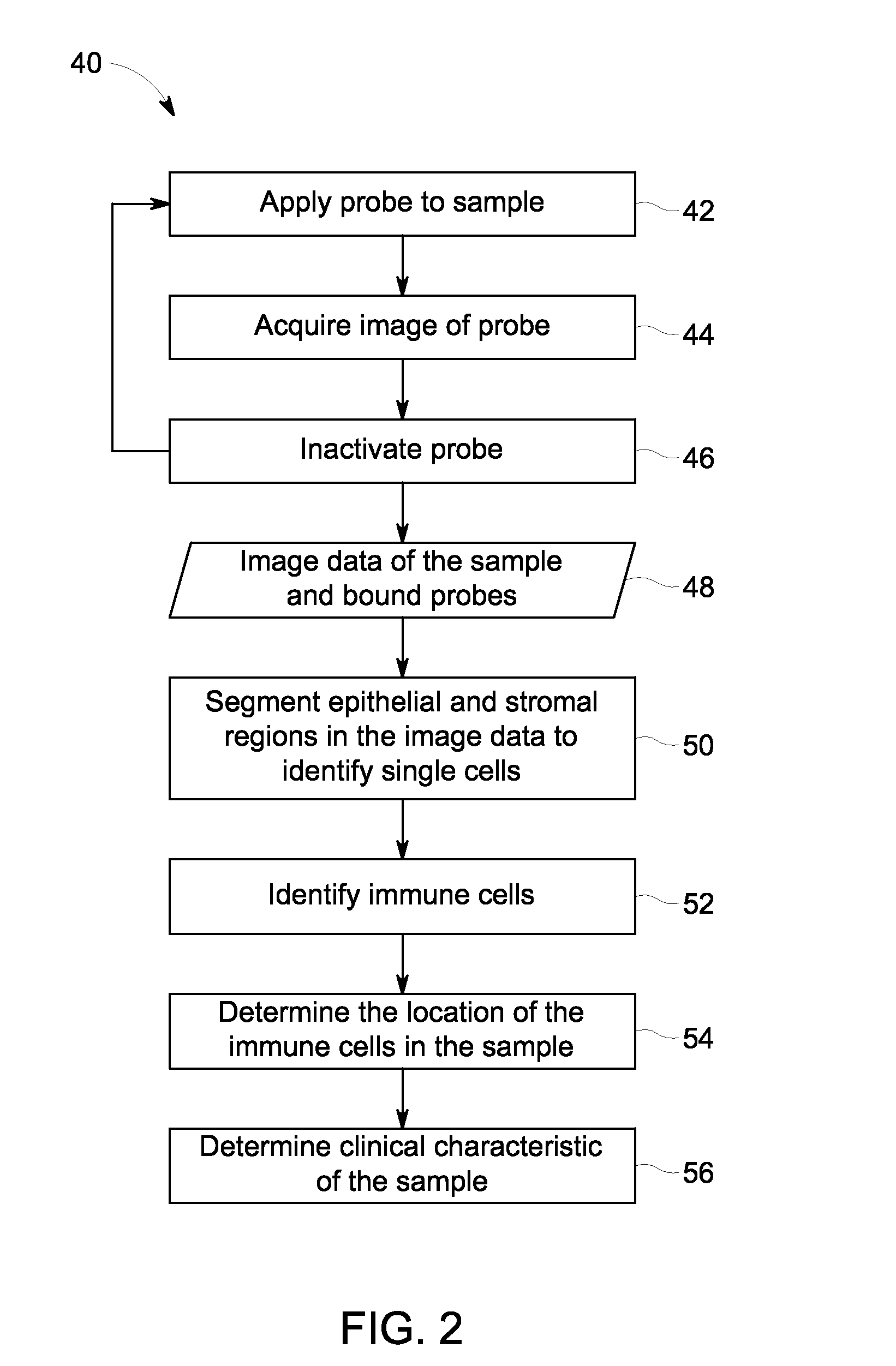
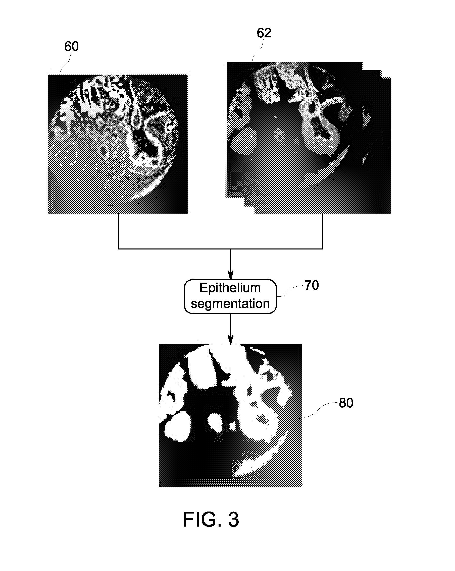
[0004]In one embodiment, a method for determining infiltration of multiple immune cell populations in a biological sample is provided. The method includes applying a plurality of probes to a biological sample in a sequential manner; acquiring image data of the biological sample representative of the respective plurality of probes bound to a respective plurality of targets in the biological sample, wherein at least one of the plurality of probes comprises an epithelium probe, a membrane probe, a cytoplasm probe, or nuclear probe specific for a cell nucleus and wherein at least one of the plurality of probes comprises an immune probe specific for an immune marker; segmenting epithelial and stromal regions of the sample to identify single cells within each region, wherein identifying individual cells in the epithelial region or the stromal region comprises using image data representative of the epithelium probe, the membrane probe, the cytoplasm probe, or the nuclear probe and wherein identification of cells in the stromal region comprises segmenting the epithelial region of the sample to generate an epithelial mask and classifying regions not contained within the epithelial mask as one or more of the stromal region or background; identifying immune cells among the single cells based on the image data representative of the immune marker, wherein identifying comprises reclassifying single cells in the sample as immune cells based on a signal from the immune probe above a threshold value; and determining a distribution, location, and type of a plurality of immune cells in the biological s...

Problems solved by technology

Many of the current techniques may detect a presence or concentration of biological targets without maintaining information about original location of those targets within the sample.
For example, certain techniques involve processing the sample in such a way that the original location information is lost.
Other techniques may involve assessing only a limited number of targets from a given sample Further analysis of targ...

Method used


Image

  • Quantitative in situ characterization of biological samples
  • Quantitative in situ characterization of biological samples
  • Quantitative in situ characterization of biological samples

Examples


examples

[0047]Multiplexing technology from GE was applied on slides consisting of colon, lung or melanoma tissue microarray (TMA) to map multiple protein expressions of non-neoplastic cells (panel of immune biomarkers and segmentation markers) in the tumor microenvironment. Multiple targets were applied on a single tissue section.

[0048]Lung TMA: Tristar 69571118 NSCLC prognosis tissue array was used which had tissues from 144 European male smokers along with patient information. Slides (#1167) containing various clinic pathological information like histologic types, stage, age, sex, predominantly from squamous and adenocarcinoma were selected for immune profiling. Melanoma TMA: Pantomics MEL961 Melanoma tissue array contained 48 cases of primary and metastatic cancer in duplicates. Colorectal Cancer TMA: Colorectal cancer tissue microarrays were provided by Clarient INC. The colorectal cancer cohort was collected from the Clearview Cancer Institute of Huntsville, Ala. from 1993 until 2002, ...


PUM


Abstract

The present disclosure relates to characterization of biological samples. By way of example, a biological sample may be contacted with a plurality of probes specific for targets in the sample, such as probes for immune markers and segmenting probes. Acquired image data of the sample may be used to segment the images into epithelial and stromal regions to characterize individual cells in the sample based on the binding of the probes. Further, the biological sample may be characterized by a distribution, location, and type of a plurality of the characterized cells.

Description

BACKGROUND[0001]The subject matter disclosed herein relates to immune profiling of biological samples. More particularly, the disclosed subject matter relates to determining one or more immune cell characteristics of the biological sample, including a distribution, type and / or location of immune cells within the sample.[0002]Various methods may be used in biology and in medicine to observe different targets in a biological sample. For example, analysis of proteins in histological sections and other cytological preparations may be performed using the techniques of histochemistry, immunohistochemistry (IHC), or immunofluorescence.[0003]Many of the current techniques may detect a presence or concentration of biological targets without maintaining information about original location of those targets within the sample. For example, certain techniques involve processing the sample in such a way that the original location information is lost. Other techniques may involve assessing only a l...

Claims


Application Information

Patent Timeline
IPC IPC(8): G01N33/574
CPCG01N33/57492G01N1/30G01N33/54393G06T2207/30024G06T7/0012G06T2207/10024G06V20/698G01N33/56977G01N33/56972
Inventor BHAUMIK, SRABANIGERDES, MICHAEL JOHNPANG, ZHENGYUGINTY, FIONASEVINSKY, JR., CHRISTOPHER JAMESLI, QINGSANTAMARIA-PANG, ALBERTOSUI, YUNXIADESAI, KEYUR HEMANTKUMAR
Owner LEICA MICROSYSTEMS CMS GMBH
Why Eureka
Try Eureka