
Wearable Electronic Device for Enhancing Visualization During Insertion of an Invasive Device

a technology of enhancing visualization and wearable electronic devices, which is applied in the direction of ultrasonic/sonic/infrasonic image/data processing, diagnostics using spectroscopy, applications, etc., can solve the problems of inability to accurately and accurately perform incomplete or incorrect procedures, loss of patient data, and increased so as to reduce the risk of medication infusion and improve clinical workflow

Active Publication Date: 2015-07-30
BECTON DICKINSON & CO
View PDF10 Cites 40 Cited by
  • Summary
  • Abstract
  • Description
  • Claims
  • Application Information

AI Technical Summary

Benefits of technology

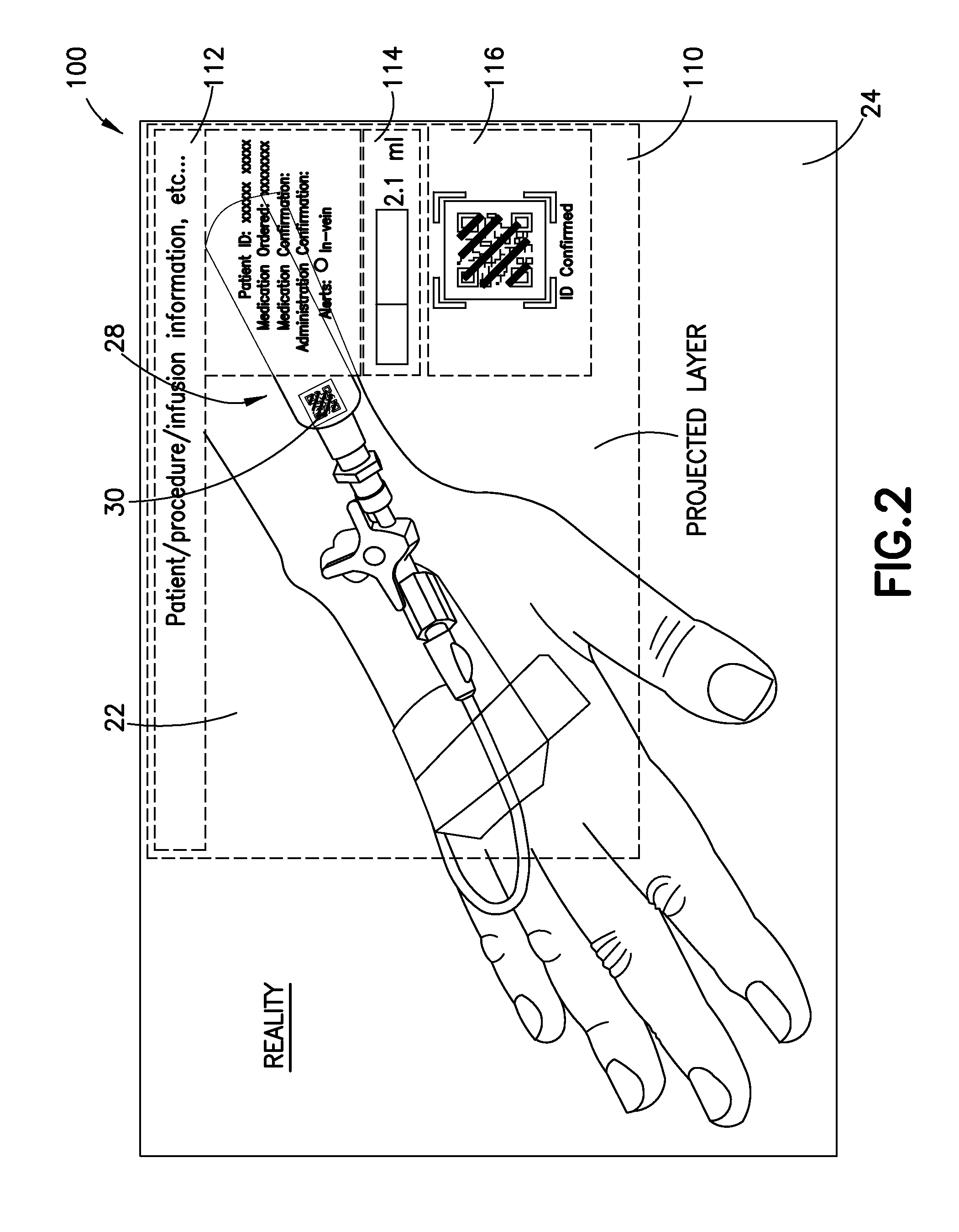
The system and method described in this patent reduce the risk of errors when giving medication to patients. It helps healthcare professionals quickly and accurately identify, confirm, and document the delivery of fluids to patients in real-time. The system is designed to be used hands-free and can help establish a chain of custody for specimens collected from patients. It also provides enhanced visualization to improve procedures that require guidance, such as insertion of a vascular access device. The system includes a virtual trace that can be seen over the actual access site and a sub-dermal illuminator to increase visibility of sub-dermal structures.

Problems solved by technology

However, even such electronic databases still require manual entry of data either by the clinician or technician, or later data entry based on contemporaneous notes taken by the clinician or technician.
The numerous manual steps required before, during, and after fluid sampling or fluid delivery procedures introduce opportunities for user error.
User errors may lead to incomplete or incorrect procedures being performed or may result in lost patient data.
For example, the clinician or technician may inject an incorrect fluid volume, incorrect fluid type or concentration, or may not obtain a sufficient volume of fluid sample for the tests being performed.
The medical clinician or technician may also forget to correctly document that a fluid sample was obtained or under what conditions the sample was obtained.
Furthermore, the clinician or technician may fail to correctly record which patient provided a particular fluid sample.
These problems may harm the patient or, at minimum, may require that certain fluid sample procedures must be repeated.

Method used


Image

  • Wearable Electronic Device for Enhancing Visualization During Insertion of an Invasive Device
  • Wearable Electronic Device for Enhancing Visualization During Insertion of an Invasive Device
  • Wearable Electronic Device for Enhancing Visualization During Insertion of an Invasive Device

Examples


Embodiment Construction

[0039]The following description is provided to enable those skilled in the art to make and use the described embodiments contemplated for carrying out the invention. Various modifications, equivalents, variations, and alternatives, however, will remain readily apparent to those skilled in the art. Any and all such modifications, variations, equivalents, and alternatives are intended to fall within the spirit and scope of the present invention. However, it is to be understood that the invention may assume various alternative variations and step sequences, except where expressly specified to the contrary. It is also to be understood that the specific devices and processes illustrated in the attached drawings, and described in the following specification, are simply exemplary embodiments of the invention. Hence, specific dimensions and other physical characteristics related to the embodiments disclosed herein are not to be considered as limiting. For the purpose of facilitating underst...


PUM


Abstract

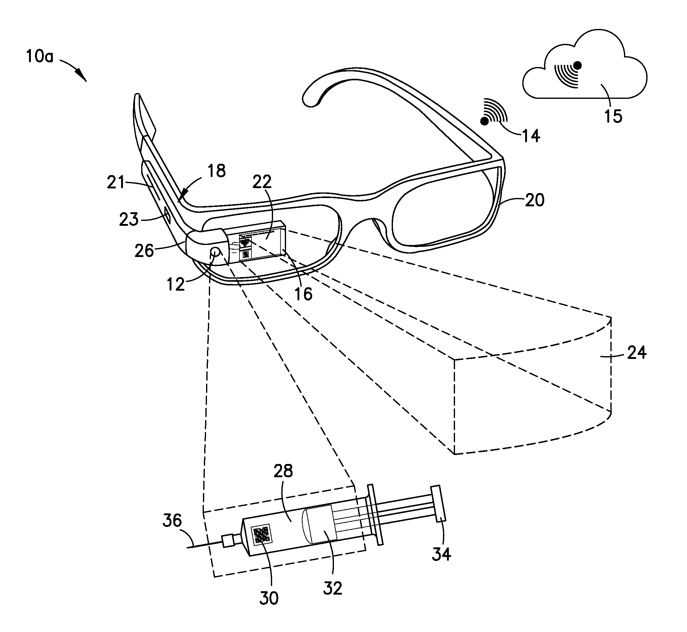
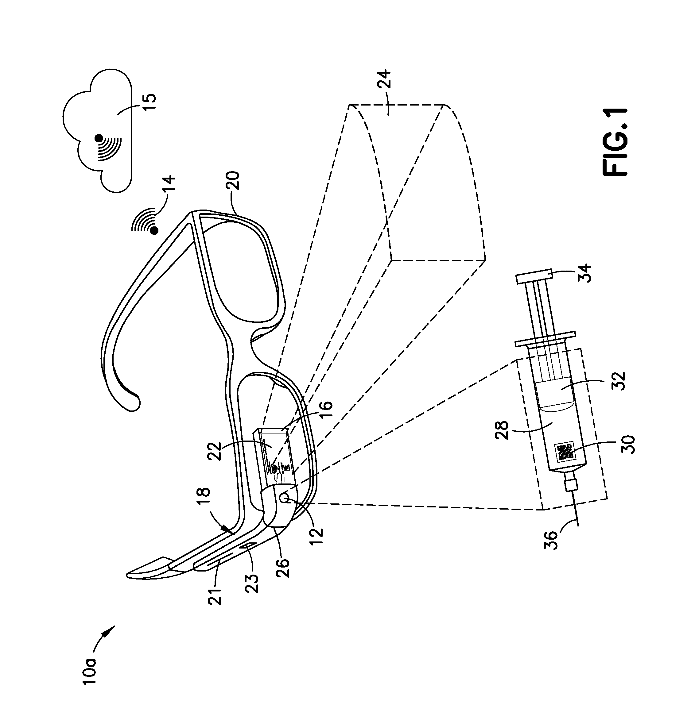
A wearable electronic device configured to be worn by a user while performing an invasive procedure for enhancing visualization of desired anatomical structures is provided. The wearable electronic device includes: a housing; at least one imaging sensor associated with the housing; and a visual display integrally formed with or associated with the housing. The device is configured to acquire an image of an invasive access site of a patient with the at least one imaging sensor, process the image to determine a location of a desired anatomical structure, and display a virtual trace of the location to the user via the visual display.

Description

CROSS-REFERENCE TO RELATED APPLICATION[0001]The present application claims priority to U.S. Provisional Application 61 / 933,049 entitled “Wearable Electronic Device for Enhancing Visualization During Insertion of a Vascular Access Device” filed Jan. 29, 2014, the entire disclosure of which is hereby incorporated by reference in its entirety.BACKGROUND OF THE INVENTION[0002]1. Field of the Invention[0003]The present invention relates to systems and methods for enhancing visualization of an invasive procedure requiring procedural guidance, such as providing enhanced visualization of a vein during an insertion procedure using a vascular access device, and, more particularly, to systems and methods that operate in a hands-free manner using a wearable electronic device.[0004]2. Description of Related Art[0005]Blood sampling is a common health care procedure involving the withdrawal of at least a drop of blood from a patient. Blood samples are commonly taken from hospitalized, homecare, an...

Claims


Application Information

Patent Timeline
IPC IPC(8): A61B19/00A61B8/08A61B5/055A61B17/00A61B5/00
CPCA61B19/26A61B2019/5287A61B19/56A61B19/44A61B19/5202A61B17/00234A61B5/489A61B5/746A61B5/055A61B5/0035A61B5/0075A61B8/0841A61B2019/262A61B2019/5274A61B2019/5265A61B2019/5293A61B2019/444A61B2019/521A61B2017/00207A61B2019/5255A61B2017/00292A61B2019/5276A61B2019/5231A61B2019/5236A61B2019/442A61B2019/448A61B2019/446A61B19/5244A61B5/15003A61M5/427A61B5/150748A61B5/150786A61B5/150793A61B5/15087A61B5/743A61B5/153A61M5/31568A61M2205/3306A61M2205/3379A61M2205/3553A61M2205/3561A61M2205/507A61M2205/52A61M2205/8206G16H40/63G16H20/40A61B90/35
Inventor BURKHOLZ, JONATHAN KARLO'BRYAN, JEFFSHEVGOOR, SIDDARTH K.MA, YIPING
Owner BECTON DICKINSON & CO
Learn More