Multiplex assay for improved scoring of tumor tissues stained for pd-l1

a tumor tissue and multi-assay technology, applied in material analysis, instruments, library creation, etc., can solve the problems of difficult differentiation of these two cell types, difficult to achieve anti-pd-1 and anti-pd-l1 directed therapies, etc., to improve the ability of samples to be scored more quickly, improve the degree of reproducibility, and improve the effect of scoring accuracy

Inactive Publication Date: 2015-12-03
VENTANA MEDICAL SYST INC
View PDF0 Cites 23 Cited by
  • Summary
  • Abstract
  • Description
  • Claims
  • Application Information

AI Technical Summary

Benefits of technology

[0008]The present invention features multiplex assays for improved scoring of tumor tissues stained with PD-L1. The assays feature PD-L1 staining in a first color plus staining of a differentiating marker specific for tumor cells or immune cells, in a second color, and optionally, a second differentiating marker specific for tumor cells or immune cells, in a third color. The assays of the present invention help to differentiate between the PD-L1 positive tumor cells and the PD-L1 positive immune cells. This may improve the ability of samples to be scored more quickly, accurately, and with a greater degree of reproducibility as compared to scoring samples stained with PD-L1 alone.

Problems solved by technology

However, the use of PD-L1 protein expression as an accurate predictor for cancer DM #69976 and / or the efficacy of anti-PD-1 and anti-PD-L1 directed therapies remain challenging.
Differentiation of these two cell types may be difficult for pathologists, especially when both are present in the same sample.

Method used


Image

  • Multiplex assay for improved scoring of tumor tissues stained for pd-l1
  • Multiplex assay for improved scoring of tumor tissues stained for pd-l1

Examples


example 1

Protocol for Multiplex Assay

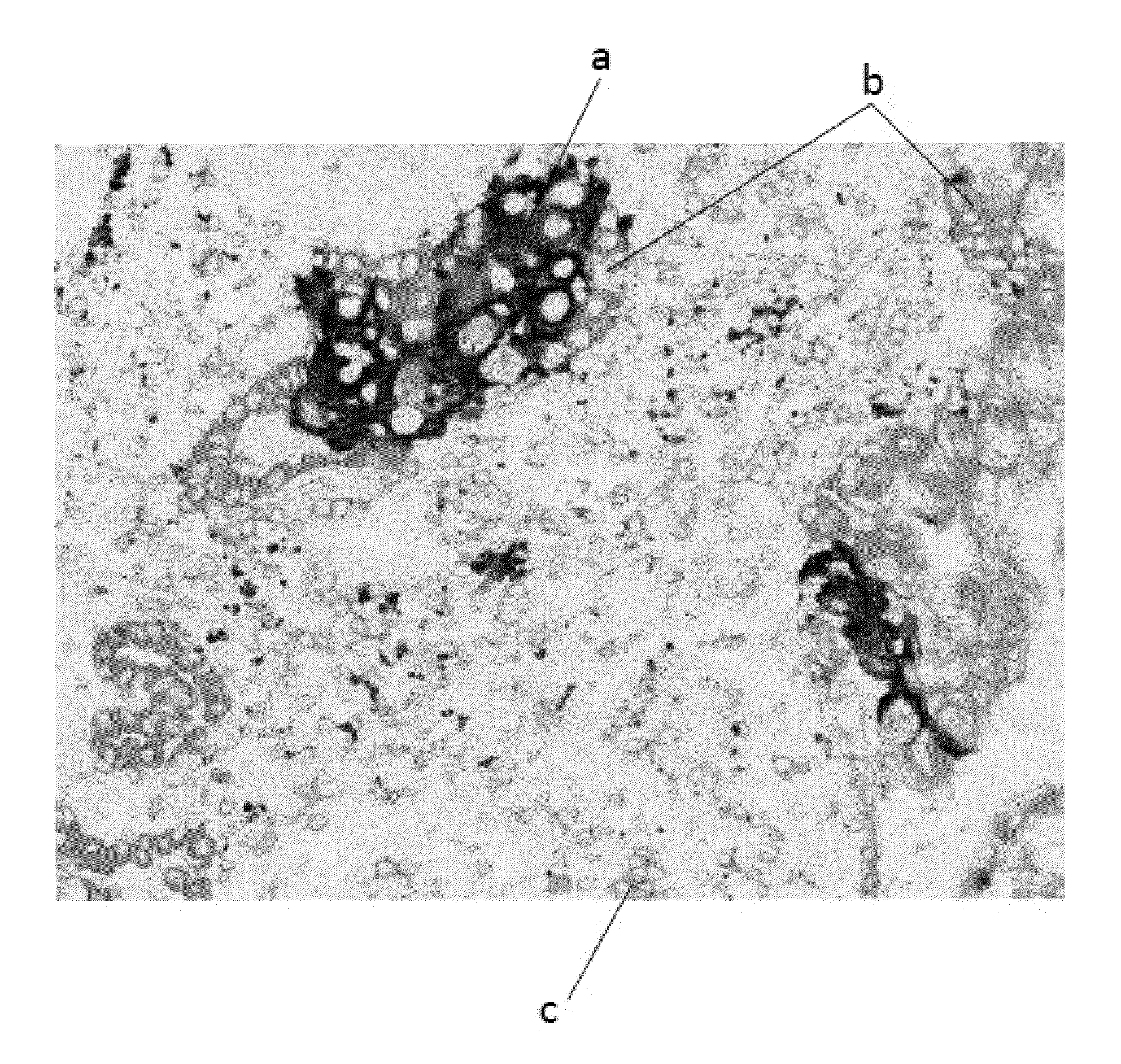
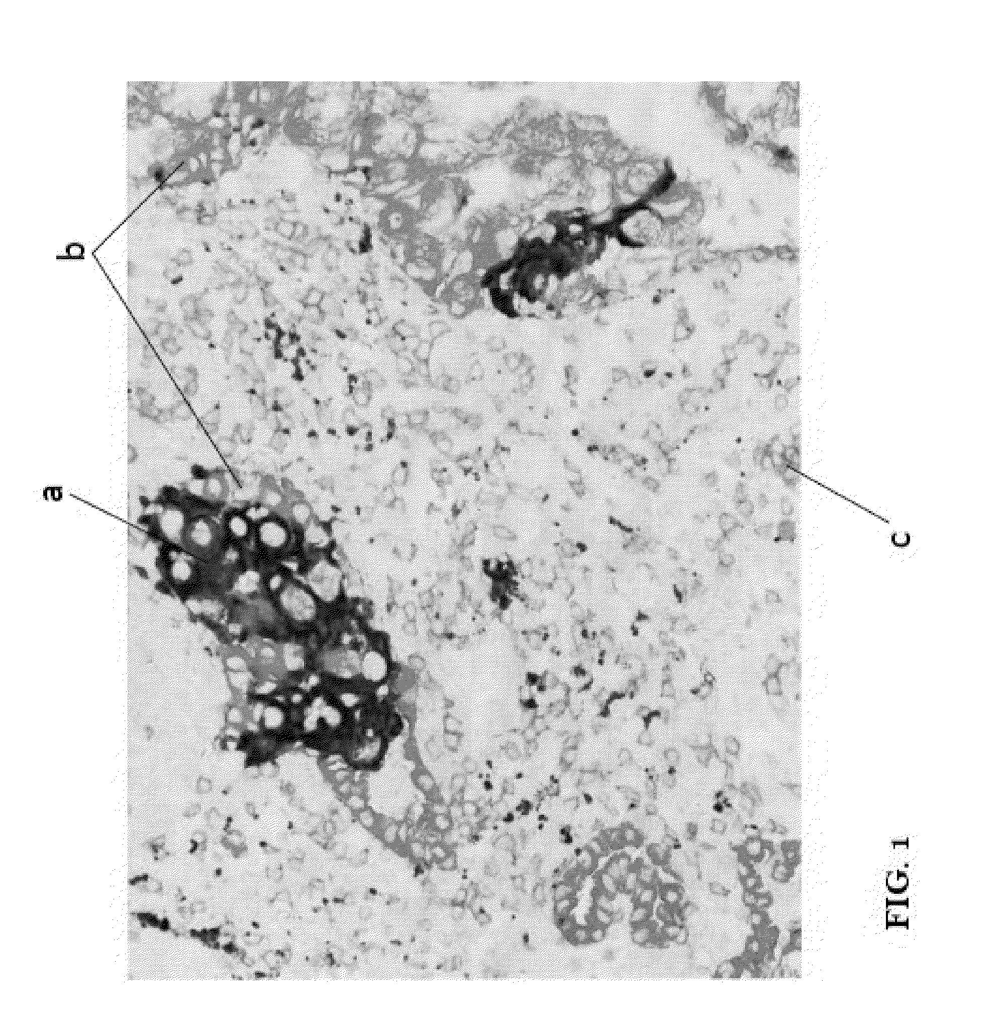
[0082]Example 1 describes a non-limiting example of a multiplex IHC assay of the present invention. A NSCLC sample slide is prepared according to standard protocols.

[0083]1. Apply 1 drop of PD-L1 SP142 antibody (Ventana Medical System, Tucson, Ariz.) to the slide and incubate for 16 minutes. Rinse slide with reaction buffer.

[0084]2. Apply 1 drop of OptiView HQ Universal Linker (Catalog No. 760-700, Ventana Medical Systems, Tucson, Ariz.) and incubate for 8 minutes. Rinse slide with Reaction Buffer.

[0085]3. Apply 1 drop of OptiView HRP Multimer (Catalog No. 760-700, Ventana Medical System, Tucson, Ariz.) and incubate for 8 minutes. Rinse slide with Reaction Buffer.

[0086]4. Apply 1 drop each of OptiView Amplifier H202 and OptiView Amplifier (Catalog No. 760-700, Ventana Medical System, Tucson, Ariz.) and incubate for 8 minutes. Rinse slide with Reaction Buffer.

[0087]5. Apply 1 drop of OptiView Amplifier Multimer (Catalog No. 760-700, Ventana Medical System,...

example 2

Signaling Conjugates

[0099]The following example describes alternative signaling conjugates described in WO Patent Application No. 2013148498, the disclosure of which is incorporated in its entirety herein by reference.

[0100]In some embodiments, methods of detecting a target in a biological sample include contacting the biological sample with a detection probe, contacting the biological sample with a labeling conjugate, and contacting the biological sample with a signaling conjugate. The labeling conjugate includes an enzyme. The signaling conjugate includes a latent reactive moiety and a chromogenic moiety. The enzyme catalyzes conversion of the latent reactive moiety into a reactive moiety, which covalently binds to the biological sample proximally to or directly on the target. The method further includes illuminating the biological sample with light and detecting the target through absorbance of the light by the chromogenic moiety of the signaling conjugate. In one embodiment, the...

example 3

Scoring

[0105]The following example describes various calculations (3A-3E) for determining PD-L1 positivity.


PUM

PropertyMeasurementUnit
FWHMaaaaaaaaaa
FWHMaaaaaaaaaa
FWHMaaaaaaaaaa

Abstract

Multiplex assays for improved scoring of tumor tissues stained with PD-L1 featuring PD-L1 staining in a first color plus staining of one or more differentiating markers, such as a marker specific for tumor cells and a marker specific for immune cells, are disclosed. The differentiation between the tumor cells and immune cells may improve the ease of scoring, the accuracy and speed of scoring, and the reproducibility of scoring of PD-L1 positive samples for therapy purposes.

Description

CROSS-REFERENCE TO RELATED APPLICATIONS[0001]This disclosure claims the benefit of U.S. 62 / 005,701, filed May 30, 2014, the contents of which are hereby incorporated by reference in their entirety.BACKGROUND OF THE INVENTION[0002]1. Field of the Invention[0003]The present disclosure relates to materials and methods for histochemically detecting and scoring PD-L1 expression in tumor tissues.[0004]2. Description of Related Art[0005]Programmed death 1 (PD-1) is a member of the CD28 family of receptors, which includes CD28, CTLA-4, ICOS, PD-1, and BTLA. Two cell surface glycoprotein ligands for PD-1 have been identified, PD-L1 and PD-L2, and have been shown to downregulate T cell activation and cytokine secretion upon binding to PD-1 (Freeman et al., J Exp Med 192:1027-34 (2000); Latchman et al., Nat Immunol 2:261-8 (2001); Carter et al., Eur J Immunol 32:634-43 (2002); Ohigashi et al., Clin Cancer Res 11:2947-53 (2005)). Both PD-L1 (B7-H1) and PD-L2 (B7-DC) are B7 homologs that bind to...

Claims


Application Information

Patent Timeline
Patent Type & Authority Applications(United States)
IPC IPC(8): G01N33/58G01N1/30
CPCG01N33/58G01N2333/70596G01N1/30G01N33/57423G01N33/57484
Inventor NITTA, HIROVENNAPUSA, BHARATHIDENNIS, ESLIE
Owner VENTANA MEDICAL SYST INC
Why Eureka
Try Eureka