
Animal model for evaluating performance of hemostatic agent for inducing hemorrhage in common carotid artery or superior sagittal sinus, and use thereof

a hemostatic agent and performance evaluation technology, applied in the field of animal models for evaluating hemostatic performance, can solve the problems of large amount of blood loss, difficult selection of blood vessels, and difficulty in evaluating the effect of hemostatic agents

Inactive Publication Date: 2015-12-17
INNOTHERAPY
View PDF1 Cites 0 Cited by
  • Summary
  • Abstract
  • Description
  • Claims
  • Application Information

AI Technical Summary

Benefits of technology

The present invention provides an animal model that can be used to evaluate the performance of a hemostatic agent. This invention also provides methods for screening and evaluating the effect of a hemostatic agent using the animal model.

Problems solved by technology

However, in the case in which natural hemorrhage is difficult, like the case of blood gushing, an animal model that is used to demonstrate the hemostatic effects of hemostatic agents in a state in which blood gushes out has not been reported.
In addition, in the case of the most severe hemorrhage such as arterial hemorrhage, that is, in the case in which blood gushes to a considerable height from a wound, blood flows out at high rate so that so that a large amount of blood will be lost.
In this case, the blood is hardly coagulated, and thus it is not easy to evaluate the effect of a hemostatic agent.
In addition, in the preparation of a hemostatic animal model for evaluating the effect of a hemostatic agent, it is difficult to select blood vessels, due to the features of arteries and veins.
For this reason, the preparation of the hemostatic animal model is limited.
Meanwhile, hemorrhage of the superior sagittal sinus is unavoidable and frequently occurs in sinus surgery.
However, it is difficult to screen an effective hemostatic agent, because tissue surrounding the superior sagittal sinus is hard tissue.

Method used


Image

  • Animal model for evaluating performance of hemostatic agent for inducing hemorrhage in common carotid artery or superior sagittal sinus, and use thereof
  • Animal model for evaluating performance of hemostatic agent for inducing hemorrhage in common carotid artery or superior sagittal sinus, and use thereof
  • Animal model for evaluating performance of hemostatic agent for inducing hemorrhage in common carotid artery or superior sagittal sinus, and use thereof

Examples


example 1

Preparation of Animal Model (Artery) for Evaluating Hemostatic Effects

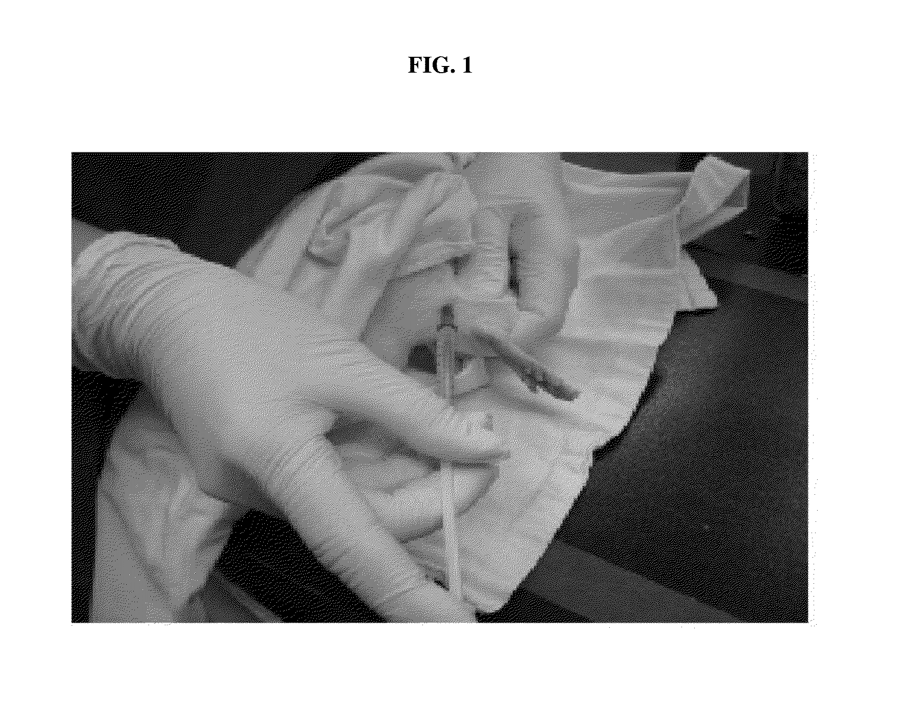
[0055]To prepare an animal model for evaluating hemostatic effects, the hind leg muscle of a rat was anesthetized by intramuscular injection of 0.1 ml / 100 g of Zoletil / Rompun (5 ml Zoletil+2.5 ml Rompun), and after 20 minutes, a surgical operation for the rat was started (FIG. 1).

[0056]The animals used in this Example were 10-week-old SD rats (weight: about 350-450 g).

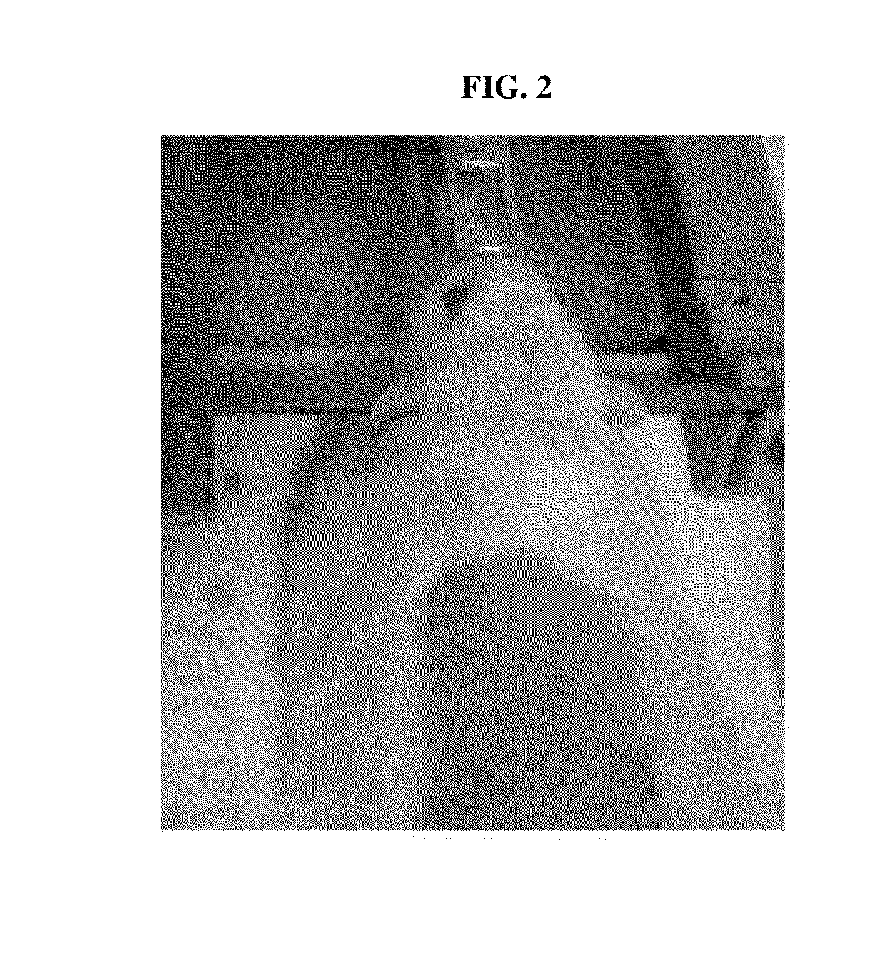
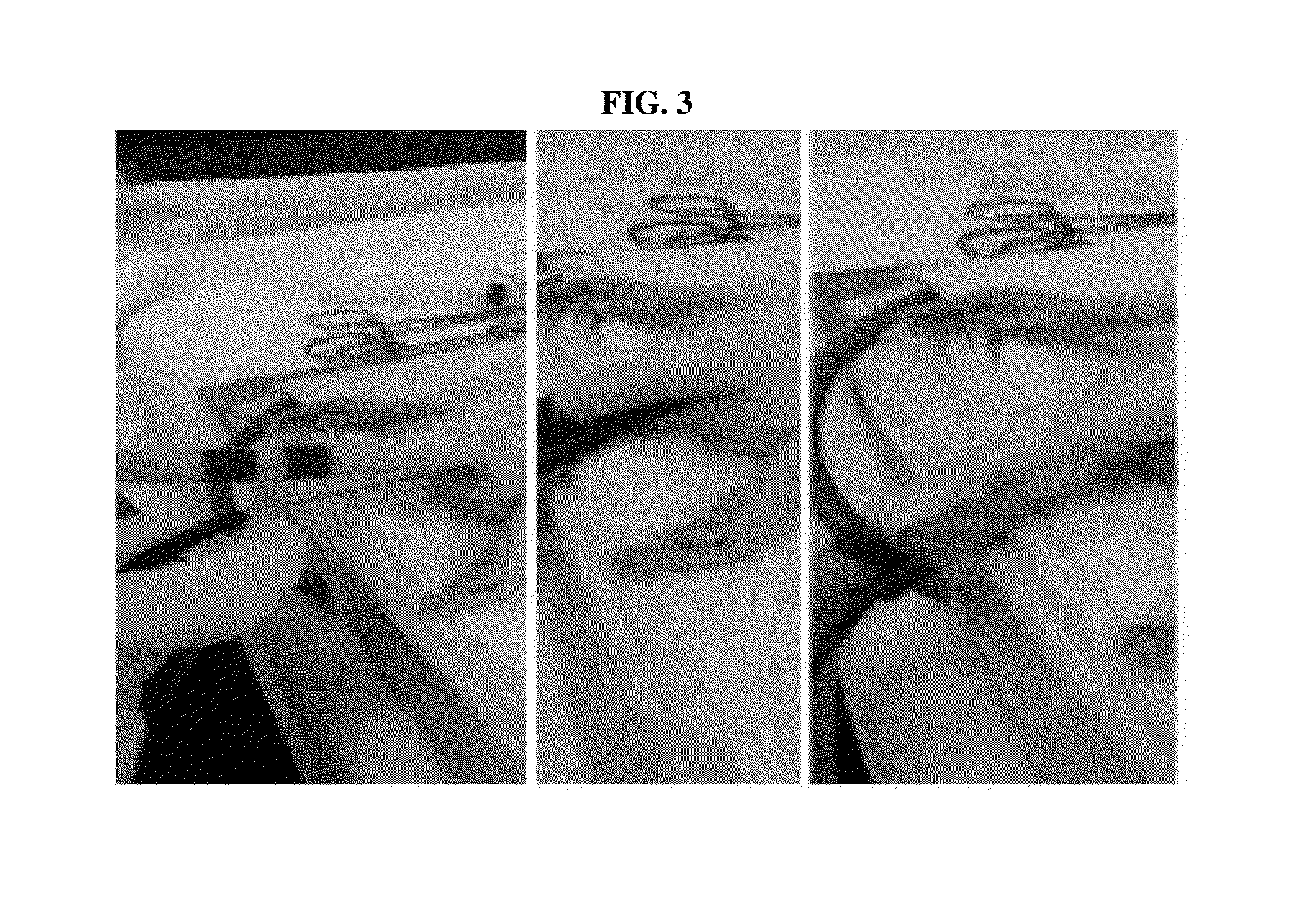
[0057]In addition, the site to be surgically operated was widely shaved, and the rat was fixed on a stereotaxic frame (FIG. 2). To maintain the body temperature at a constant level, a body temperature sensor with a warm pad was inserted into the anus of the rat and fixed (FIG. 3). The shaved portion of the animal was disinfected with betadine, and then the midline of a site ranging from the chest line to the neck portion was incised by about 3-4 cm. Next, as shown in FIG. 4, connective tissues were removed with cotton swabs, and omohyoid muscle exten...

example 2

Preparation of Animal Model (Vein) for Evaluating Hemostatic Effects

[0060]To prepare an animal model for evaluating hemostatic effects, the hind leg muscle of a rat was anesthetized by intramuscular injection of 0.1 ml / 100 g of Zoletil / Rompun (5 ml Zoletil+2.5 ml Rompun), and after 20 minutes, a surgical operation for the rat was started (FIG. 1).

[0061]The animals used in this Example were 10-week-old SD rats (weight: about 350-450 g).

[0062]In addition, the site to be surgically operated was widely shaved, and the rat was fixed on a stereotaxic frame (FIG. 2). To maintain the body temperature at a constant level, a body temperature sensor with a warm pad was inserted into the anus of the rat and fixed (FIG. 3). The shaved portion of the animal was disinfected with betadine, and then the midline of a site ranging from the middle of the forehead to the ear portion was incised by about 2-3 cm using a scalpel (No. 10). The incised portion was opened and fixed with forceps, and connecti...

example 3

Preparation of Animal Model (Liver) for Evaluating Hemostatic Effects

[0064]To prepare an animal model for evaluating hemostatic effects, the hind leg muscle of a rabbit was anesthetized by subcutaneous injection of 0.5 ml / kg of Zoletil / Rompun (5 ml Zoletil+2.5 ml Rompun), and after 20 minutes, a surgical operation for the rabbit was started.

[0065]The animals used in this Example were New Zealand White rabbits (weight: about 2.3-3.0 kg).

[0066]In addition, the site to be surgically operated was widely shaved and locally anesthetized with lidocaine, and the animal was fixed on a surgical operation table equipped with hyperhypo thermia.

[0067]The thorax was opened by incision, and then the liver central lobe was exposed onto a gauze wet with sterile physiological saline. The middle portion of the liver (central lobe) was pierced to a depth of about 2-3 mm using a biopsy punch (d=6 mm) to induce hemorrhage, thereby preparing an animal model.


PUM


Abstract

The present invention relates to an animal model for evaluating hemostatic performance, and the use thereof, and more particularly, to an animal model for evaluating the performance of a hemostatic agent, which has hemorrhage induced in the common carotid artery (CCA) or superior sagittal sinus (SSS) of the animal, a method of screening a hemostatic agent using the animal model, and a method of evaluating the effect of a hemostatic agent using the animal model. The animal model according to the present invention makes it possible to observe the hemostatic effect of a hemostatic agent in a rapid and accurate manner without causing side effects. Thus, the animal model is useful for screening a hemostatic agent and evaluating the effect of a hemostatic agent.

Description

TECHNICAL FIELD[0001]The present invention relates to an animal model for evaluating hemostatic performance, and the use thereof, and more particularly, to an animal model for evaluating the performance of a hemostatic agent, which has hemorrhage induced in the common carotid artery (CCA) or superior sagittal sinus (SSS) of the animal, a method of screening a hemostatic agent using the animal model, and a method of evaluating the effect of a hemostatic agent using the animal model.BACKGROUND ART[0002]As used herein, the term “medical instant adhesive” means, in a broad sense, medical supplies, including adhesive plasters, surgical adhesives and hemostatics, and in a narrow sense, adhesives that are used directly in medical fields, including dermatology, vascular surgery, gastroenterology and plastic surgery. Because the medical instant adhesive comes into contact with the skin, it should be biocompatible, should not be toxic and harmful to the body, should be biocompatible, and shou...

Claims


Application Information

Patent Timeline
IPC IPC(8): A01K67/027A61K49/00
CPCA01K67/027A61K49/0008A01K2267/0375A01K2227/10A01K2207/30A01K2227/105G01N33/15
Inventor LEE, MOON SUEKOH, MI YOUNGKIM, KEUMYEON
Owner INNOTHERAPY
Learn More