Mapping cardiac tissue architecture systems and methods

a cardiac tissue and architecture technology, applied in the field of magnetic resonance imaging, can solve the problems of limited ability of these metrics to fully characterize the structural dynamics of the heart, and inability to reproduce in the human heart in vivo

Active Publication Date: 2016-03-03
THE GENERAL HOSPITAL CORP
View PDF2 Cites 7 Cited by
  • Summary
  • Abstract
  • Description
  • Claims
  • Application Information

AI Technical Summary

Problems solved by technology

However, the ability of these metrics to fully characterize structural dynamics during he

Method used


Image

  • Mapping cardiac tissue architecture systems and methods
  • Mapping cardiac tissue architecture systems and methods
  • Mapping cardiac tissue architecture systems and methods

Examples


Embodiment Construction

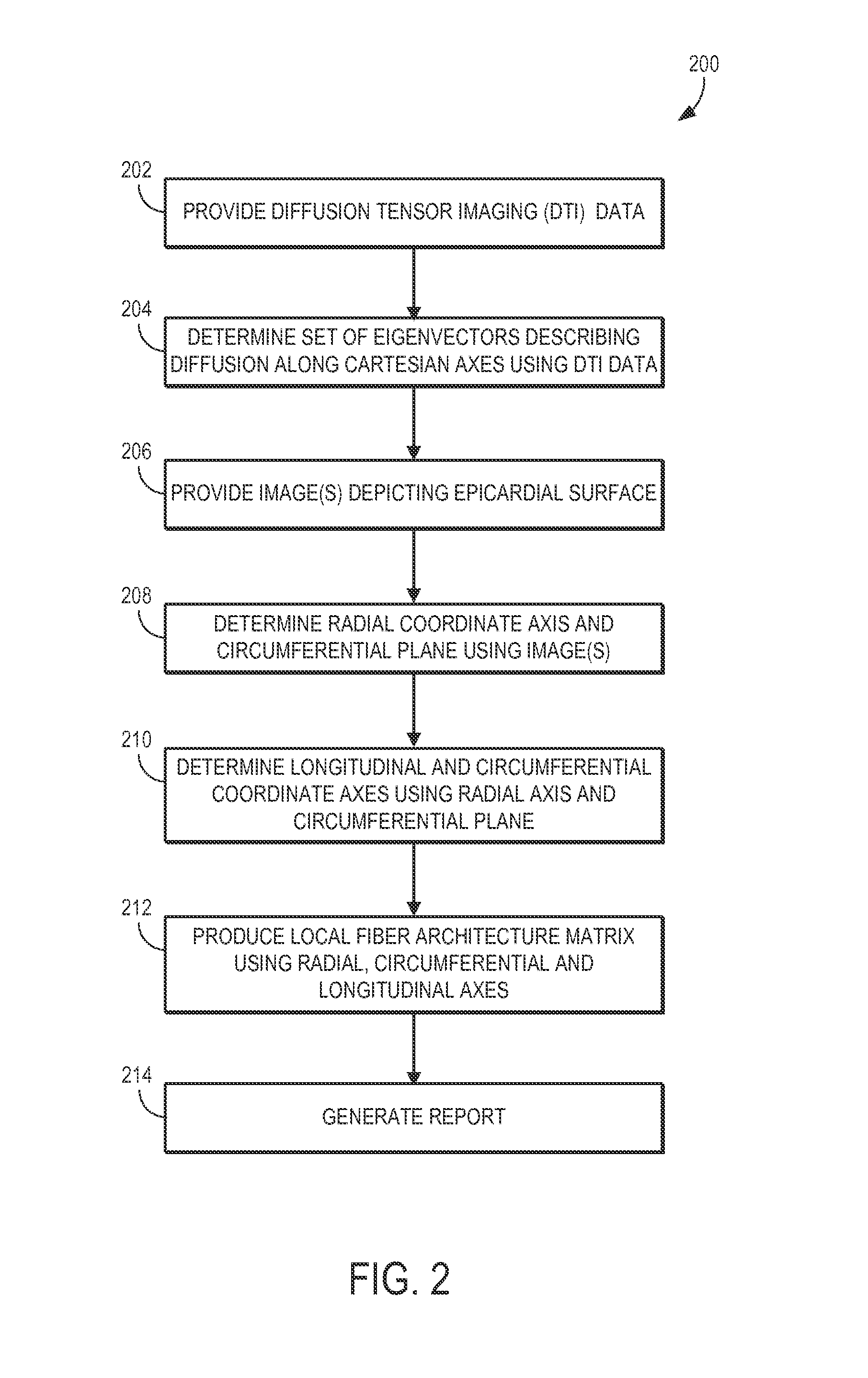
[0021]Traditional diffusion tensor imaging (“DTI”) studies of the heart generally rely on a global cardiac coordinate system that does not take local changes in cardiac morphology into account. The systems and methods described here, however, provide a solution to this problem by mapping myocardial tissue architecture based on projecting information derived from a diffusion tensor into a local, subject-specific cardiac coordinate system. More particularly, the systems and methods described here include computing a fiber architecture matrix (“FAM”) based on projections of eigenvectors, such as those derived from a diffusion tensor, onto a local coordinate system that more accurately characterizes diffusion in the myocardial tissue.

[0022]The FAM serves as a function of the complete diffusion tensor eigensystem, and is designed to characterize myocardial tissue dynamics both in vivo and ex vivo. The relationship between the eigensystem and the local cardiac coordinate system, provided ...


PUM


Abstract

Systems and methods are provided for mapping myocardial tissue architecture based on diffusion tensor imaging (DTI). A set of eigenvectors is derived from diffusion tensor data, where each eigenvector describes the diffusion of spins along one of the Cartesian directions. A radial coordinate axis and a circumferential plane are determined based on anatomical information of the subject, such as from an image depicting the epicardial surface of the subject's heart. A longitudinal coordinate axis and a circumferential coordinate axis are determined based on the radial coordinate axis and circumferential plane. A fiber architecture matrix (FAM) is then computed for locations in the subject's heart based on projecting the set of eigenvectors onto a local coordinate system defined by the circumferential, radial, and longitudinal axes, Maps that represent myocardial tissue architecture can then be generated using the FAM for locations within the subject's heart.

Description

CROSS-REFERENCE TO RELATED APPLICATIONS[0001]This application claims the benefit of U.S. Provisional Patent Application Ser. No. 61 / 807,996, filed on Apr. 3, 2013, and entitled “Fiber Architecture Matrix.”STATEMENT REGARDING FEDERALLY SPONSORED RESEARCH[0002]This invention was made with government support under HL093038 and RR14075 awarded by the National Institutes of Health. The government has certain rights in the invention.BACKGROUND[0003]The present disclosure relates generally to systems and methods for magnetic resonance imaging (“MRI”) and, in particular, to systems and methods for mapping cardiac fiber architecture using diffusion-weighted imaging techniques, such as diffusion tensor imaging.[0004]For diffusion MRI techniques, motion sensitizing magnetic field gradients are applied using diffusion weighted imaging (“DWI”) pulse sequences so that the magnetic resonance images include contrast related to the diffusion of water or other fluid molecules. Since microscopic arran...

Claims


Application Information

Patent Timeline
IPC IPC(8): G01R33/56A61B5/00A61B5/055G06T7/00G06T11/00
CPCA61B5/055A61B2576/023G01R33/56341G06T2207/10088G06T2207/30048G06T7/0081G06T7/0012G01R33/5602A61B5/0044G06T11/003G06T7/11G16H30/40
Inventor MEKKAOUI, CHOUKRI
Owner THE GENERAL HOSPITAL CORP
Why Eureka
Try Eureka