Biomarkers for differentiating melanoma from benign nevus in the skin

a biomarker and melanoma technology, applied in the field of detection of melanoma, can solve the problems of adversely affecting patient outcome and quality of life, poor interobserver reproducibility, and different approaches to translate differential gene expression into clinical diagnostics, and achieve the effect of increasing the expression of the marker

Inactive Publication Date: 2016-04-28
ADVANCED CELL DIAGNOSTICS INC
View PDF0 Cites 11 Cited by
  • Summary
  • Abstract
  • Description
  • Claims
  • Application Information

AI Technical Summary

Problems solved by technology
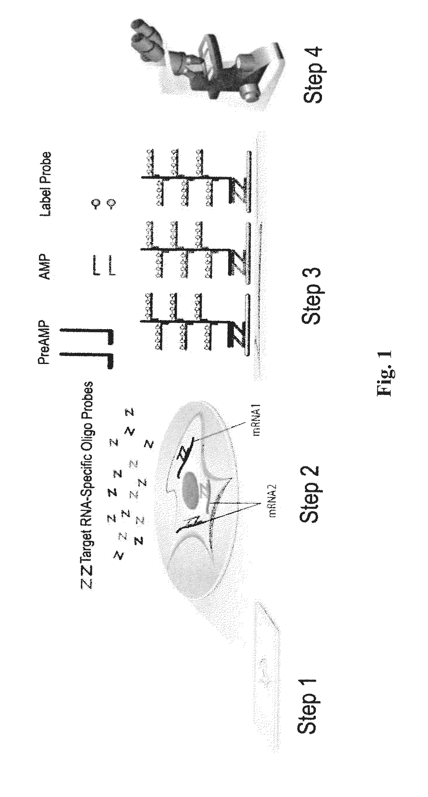
Misdiagnosis can lead to under-treatment or over-treatment for the suspected lesion, adversely impacting patient outcome and quality of life.
Furthermore, the recognition of the many histological features used in diagnosis can be subjective and dependent on experience and thus cause poor inter-observer reproducibility.
These different approaches of translating differential gene expression into clinical diagnostics all have significant limitations.
Careful microdissection can help, but it is too cumbersome for routine practice.
IHC is an attractive approach to provide cellular resolution, but translating differential gene expression at the RNA level to IHC can be difficult, because the best candidate markers may not have suitable antibodies for IHC.
However, existing RNA ISH methods have poor sensitivity and specificity and are technically demanding, making them impractical for most of the genes identified in microarray analysis.
Its implementation as a microarray- or RT-PCR-based test for clinical practice will be challenging since it will require careful microdissection of the stroma from the tumor and it cannot differentiate the many stromal cell types.

Method used


Image

  • Biomarkers for differentiating melanoma from benign nevus in the skin
  • Biomarkers for differentiating melanoma from benign nevus in the skin
  • Biomarkers for differentiating melanoma from benign nevus in the skin

Examples


example 1

Selection of PHACTR1, SPP1, and MLANA as Biomarkers for Detecting Melanoma

[0216]To identify the molecular events associated with melanoma progression and potential novel markers for early melanoma diagnosis, genome-wide gene expression microarrays were performed by several groups, and several datasets were made available through the Gene Expression Omnibus (GEO) database (http: / / (www.)ncbi.nlm.nih.gov / geo / ). Although bioinformatic and statistical analysis had already been performed on the these datasets by the original authors, it was reasoned that combining results from multiple datasets, especially those from different microarray platforms, may lead to consistent, cross-validated candidate markers. Consequently, two different datasets comparing normal skin or benign nevi and malignant melanoma, GSE3189 (Affymetrix® GeneChip® HG-U133A, 22,000 genes) and GSE12391 (Agilent® Human Whole Genome Oligo Microaary, 41,000 genes) were selected, for candidate identification. GSE3189 contains...

example 2

Validation of PHACTR1, SPP1, and MLANA as Biomarkers for Detecting Melanoma

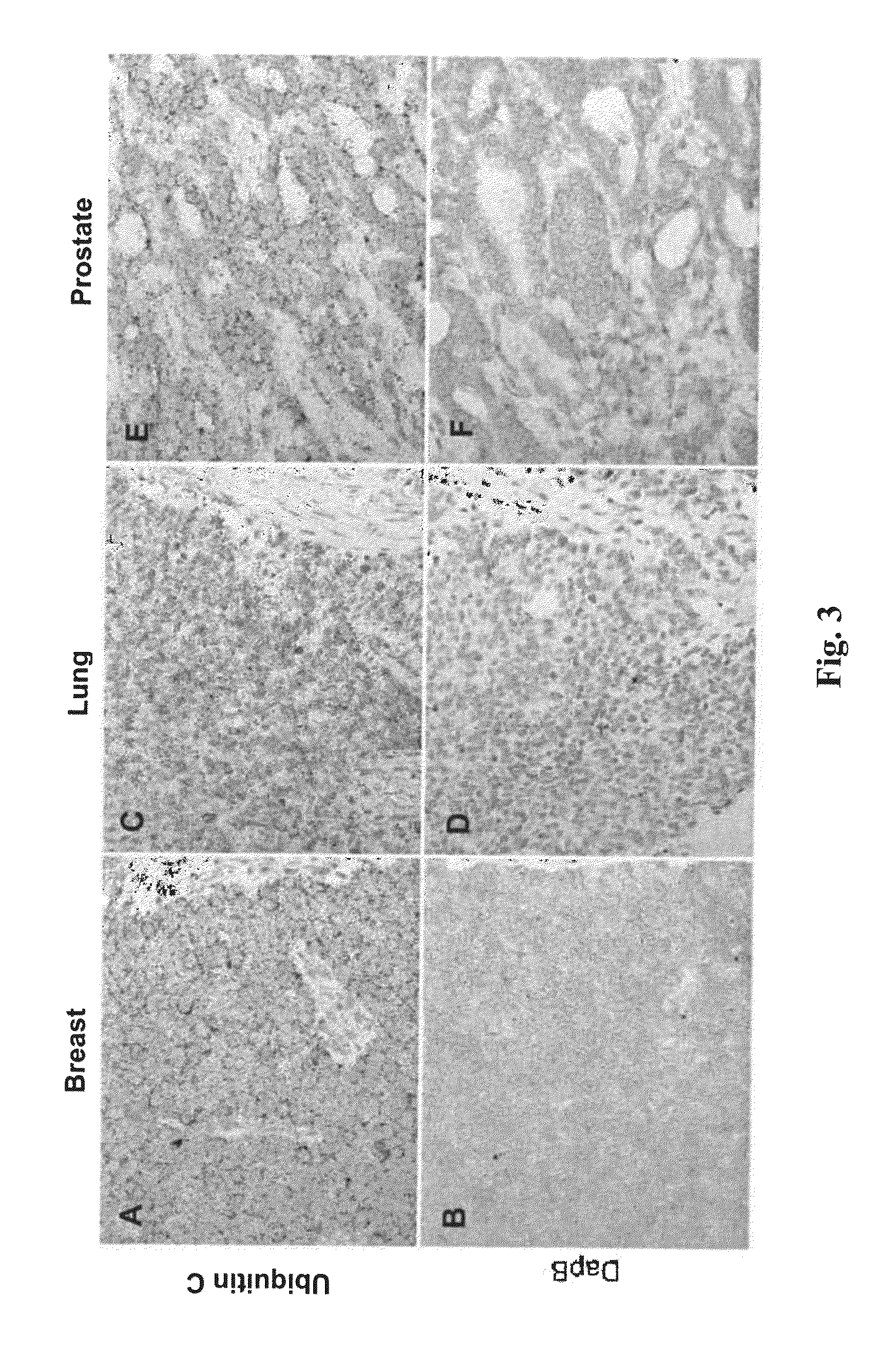
[0219]RNAscope® assays using gene-specific probes against these eight selected candidate genes were performed on a series of FFPE tissue microarrays (TMAs) obtained from commercial vendors. The bacterial gene DapB was used as negative control. These TMAs contained normal skin, nevi and melanomas of different stages. TMAs are efficient means to test many candidate genes in multiple samples.

[0220]In these TMA experiments, MLANA had higher and more consistent expression in benign nevi and melanomas than TYR. Thus, MLANA was chosen as the marker for melanocytes and melanoma cells. It also served as a positive control for RNA integrity, since these TMAs were constructed from archival tissues from various times and sources and had variable quality in RNA preservation. Together, 70% of the melanocytic nevi and melanomas on the TMAs had MLANA-staining intensity scores above 2+ on a semi-quantitative scale and were co...

example 3

Selection of Additional Biomarkers for Detecting Melanoma

[0224]Further improvements in RNAscope® Mela performance are highly desirable. First, MLANA was used as a positive control to help identify melanocytes (benign or malignant) and to assess quality of specimens. However, desmoplastic melanoma, representing 2-3% of all melanomas, is known to be negative for MLANA expression. A small fraction of other melanomas may also be negative for MLANA. Thus a negative MLANA staining result may be difficult to interpret. Second, PHACTR1 and SPP1 were negative in 5% of the melanomas, resulting in false negative classifications.

[0225]Therefore, it was sought to add additional markers to the 3-gene panel. First, two S100 genes, S100A6 and S100B, were added to MLANA to serve as positive control for assay procedure and sample quality control. S100A6 and S100B are known to be expressed in desmoplastic melanomas which are usually nelan-A-negative. An example of RNAscope® detection of S100 in desmop...


PUM

PropertyMeasurementUnit
Nanostringaaaaaaaaaa
multicolor fluorescence in situ hybridizationaaaaaaaaaa
Fluorescence in situ hybridizationaaaaaaaaaa

Abstract

Disclosed is a method for diagnosing melanoma in a human subject, as well as a method for providing a prognosis to a human subject who is at risk of developing melanoma recurrence, and a method for determining the stage of melanoma in a human subject, comprising the step of determining the level of expression of phosphatase and actin regulator 1 (PHACTR1) gene, or fragments thereof, either alone or in combination with the level of expression of secreted integrin-binding phosphoprotein (SPP1), preferentially expressed antigen in melanoma (PRAME), growth differentiation factor 15 (GDF15), and chemokine C-X-C motif ligand 10 (CXCL10) genes. Further, the invention relates to a diagnostic kit, comprising at least one substance for detection of the expression of PHACTR1, or fragments thereof, either alone or in combination with the detection of SPP1, PRAME, GDF15, and CXCL10, for the diagnosis or prognosis of melanoma.

Description

CROSS-REFERENCE TO RELATED APPLICATIONS[0001]This application claims priority to and benefit of U.S. Provisional Application No. 61 / 384,707, filed Sep. 20, 2010, entitled “BIOMARKER FOR DIFFERENTIATING MELANOMA FROM BENIGN NEVUS IN THE SKIN” and U.S. Provisional Application No. 61 / 470,682, filed Apr. 1, 2011, entitled “METHODS FOR DIFFERENTIATING MELANOMA FROM BENIGN NEVUS IN THE SKIN”.[0002]Each of the aforementioned applications is incorporated herein by reference in its entirety for all purposes.FIELD OF INVENTION[0003]Provided herein is a method for detection of melanoma by monitoring the presence and levels of specific biomarkers in samples obtained from patients suspected of melanoma. Also provided herein are methods and kits for diagnosing melanoma and providing prognosis of melanoma development in patients based on the presence and levels of specific biomarkers. Further provided herein are methods for determining the stage of melanoma based on the presence of malignant melan...

Claims


Application Information

Patent Timeline
Patent Type & Authority Applications(United States)
IPC IPC(8): C12Q1/68
CPCC12Q1/6886C12Q2600/16C12Q2600/158C12Q2600/112C12Q2600/118
Inventor MA, XIAO-JUNWU, XINGYONGLUO, YULING
Owner ADVANCED CELL DIAGNOSTICS INC
Why Eureka
Try Eureka