Simultaneous Capture of Filtered Images of the Eye

a technology of filtered images and eye, applied in the field of eye imaging, can solve the problems of limiting the diagnostic ability of many diseases, difficult or impossible to assess absolute measurements of area, depth or volume for three-dimensional anatomy, and the drawbacks of conventional fundus cameras

Active Publication Date: 2016-09-29
RICOH KK
View PDF7 Cites 21 Cited by
  • Summary
  • Abstract
  • Description
  • Claims
  • Application Information

AI Technical Summary

Benefits of technology

[0010]Another aspect is an after-market conversion kit for converting a co

Problems solved by technology

However, these images are limited by brightness, field-of-view, motion blur, and resolution, which restrict their diagnostic abilities for many diseases.
However, conventional fundus cameras have several drawbacks.
This makes it difficult or impossible to assess absolute measurements of area,

Method used


Image

  • Simultaneous Capture of Filtered Images of the Eye
  • Simultaneous Capture of Filtered Images of the Eye
  • Simultaneous Capture of Filtered Images of the Eye

Examples


Embodiment Construction

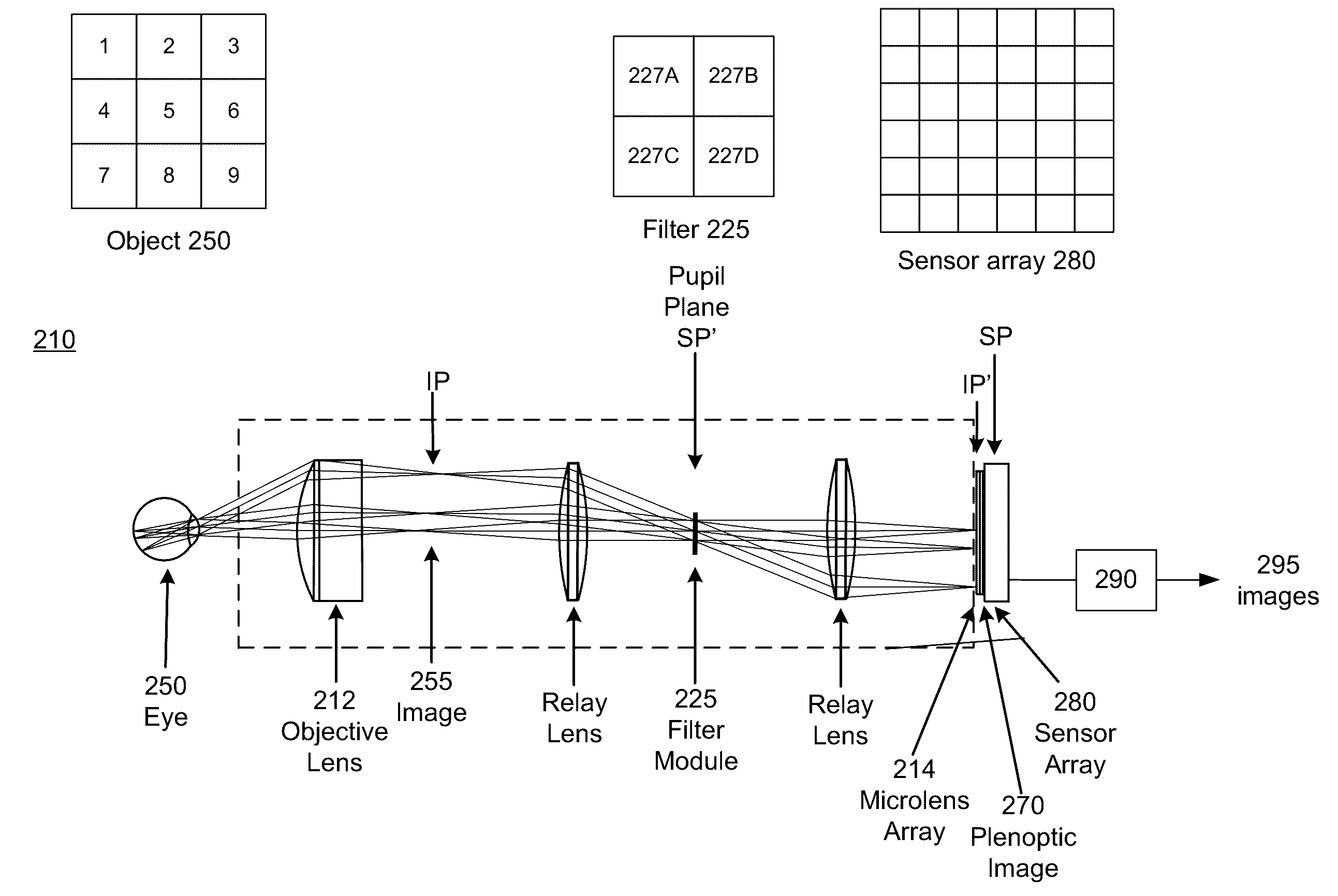
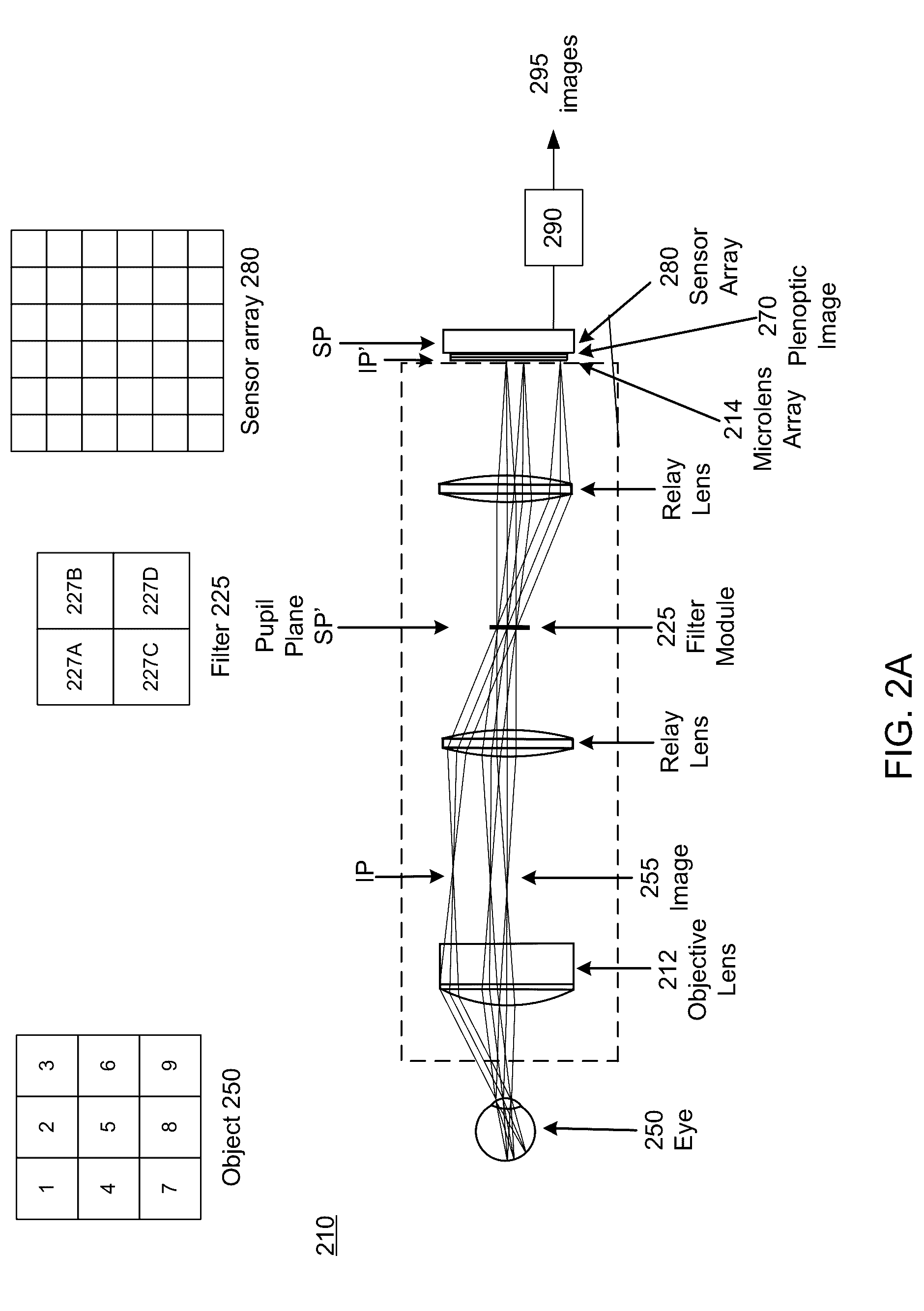
[0025]The figures and the following description relate to preferred embodiments by way of illustration only. It should be noted that from the following discussion, alternative embodiments of the structures and methods disclosed herein will be readily recognized as viable alternatives that may be employed without departing from the principles of what is claimed. To facilitate understanding, identical reference numerals have been used where possible, to designate identical elements that are common to the figures.

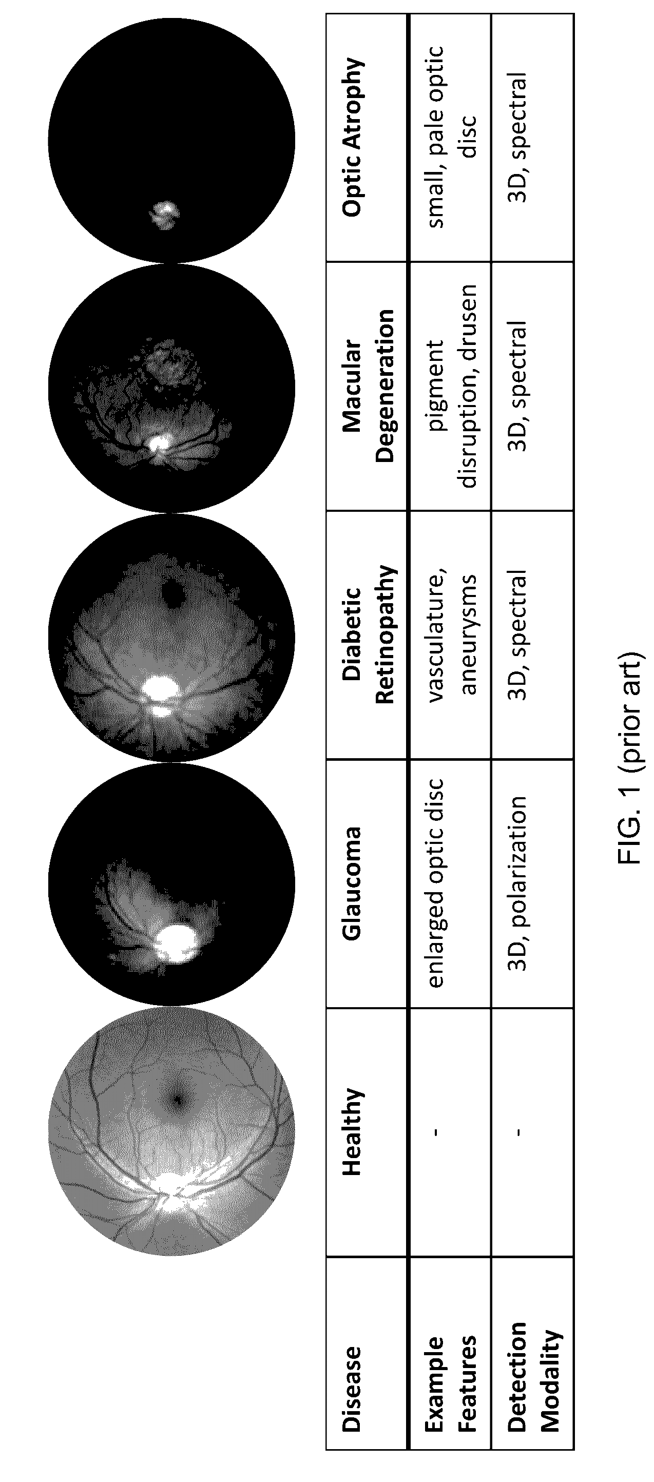
[0026]FIG. 1 (prior art) are example images showing different conditions of the eye. The images are images of the retina. The entries below each retina image list the disease condition, the features that can be used to diagnose the disease, and the modalities that would be useful to image the features. The leftmost column is a healthy eye.

[0027]The first disease listed is glaucoma, which has a clinical presentation of an enlarged optic disc. In order to assess enlargement of t...


PUM


Abstract

A multimode fundus camera enables three-dimensional and/or spectral/polarization imaging of the interior of the eye to assist in improved diagnosis. In one aspect, the multimode fundus camera includes a first imaging subsystem, a filter module, and a second imaging subsystem. The first imaging subsystem is positionable in front of an eye to form an optical image of an interior of the eye. The filter module is positioned at a pupil plane of the first imaging subsystem or at a conjugate thereof. The second imaging subsystem include a microimaging array and a sensor array. The microimaging array is positioned at the image plane or a conjugate thereof, and the sensor array is positioned at the pupil plane or a conjugate thereof.

Description

CROSS-REFERENCE TO RELATED APPLICATION(S)[0001]This application is a continuation-in-part of U.S. patent application Ser. No. 14 / 542,064, “Simultaneous Capture of Filtered Images of the Eye,” filed Nov. 14, 2014.BACKGROUND OF THE INVENTION[0002]1. Field of the Invention[0003]This invention relates generally to imaging of the eye, for example spectral, polarization and / or three-dimensional imaging of the retina.[0004]2. Description of the Related Art[0005]Specialized cameras are used by optometrists, ophthalmologists, and other medical professionals to record images of the interior surface of the eye. During a routine physical examination, a handheld ophthalmoscope is often used to quickly view the fundus. Additional camera attachments can be used to record digital images from handheld ophthalmoscopes, allowing acquired images to be saved, manipulated, and reevaluated at future examinations. However, these images are limited by brightness, field-of-view, motion blur, and resolution, ...

Claims


Application Information

Patent Timeline
IPC IPC(8): A61B3/14A61B3/00H04N5/247A61B3/12H04N5/232H04N13/02G02B5/20A61B3/10
CPCA61B3/14G02B5/201A61B3/0025H04N5/247A61B3/12H04N5/23245H04N13/0203A61B3/1015H04N13/204H04N23/90H04N23/667
Inventor GAO, LIANGMENG, LINGFEIBEDARD, NOAH
Owner RICOH KK
Why Eureka
Try Eureka