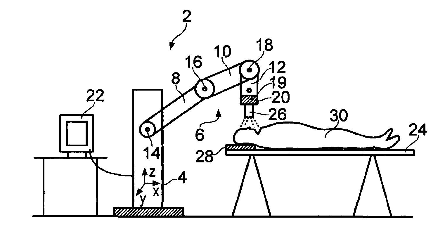
[0008]Apparatus of the present invention, in which the moveable arm of the surgical robot can also perform an x-ray imaging function, has a number of advantages. As explained above, the first step in any surgical robot based procedure is to “register” the robot to the subject. The registration process involves establishing the position of the relevant part of the subject's anatomy (e.g. the position of a target position within the brain that has been identified by a pre-operative medical scan) in the coordinate system of the surgical robot. Once registered, the surgical robot can then drive instruments (e.g. neurosurgical catheters, shunts, electrodes etc) to the pre-identified target position. The accuracy of a surgical robot based procedure is thus governed not only by the mechanical accuracy of the surgical robot, but also by the accuracy with which the robot is registered to the subject.
[0009]It has been found that surgeons implementing prior art registration techniques for surgical robots, such as the technique described with reference to FIG. 5 in WO2012 / 085511, can spend a significant amount of time in the operating theatre ensuring that the registration of the surgical robot to the subject is performed with the necessary accuracy. In contrast, the present invention allows x-ray images to be acquired with the x-ray source and / or the x-ray detector held by the moveable arm of the robot in one or more locations that are known to the robot (e.g. in locations that are known in the local coordinate system of the surgical robot). This allows the location of features (e.g. x-ray visible fiducial markers) within acquired x-ray images to be directly established in the robot co-ordinate system without the introduction of any errors that may arise from registering the coordinate system of a totally separate intra-operative x-ray imaging system (e.g. a C-arm or O-arm) with the robot coordinate system. Potential problems associated with ensuring there is no unintended relative movement between the surgical robot and a separate x-ray imaging system during use are also overcome. Furthermore, surgical robots typically have a higher positional accuracy than intra-operative CT scanners thereby allowing higher accuracy position data to be extracted from the acquired x-ray images.
[0010]The present invention thus improves the speed and accuracy of registration, meaning that it also becomes practical to adjust the position of the patient during the surgical procedure because the registration procedure can be quickly and reliably performed again. Avoiding the need for a separate CT scanner also reduces the amount of kit that is required in the operating theatre, thereby reducing clutter and improving the surgeon's access to the subject.
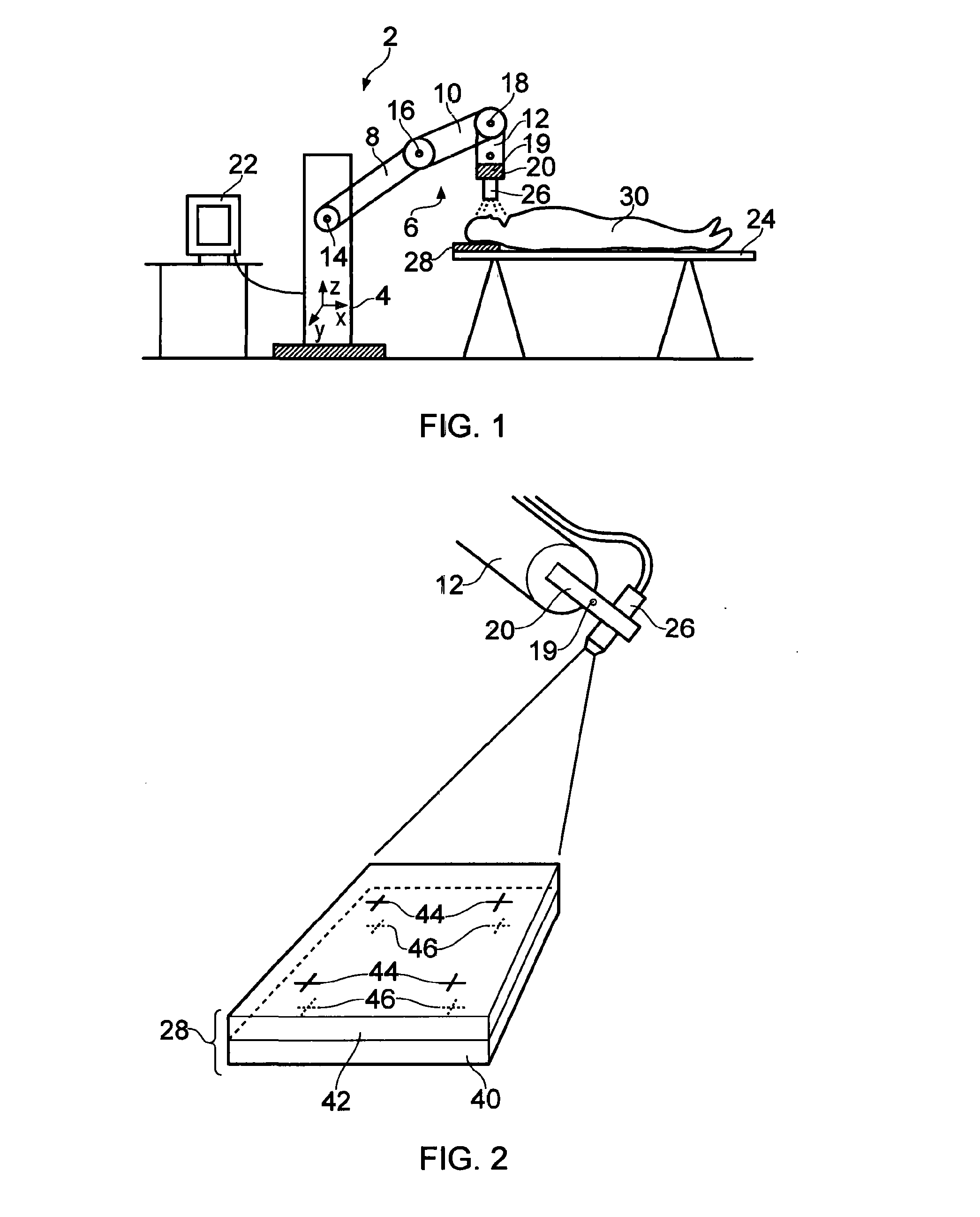
[0021]As described above, the position of an x-ray detector may be known (e.g. by calibration) in the robot coordinate system. It is also described above how the position of an x-ray detector in the robot coordinate system can be measured using one or more x-ray visible fiducial markers that have a known position relative to the x-ray detector. It should, however, be noted that it is not always necessary to establish the position of the x-ray detector. In particular, one or more x-ray visible fiducial markers may be provided that have a known position in the robot coordinate system. For example, one or more x-ray visible fiducial markers may be fitted to a head frame or the like that have a known (calibrated) position in the robot coordinate system. The x-ray detector may then be used to measure the position of targets relative to the x-ray visible fiducial markers without having to establish the position of the x-ray detector in the robot coordinate system.
[0022]Alternatively, the moveable arm of the surgical robot may be configured to hold one or more x-ray visible fiducial markers. For example, there may be one or more x-ray visible fiducial markers removeably attached to the moveable arm. Each x-ray visible fiducial marker may be attached to the moveable arm in a repeatable position; e.g. each x-ray visible fiducial marker may have a known (e.g. calibrated) position relative to a datum point on the moveable arm. The moveable arm may also carry an x-ray source and the one or more x-ray visible fiducial markers may be located between the x-ray source and an x-ray detector. In this manner, the x-ray visible fiducial markers visible in the image acquired by the x-ray detector have known positions in the robot coordinate system. It is then possible to establish the position of other features present in x-ray images acquired by the x-ray detector relative to the known positions of the x-ray visible fiducial markers. Again, this process does not require any knowledge of the position of the x-ray detector in the robot coordinate system. A further advantage of this arrangement is that the need for patient mounted fiducials can be removed.
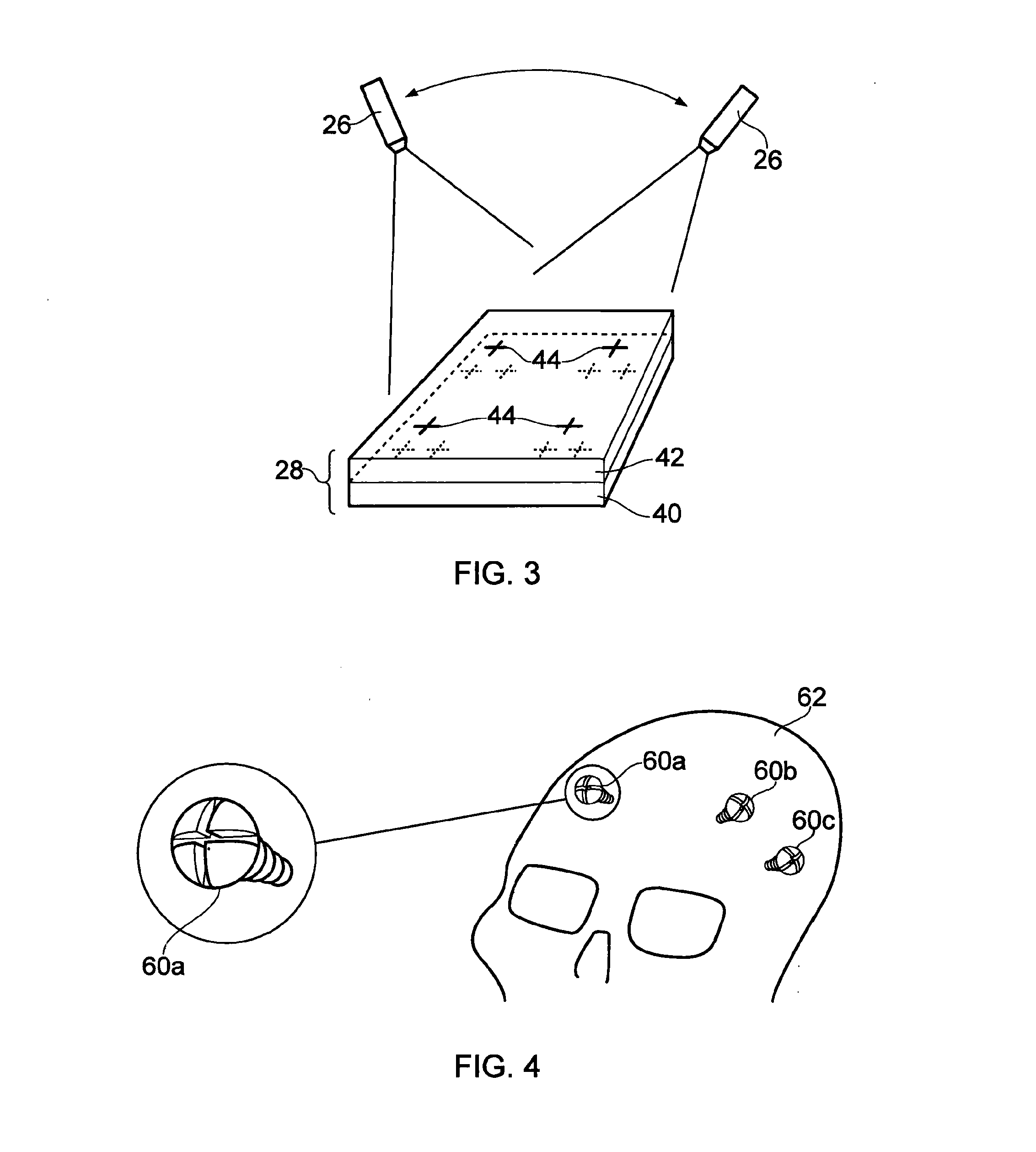
[0030]As explained above, the apparatus preferably includes one or more x-ray visible fiducial markers and an analyser that is arranged to analyse one or more acquired x-ray images to determine the position of the x-ray visible fiducial markers in a coordinate system of the surgical robot. A plurality of x-ray images may be acquired with the x-ray source and / or x-ray detector in different positions to allow x-ray images of the subject to be taken from different perspectives. Analysis of such a plurality of x-ray images by the analyser allows the position of the position of the x-ray visible fiducial markers to be measured in three dimensions in the robot coordinate system. Advantageously, at least some of the plurality of x-ray visible fiducial markers have a predetermined positional relationship. For example, the relative position of at least some of the x-ray visible fiducial markers may be predetermined using, for example, a pre-operative (CT or MM) scan. The additional information about the relative position of at least some of the x-ray visible fiducial markers introduces known values in the mathematical analysis such that the accuracy of fiducial position information can be increased and / or fewer x-ray images are required to obtain position information with a certain accuracy.
