Identification of inflammation in tissue images

a tissue image and image technology, applied in the field of medical images, can solve the problems of difficult segmentation of histology tissue images, time-consuming and laborious,

Inactive Publication Date: 2017-03-16
RGT UNIV OF CALIFORNIA +1
View PDF1 Cites 15 Cited by
  • Summary
  • Abstract
  • Description
  • Claims
  • Application Information

AI Technical Summary

Benefits of technology

[0007]In accordance with yet another aspect of the present invention, a method is provided for identifying glands and villi within a tissue image. A superpixel segmentation of the image, comprising a plurality of superpixels, is generated. Each of the plurality of superpixels is classified as one of epithelium, lumen, and extracellular material. An initial pseudo-probability map is generated from the classified plurality of superpixels. A plurality of candidate objects in the tissue image is generated according to the generated initial pseudo-probability map. The pseudo-probability map is updated according to the newly generated candidate objects so that future candidates generated from the updated pseudo-probability map are more accurate. Each candidate object is classified by an associated pattern recognition classifier.

Problems solved by technology

Conventionally, in order to diagnose inflammatory diseases, H&E stained histology tissue images are manually analyzed by a trained pathologist, which can be tedious and time consuming.
However, segmentation in histology tissue images is very challenging, due to intra-class variations and inter-class similarity, complex and crowded background, and noisy data.

Method used


Image

  • Identification of inflammation in tissue images
  • Identification of inflammation in tissue images
  • Identification of inflammation in tissue images

Examples


Embodiment Construction

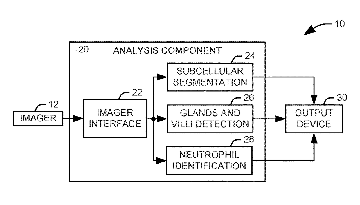
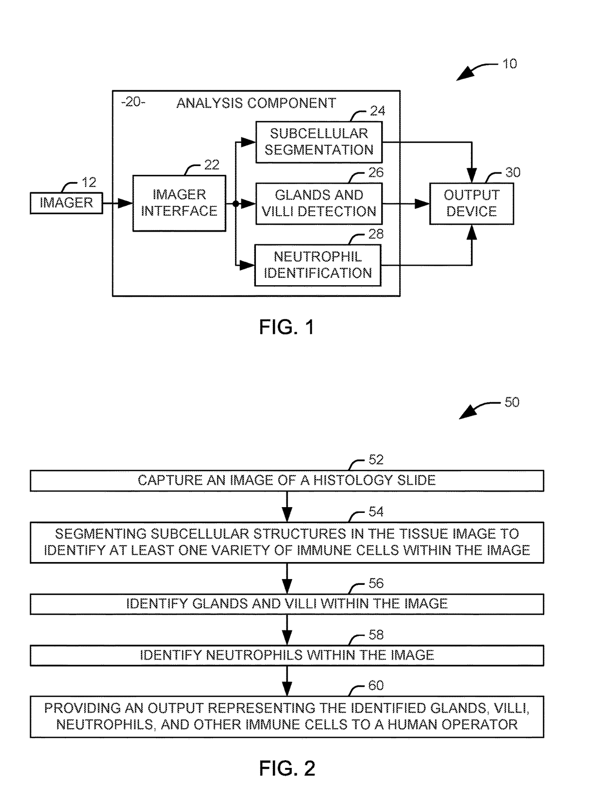
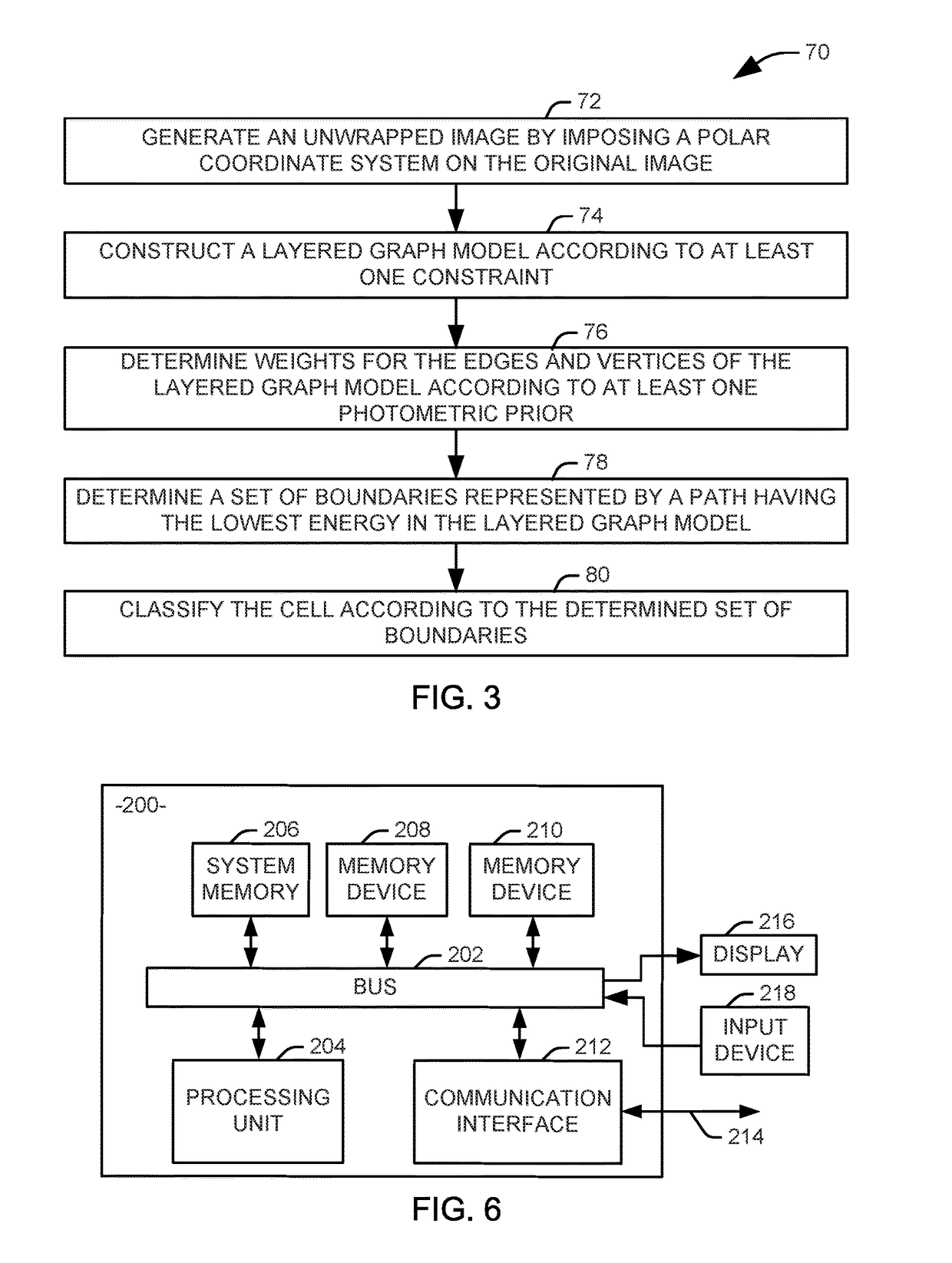
[0015]In general, this disclosure describes techniques for analyzing tissue images, such as hematoxylin and eosin (H&E) stained tissue images, to diagnose inflammatory diseases. There are four types of immune cells commonly analyzed in the diagnosis of inflammatory diseases: neutrophils, plasma cells, lymphocytes, and eosinophils, and the techniques described herein may be used to identify and classify these types of immune cells and provide analytic information that may assist diagnostic pathologists. Pathologists often rely on features of the subcellular structures to differentiate different types of immune cells, and accurate segmentation of the subcellular structures can help classify cell types. Further, architectural distortions of glands and villi are indicators of chronic inflammation, but the “duality” nature of these two structures causes ambiguity for their detection in H&E histology tissue images, especially when multiple instances are clustered closely together. In one ...


PUM


Abstract

Systems and methods are provided for identifying markers for inflammation in a tissue image. The tissue image is captured as an image of a histology slide. Subcellular structures in the tissue image are segmented via a first automated process to identify at least one variety of immune cells within the image. Glands and vilii are identified within the tissue image via a second automated process. Neutrophils are identified within the tissue image via a third automated process. An output representing the identified glands, villi, neutrophils, and other immune cells is provided to a human operator.

Description

RELATED APPLICATIONS[0001]This application claims priority from U.S. Patent Application Ser. No. 62 / 218,351, filed 14 Sep. 2015, which is incorporated herein in its entirety.GOVERNMENT SUPPORT[0002]This invention was made with government support under Grant No. CCF-1217906 awarded by the National Science Foundation (NSF), and Grant No. K08-AR061412-02 awarded by the National Institute of Health (NIH). The government has certain rights in the invention.TECHNICAL FIELD[0003]This invention relates to medical images, and more particularly, to identifying inflammation in tissue images.BACKGROUND[0004]Human or mammalian tissue samples (e.g., samples taken during a biopsy procedure) may be processed using hematoxylin and eosin (H&E) staining techniques in order to generate H&E stained histology tissue images. H&E stained histology tissue images may be analyzed to diagnose diseases, including inflammatory diseases. Conventionally, in order to diagnose inflammatory diseases, H&E stained hist...

Claims


Application Information

Patent Timeline
Patent Type & Authority Applications(United States)
IPC IPC(8): G06T7/00G06K9/00G06K9/62G06V10/46
CPCG06T7/0012G06T7/0093G06T7/0087G06K9/6297G06T2207/10056G06K9/00147G06K9/0014G06T2207/30088G06T2207/30024G06T7/0083G06T2207/10024G06T7/12G06T7/162G06V20/695G06V20/698G06V10/46
Inventor CHEN, DANNY ZIYIWANG, JIAZHUOMACKENZIE, JOHN DEWOLFERAMACHANDRAN, RAGESHREE
Owner RGT UNIV OF CALIFORNIA
Why Eureka
Try Eureka