

Self-calibrating projection geometry for volumetric image reconstruction
a volumetric image and projection geometry technology, applied in the field of medical imaging, can solve the problems of geometric calibration errors, blurry images, geometric distortion, other artifacts, etc., and achieve the effect of improving the accuracy of geometric calibration
- Summary
- Abstract
- Description
- Claims
- Application Information
AI Technical Summary
Benefits of technology
Problems solved by technology
Method used
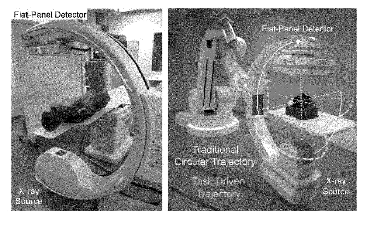
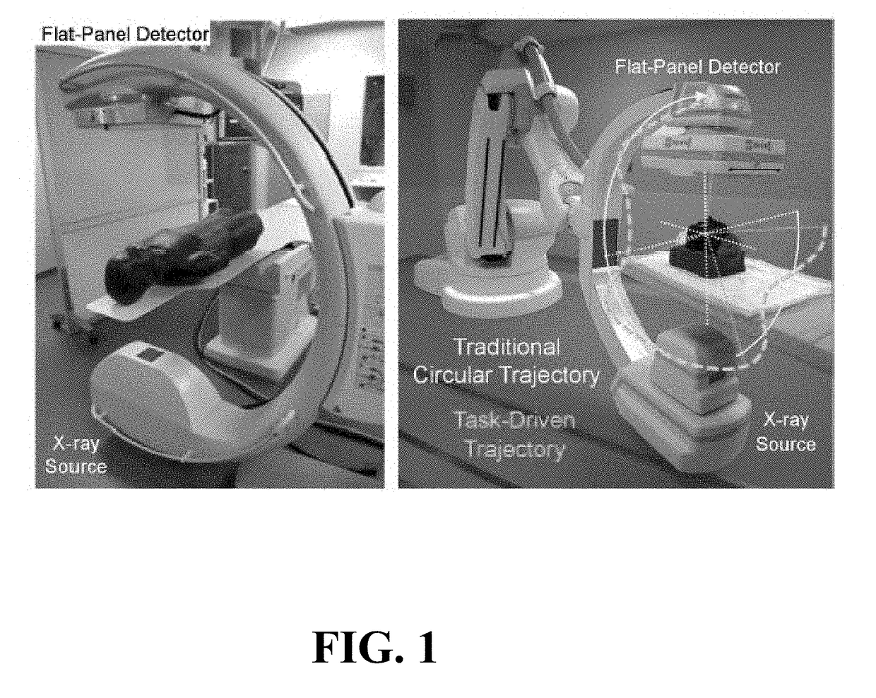
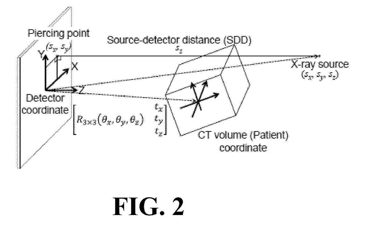
Image

Examples
experiment 1
[0084]
[0085]Cylinder phantom on imaging bench. Experiment 1 involved the cylinder phantom imaged on the CBCT bench to test the feasibility of the self-calibration method and obtain quantitative analysis of basic performance. A circular orbit was used, with the nominal scan technique described above. A previous CBCT scan of the phantom formed the 3D image input to the self-calibration method, with the previous scan acquired with an angular offset in projection views so that the projections used in 3D reconstruction were not identical to those in 3D-2D registration.
experiment 2
[0086]
[0087]Anthropomorphic head phantom on imaging bench. Experiment 2 involved the anthropomorphic head phantom imaged on the CBCT bench to test the robustness of 3D-2D registration under more clinically / anatomically realistic conditions of x-ray scatter, image noise, and complexity of the subject. A previous scan of the head phantom on a diagnostic CT scanner (Siemens Somatom Definition, 120 kVp, 227 mAs, (0.46×0.46×0.40 mm3) voxels) formed the 3D image input to the self-calibration method.
experiment 3
[0088]
[0089]Anthropomorphic head phantom on robotic C-arm. Experiment 3 involved the anthropomorphic head phantom imaged on the Artis Zeego to test the method in a clinically realistic system geometry and orbit. A previous CBCT scan of the head phantom acquired using the Zeego formed the 3D image input to the self-calibration method. To challenge the method further, a realistic, pronounced change in image content was produced between the previous 3D image and the projection images acquired in the current CBCT scan—viz., a 2 mm diameter steel biopsy needle placed in the nasal sinuses and positioning the head with a strong) (˜30° canthomeatal tilt to mimic a typical clinical setup. The reference calibration (using the spiral BB phantom mentioned above) was performed by the system service engineer as part of regular preventative maintenance within 6 months of the current scan in accordance with standard clinical practice.
PUM
 Abstract
Description
Claims
Application Information