Method and microscope for measuring and calculating heights on curved surface of microscope slide
a technology of curved surface and microscope, which is applied in the direction of microscopes, measurement devices, instruments, etc., can solve the problems of inability to achieve auto-focus fast photographing and scanning, and achieve the effects of accurate measurement, high imaging definition, and fast micro-image scanning of sample objects
- Summary
- Abstract
- Description
- Claims
- Application Information
AI Technical Summary
Benefits of technology
Problems solved by technology
Method used
Image

Examples
embodiment 1
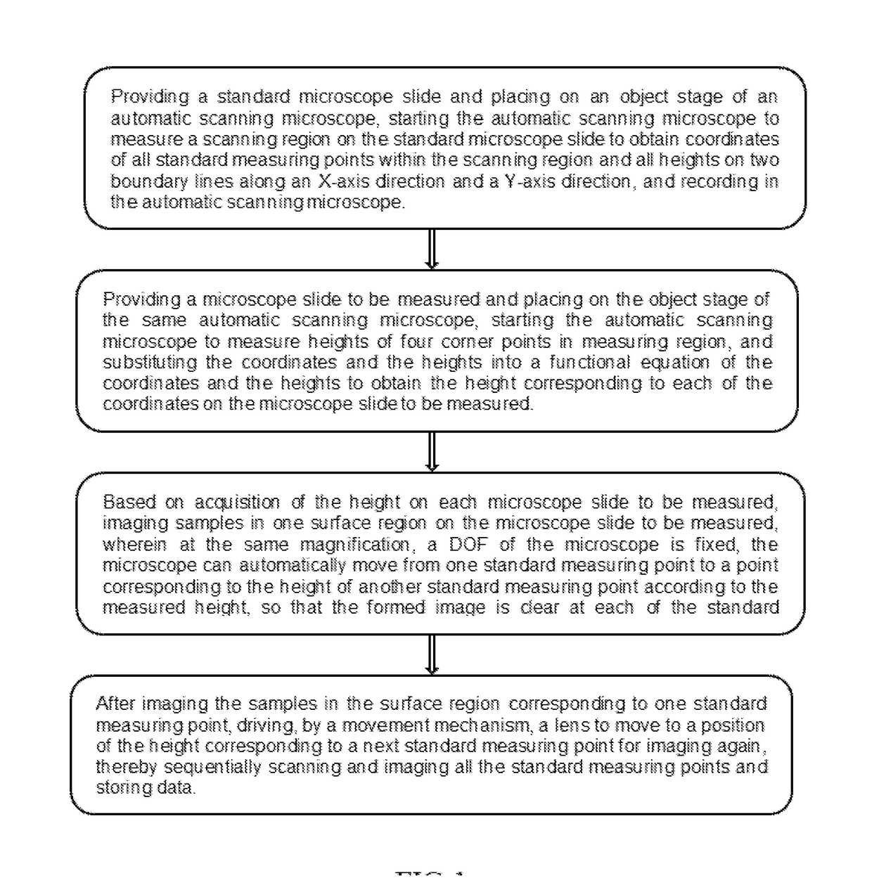
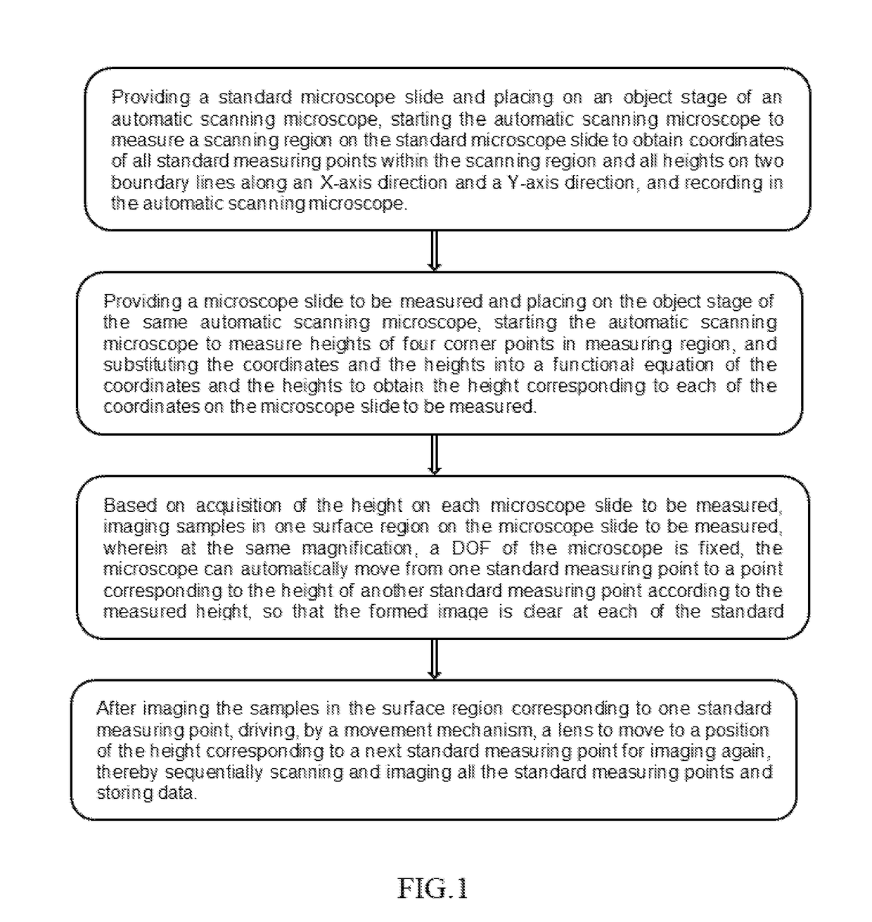
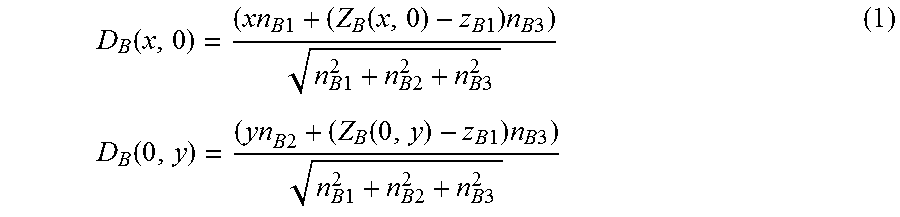
[0038]Scanning of standard microscope slides: preparing a plurality of standard microscope slides; taking a plurality of blank microscope slides; uniformly coating a single layer of the sample on the microscope slides; accurately placing the microscope slides on a sample rack of a scanning instrument; setting a software to define a scanning region of 2.5 cm×1.5 cm in length and width; sampling scanning the region with 27 visual fields scanned at equal intervals in an X-axis direction, 13 visual fields scanned at equal intervals in a Y-axis direction and 351 visual fields sampled in the overall region; and after dropping oil onto the sample, using an NA 1.25 100× lens of olympus company to focus a first line x(i, 0) (a value of i is 0-26) so as to obtain 27 focused heights ZB(i, 0) on an x axis:
0.5, 0.684568671, 0.913734812, 0.960046768, 1.024200047, 0.785085559, 0.871537623, 0.873252109, 0.464664036, 0.591254547, 0.601890489, 0.313716038, 0.444767363, 0.040638322, 0.055733448, −0.00...
embodiment 2
[0053]A Microscopic Scanning Method of the Microscope:
[0054]Scanning patterns of the samples on the microscope slides by applying the heights on the curved surfaces of the microscope slides obtained in the embodiment 1.
[0055]A single layer of samples (which are microspheres uniform in size in the present embodiment) are uniformly coated on the microscope slides. The software is set to define the scanning region of 2.5 cm×1.5 cm in length and width. The region is sampling scanned by the microscope with 27 visual fields scanned at equal intervals in the X-axis direction, 13 visual fields scanned at equal intervals in the Y-axis direction, and 351 visual fields sampled in the overall region. The microspheres are uniformly distributed in the 351 visual fields.
[0056]The heights of the focused points within the 351 visual fields are obtained by the method of the embodiment 1. Under the NA 1.25 100× lens, the DOF of the microscope lens is fixed. Therefore, it only needs to adjust a distanc...
PUM
 Abstract
Description
Claims
Application Information
 - R&D
- Intellectual Property
- Life Sciences
- Materials
- Tech Scout
- Unparalleled Data Quality
- Higher Quality Content
- 60% Fewer Hallucinations
Browse by: Latest US Patents, China's latest patents, Technical Efficacy Thesaurus, Application Domain, Technology Topic, Popular Technical Reports.
© 2025 PatSnap. All rights reserved.Legal|Privacy policy|Modern Slavery Act Transparency Statement|Sitemap|About US| Contact US: help@patsnap.com