
Medical device for detecting at least one analyte in a body fluid

Pending Publication Date: 2019-01-24
ROCHE DIABETES CARE INC
View PDF0 Cites 3 Cited by
  • Summary
  • Abstract
  • Description
  • Claims
  • Application Information

AI Technical Summary

Benefits of technology

This patent describes an analyte sensor that can be placed under the skin of a patient or user. The sensor has a part that is inserted into the body tissue and is covered by a biocompatible membrane. The sensor is designed to be small and easily inserted into the skin, using an insertion aid, and can be connected to an evaluation device without needing to open a sterile range. This makes the system compact and easy to use. The technical effect is that the invention provides a simple and effective way to measure certain substances in the body.

Problems solved by technology

Despite the advantages and the progress achieved by the above-mentioned developments, specifically in the field of continuous monitoring technology, some significant technical challenges remain.
Thus, generally, known techniques for protecting an electrical contact between a sensor and a control part generally are rather complex.
An assembly of a plurality of components is generally required, which typically implies a complex and costly manufacturing process.
Further, known techniques generally require voluminous components, which is an issue, specifically considering the fact that miniaturizing the sensor systems is a factor contributing to the convenience of use.
Specifically in case complex encapsulation parts manufactured by plastic molding techniques are required for protecting the electrical contacts, a rising of costs and sensor volume typically has to be taken into account.
Further, cleaning of complex protective covers, such as protections including O-rings or other seals, turns out to be difficult.

Method used


Image

  • Medical device for detecting at least one analyte in a body fluid
  • Medical device for detecting at least one analyte in a body fluid
  • Medical device for detecting at least one analyte in a body fluid

Examples


Embodiment Construction

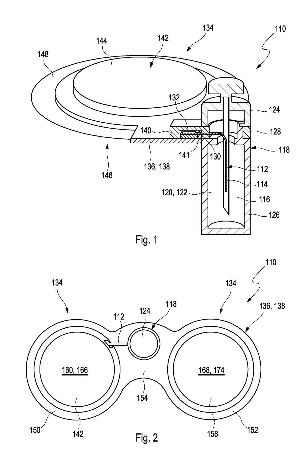
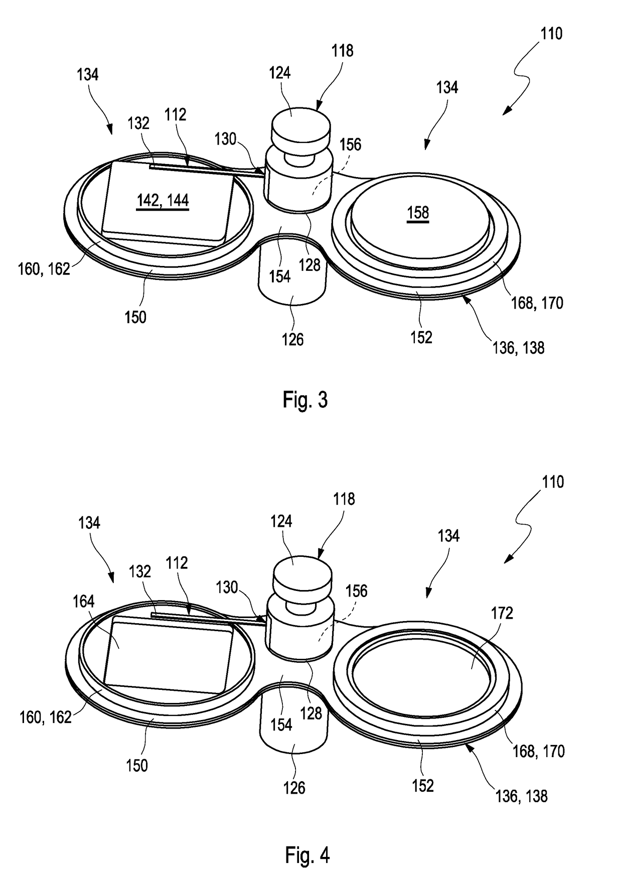
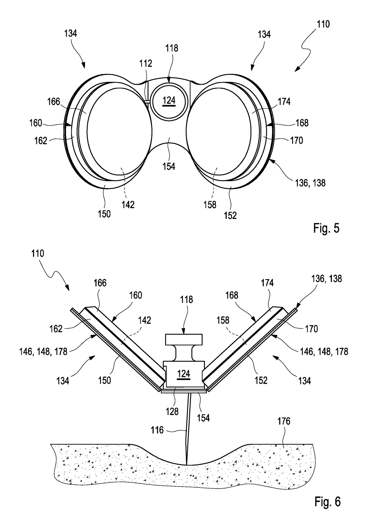
[0159]In FIG. 1, a cross-sectional view of a first embodiment of a medical device 110 for detecting at least one analyte in a body fluid is shown. The medical device 110 comprises an analyte sensor 112, which, as an example, may be embodied as flexible analyte sensor and which preferably is embodied as an electrochemical analyte sensor. The analyte sensor 112 comprises an insertable portion 114 which is configured for insertion into a body tissue of a user.

[0160]The medical device 110 further comprises an insertion cannula 116, which, as an example, may be embodied as a slotted insertion cannula 116. Other types of insertion cannulae are feasible, too. The insertable portion 114 of the analyte sensor 112 is located inside the insertion cannula 116.

[0161]The medical device 110 further comprises a housing 118, enclosing a sensor compartment 120. The sensor compartment 120 forms a sealed compartment 122 which is sealed against an environment of the medical device 110. The sealed compar...


PUM


Abstract

A medical device for detecting at least one analyte in a body fluid is disclosed. The medical device comprises:at least one analyte sensor having an insertable portion adapted for at least partially being inserted into a body tissue of a user,at least one insertion cannula, wherein the analyte sensor at least partially is placed inside the insertion cannula;at least one housing, wherein the housing comprises at least one sensor compartment, wherein the sensor compartment forms a sealed compartment receiving at least the insertable portion of the analyte sensor, wherein the sealed compartment comprises at least one detachable upper cap and at least one detachable lower cap, wherein the detachable lower cap is configured for detachment before insertion, thereby opening the insertable portion for insertion, wherein the insertion cannula is attached to the detachable upper cap, wherein the detachable upper cap is configured for detachment after insertion, thereby removing the insertion cannula; andat least one electronics unit, wherein the analyte sensor is operably connected to the electronics unit, wherein the electronics unit comprises at least one interconnect device with at least one electronic component attached thereto, wherein the interconnect device fully or partially surrounds the housing.

Description

FIELD OF THE INVENTION[0001]The invention relates to a medical device for detecting at least one analyte in a body fluid, a method for assembling a medical device and a method of using a medical device. The device and methods according to the present invention may mainly be used for long-term monitoring of an analyte concertation in a body fluid, such as for long-term monitoring of a blood glucose level or of the concentration of one or more other types of analytes in a body fluid. The invention may both be applied in the field of home care as well as in the field of professional care, such as in hospitals. Other applications are feasible.RELATED ART[0002]Monitoring certain body functions, more particularly monitoring one or more concentrations of certain analytes, plays an important role in the prevention and treatment of various diseases. Without restricting further possible applications, the invention will be described in the following text with reference to blood-glucose monitor...

Claims


Application Information

Patent Timeline
IPC IPC(8): A61B5/1473A61B5/145A61B5/00B08B7/00B08B5/00
CPCA61B5/1473A61B5/14532A61B5/14546A61B5/6849B08B7/0035B08B5/00A61B2560/063A61B2562/166A61B5/688A61B2562/242A61B5/14503A61B5/14865
Inventor WALTER, HELMUT
Owner ROCHE DIABETES CARE INC
Learn More