
Method for characterization of cell specific microvesicles

a cell specific and microvesicle technology, applied in the field of cell specific microvesicles characterization, can solve the problems of limiting the application of this technology, the lack of a selective characterization and quantification method of circulating cardiac-derived mv, and the leading cause of morbidity and mortality among adults

Inactive Publication Date: 2019-03-14
HUMANITAS MIRASOLE SPA
View PDF2 Cites 0 Cited by
  • Summary
  • Abstract
  • Description
  • Claims
  • Application Information

AI Technical Summary

Benefits of technology

The patent shows that measuring specific microvesicles in the body, particularly heart-derived microvesicles, can help diagnose and monitor the health of the cardiovascular system. This is useful because these microvesicles can provide a reliable indicator of stress on the heart and blood vessels.

Problems solved by technology

Cardiovascular diseases continue to be a leading cause of morbidity and mortality among adults in all economically advanced Countries.
The identification of clear biomarkers related to a high risk of cardiovascular diseases remains still largely elusive due to the heterogeneity of the pathological processes underlying these diseases.
The absence of a method for selectively characterizing and quantifying circulating cardiac-derived MV is a limiting factor for applying this technology to disease staging and prediction of acute events.

Method used


Image

  • Method for characterization of cell specific microvesicles
  • Method for characterization of cell specific microvesicles
  • Method for characterization of cell specific microvesicles

Examples


example 1

[0183]Materials and Methods

[0184]Plasma Isolation and Storage

[0185]According to published recommendations, plasma sampling and storage techniques were standardized for overall patients and healthy subjects.

[0186]Informed consent was obtained from each patient and healthy subjects.

[0187]A 5 ml sample of peripheral blood was collected in EDTA-containing Vacutainer tubes. The vials were processed within 2 h of collection by centrifugation at 1200×g at room temperature in a bench top centrifuge for 20 minutes to eliminate all blood cells. To further reduce leukocyte and red cells contamination, the top third of the plasma was aspirated and placed in fresh tubes and frozen at −80° C.

[0188]MV Isolation from Plasma.

[0189]Human plasma was diluted with filtered PBS− / − (no calcium and magnesium) and centrifuged at 500 g×30 minutes at 4° C. The obtained Platelet Free Plasma (PFP) was centrifuged at 12000 g×45 minutes at 4° C. Finally, the supernatant was removed leaving 25 μl of a MV-enriched ...

example 2

[0237]MV Isolation and Storage.

[0238]According to published recommendations, plasma samples and storage techniques were standardized. A 5 ml sample of peripheral blood was collected in EDTA-containing Vacutainer tubes. The vials were processed within 2 h of collection by centrifugation at 1,200 g×20 min. at room temperature (RT) to eliminate all blood cells. To further reduce leukocyte and red cell contamination, the top third of the plasma was aspirated and placed in a fresh tube. According to the literature, isolated plasma was subsequently diluted with filtered PBS and centrifuged at 500 g×30 min. at 4° C. The obtained platelet-free plasma was centrifuged at 12,000 g×45 min. at 4° C. Finally, the supernatant was removed, leaving 25 μl of an MV-enriched suspension, which was diluted with 75 μl of filtered PBS (Caby M P, et al. Int Immunol. 2005 July; 17(7):879-87).

[0239]FACS Analysis.

[0240]To limit background noise from dust and crystals, a 0.22 μm filtered sheath fluid was used f...


PUM


Abstract

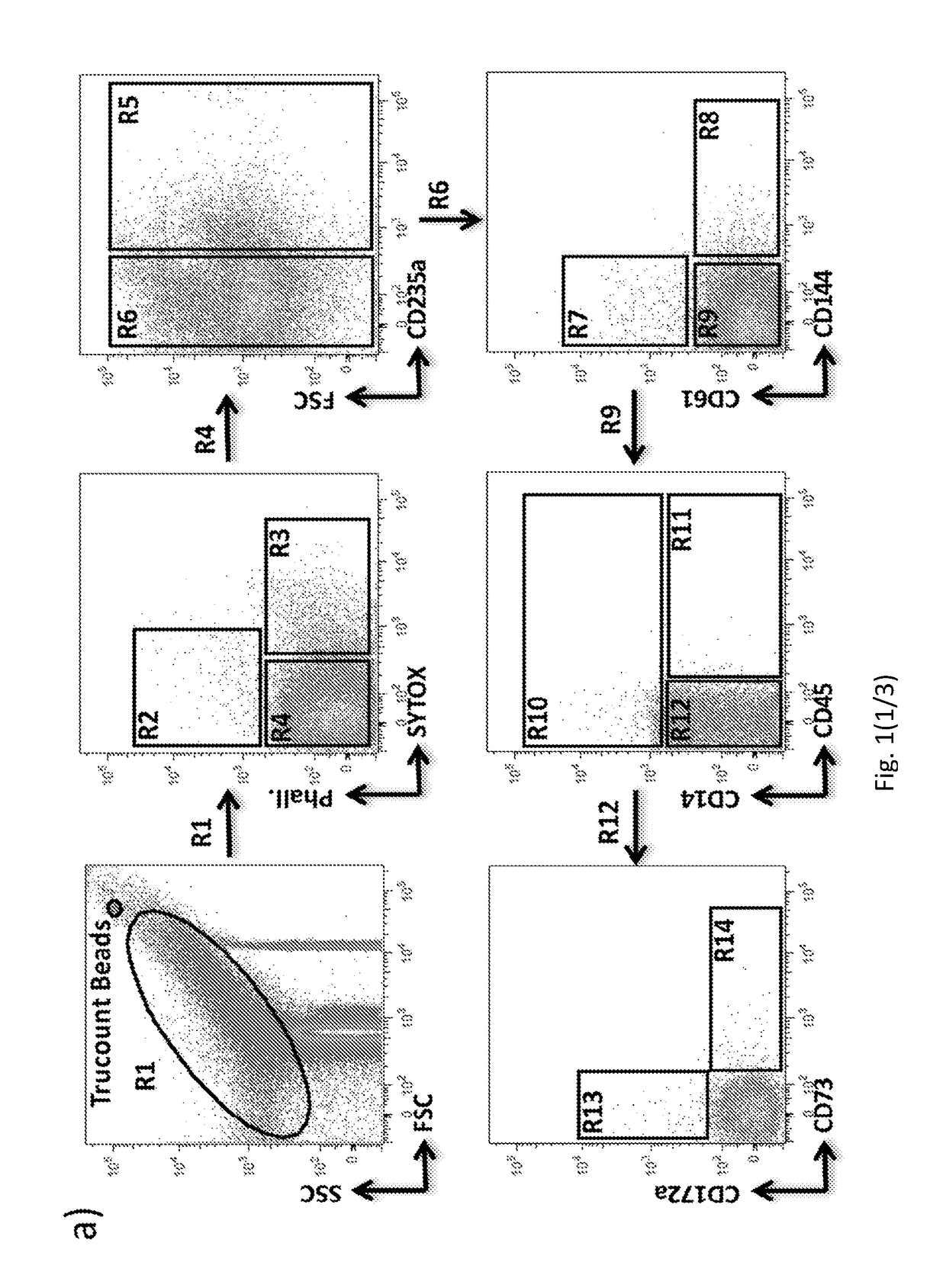
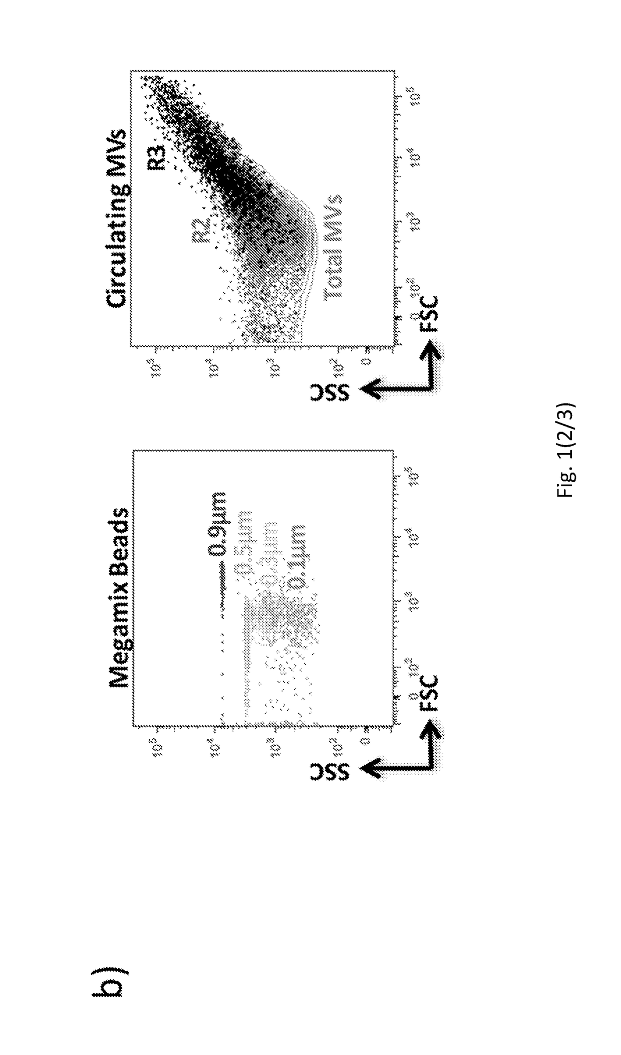
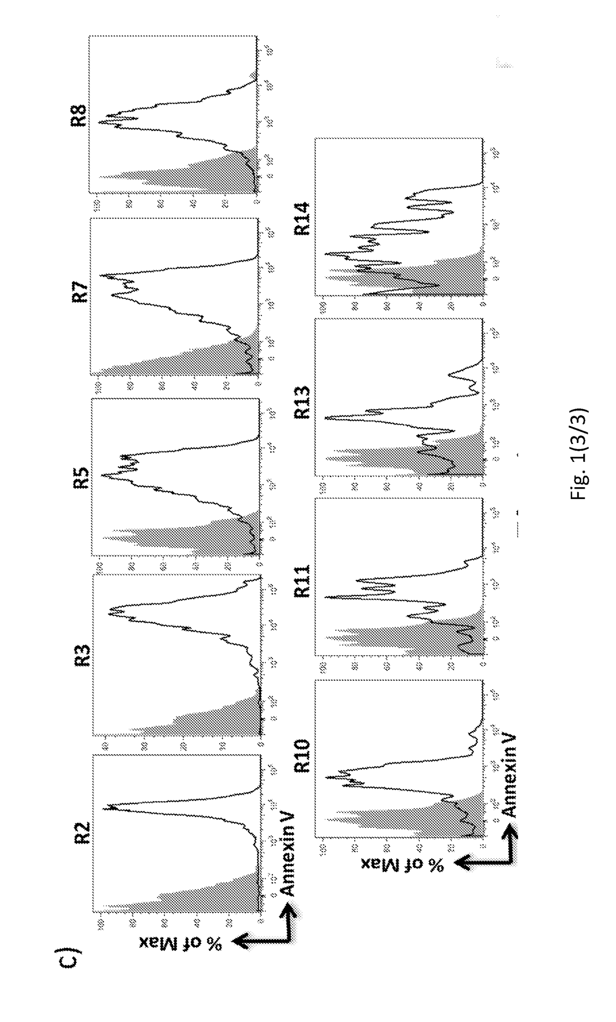
The present invention refers to a method for characterizing and / or measuring and / or sorting the amount of cardiac-derived microvesicles, having the step of detecting CD172a marker on the microvesicles, in an isolated biological sample obtained from the subject.

Description

FIELD OF THE INVENTION[0001]The present invention relates to a method for characterizing, quantifying and sorting the circulating cell specific microvesicles (MV), in particular the cardiac-derived MV.[0002]The present invention also refers to methods for diagnosing and / or assessing the risk of developing and / or for prognosing and / or for monitoring the progression and / or for monitoring the efficacy of a therapeutic treatment and / or for the screening of a therapeutic treatment of a disease, in particular of cardiovascular diseases, by characterizing, quantifying and sorting the circulating cardiac-derived microvesicles (MV).BACKGROUND ART[0003]Cardiovascular diseases continue to be a leading cause of morbidity and mortality among adults in all economically advanced Countries. The identification of clear biomarkers related to a high risk of cardiovascular diseases remains still largely elusive due to the heterogeneity of the pathological processes underlying these diseases. Therefore,...

Claims


Application Information

Patent Timeline
IPC IPC(8): G01N33/68G01N33/543
CPCG01N33/6893G01N33/54326G01N33/6842G01N2333/70596G01N33/56972G01N33/80G01N33/56966G01N2333/70589
Inventor CONDORELLI, GIANLUIGIANSELMO, ACHILLEVIVIANI ANSELMI, CHIARAPAPA, LAURA
Owner HUMANITAS MIRASOLE SPA
Learn More