Massively Multi-Frequency Ultrasound-Encoded Tomography

a multi-frequency, ultrasonically encoded technology, applied in tomography, instruments, active medium materials, etc., can solve the problems of not having a good solution to get and the challenge of getting a high-resolution spatial map

Inactive Publication Date: 2019-03-21
CHARLES STARK DRAPER LABORATORY
View PDF0 Cites 9 Cited by
  • Summary
  • Abstract
  • Description
  • Claims
  • Application Information

AI Technical Summary

Benefits of technology

[0008]Embodiments of the present invention are directed to computer-implemented systems for multi-frequency ultrasonically-encoded optical tomography of a target object such as a brain of a patient. One or more probe inputs are configured for generating optical probe input signals to the target object. An ultrasound transducer array is configured for placement on the outer surface of the target object and has multiple ultrasound transducers each operating at a different ultrasound frequency to generate ultrasound input signals to a target probe volume within the target object. A photorefractive crystal is configured for mixing scattered light output signals from the target probe volume with an optical reference beam input to the photorefractive crystal to produce optical tomography output signals including ultrasound sum frequencies components. A photodetector are configured for sensing the optical tomography output signals from the photorefractive crystal, in the form of intensity modulation of the scattered light beam passing through the photorefractive crystal. Data storage memory is configured for storing optical tomography software, the optical tomography output signals, and other system information. A tomography processor includes at least one hardware processor coupled to the data storage memory and configured to execute the optical tomography software including instructions to perform acousto-optic tomography analysis of the optical tomography output signals including ultrasound sum frequencies components to create a three-dimensional object map representing structural and / or functional characteristics of the target object.

Problems solved by technology

Unfortunately, red and NIR light undergoes multiple scattering which obfuscates the spatial structure of the target tissue, thus making it very challenging to get a high-resolution spatial map.
There is currently no good solution to this problem.

Method used


Image

  • Massively Multi-Frequency Ultrasound-Encoded Tomography
  • Massively Multi-Frequency Ultrasound-Encoded Tomography
  • Massively Multi-Frequency Ultrasound-Encoded Tomography

Examples


Embodiment Construction

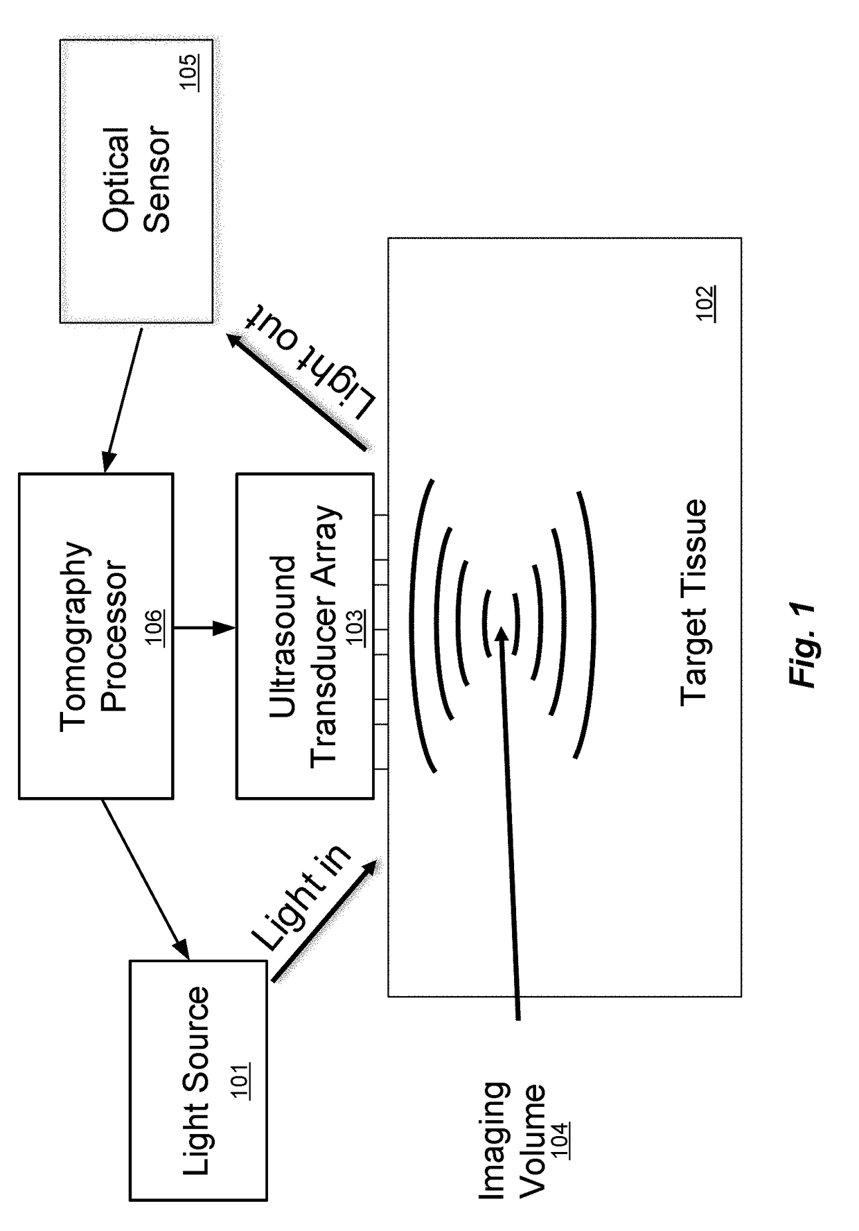
[0028]The discussion that follows is set forth in terms of examples of multi-frequency ultrasonically-encoded tomography that specifically perform ultrasonically-encoded optical tomography. But the skilled person will understand that the invention is not limited to such applications and includes other specific forms of ultrasonically-encoded tomography as explained later. In addition, the following discussion and examples are set forth in terms of red / infrared imaging of the brain. But the various discussed techniques may be useful for any medium which is highly scattering to light. Other specific applications include other tissues (e.g. breast cancer diagnostics), imaging in turbid water, generating a 3D refractive index map of water to infer its temperature profile, microwave probing of the brain and other tissues, microwave probing of pipes and other infrastructure and geological features, and so on. Also, the discussion is set forth using terms like “light” and “optical”, it wil...


PUM


Abstract

A system and corresponding method are described for multi-frequency ultrasonically-encoded tomography of a target object. One or more probe inputs generate probe input signals to the target object. An ultrasound transducer array is placed on the outer surface of the target object and has multiple ultrasound transducers each operating at a different ultrasound frequency to generate ultrasound input signals to a target probe volume within the target object. A photorefractive crystal mixes scattered light output signals from the target probe volume with an optical reference beam input to produce optical tomography output signals including ultrasound sum frequencies components. A photodetector senses the optical tomography output signals from the photorefractive crystal. A tomography analysis of the tomography output signals including the ultrasound sum frequencies components is performed to create a three-dimensional object map representing structural and/or functional characteristics of the target object.

Description

[0001]This application claims priority from U.S. Provisional Patent Application 62 / 653,646, filed Apr. 6, 2018, and U.S. Provisional Patent Application 62 / 621,100, filed Jan. 24, 2018, and U.S. Provisional Patent Application 62 / 582,391, filed Nov. 7, 2017, and U.S. Provisional Patent Application 62 / 559,779, filed Sep. 18, 2017, all of which are incorporated herein by reference in their entireties.TECHNICAL FIELD[0002]The present invention relates to multi-frequency arrangements for ultrasonically-encoded tomography.BACKGROUND ART[0003]Tomography refers to the imaging of a target object by sections using of any kind of penetrating wave. One family of tomography techniques is variously called ultrasound-encoded tomography, ultrasound-modulated tomography, or various more specific terms as discussed below. Generally this involves some form of probe input signals (e.g., an electrical signal injected by an electrode, current induced by a changing current in a magnetic coil, microwave-fre...

Claims


Application Information

Patent Timeline
Patent Type & Authority Applications(United States)
IPC IPC(8): A61B8/15G01S15/89H01S3/30H01S3/16
CPCA61B8/15G01S15/8968G01S15/8952H01S3/302H01S3/1666G16H30/20A61B5/0097A61B6/032A61B6/5205A61B8/4477G02B27/12A61B5/0073A61B8/4494G01N21/1717A61B6/501
Inventor BYRNES, STEVEN J.HOLLMANN, JOSEPH
Owner CHARLES STARK DRAPER LABORATORY
Why Eureka
Try Eureka