A novel invadopodia-specific marker of invasive cancer stem cells and the use thereof

a cancer stem cell and invadopodia technology, applied in the field of invadopodia-specific markers of invasive cancer stem cells, can solve the problems of ineffective eradication of limitations of current cancer therapies, and inability to effectively kill solid cancer stem cells, and achieve high likelihood of invading and high likelihood of causing severe damag

Inactive Publication Date: 2019-08-08
TSAI KUN CHIH +1
View PDF0 Cites 1 Cited by
  • Summary
  • Abstract
  • Description
  • Claims
  • Application Information

AI Technical Summary

Problems solved by technology

Another possibility is that most cancer cells lack the ability to form a new tumor such, that only the dissemination of rare CSCs can lead to metastatic disease.
Since current therapies are directed against the bulk population, they may be ineffective at eradicating solid cancer stem cells.
The limitations of current cancer therapies derive from their inability to effectively kill solid cancer stem cells.
Consistently, depletion of caveolin disrupts the association of essential components of invadopodia / podosomes, including Src kinases, beta1-integrin and urokinase receptor (uPAR), thereby compromising the migration of cells on ECM (Wei et al., 1999).

Method used


Image

  • A novel invadopodia-specific marker of invasive cancer stem cells and the use thereof
  • A novel invadopodia-specific marker of invasive cancer stem cells and the use thereof
  • A novel invadopodia-specific marker of invasive cancer stem cells and the use thereof

Examples


example 1

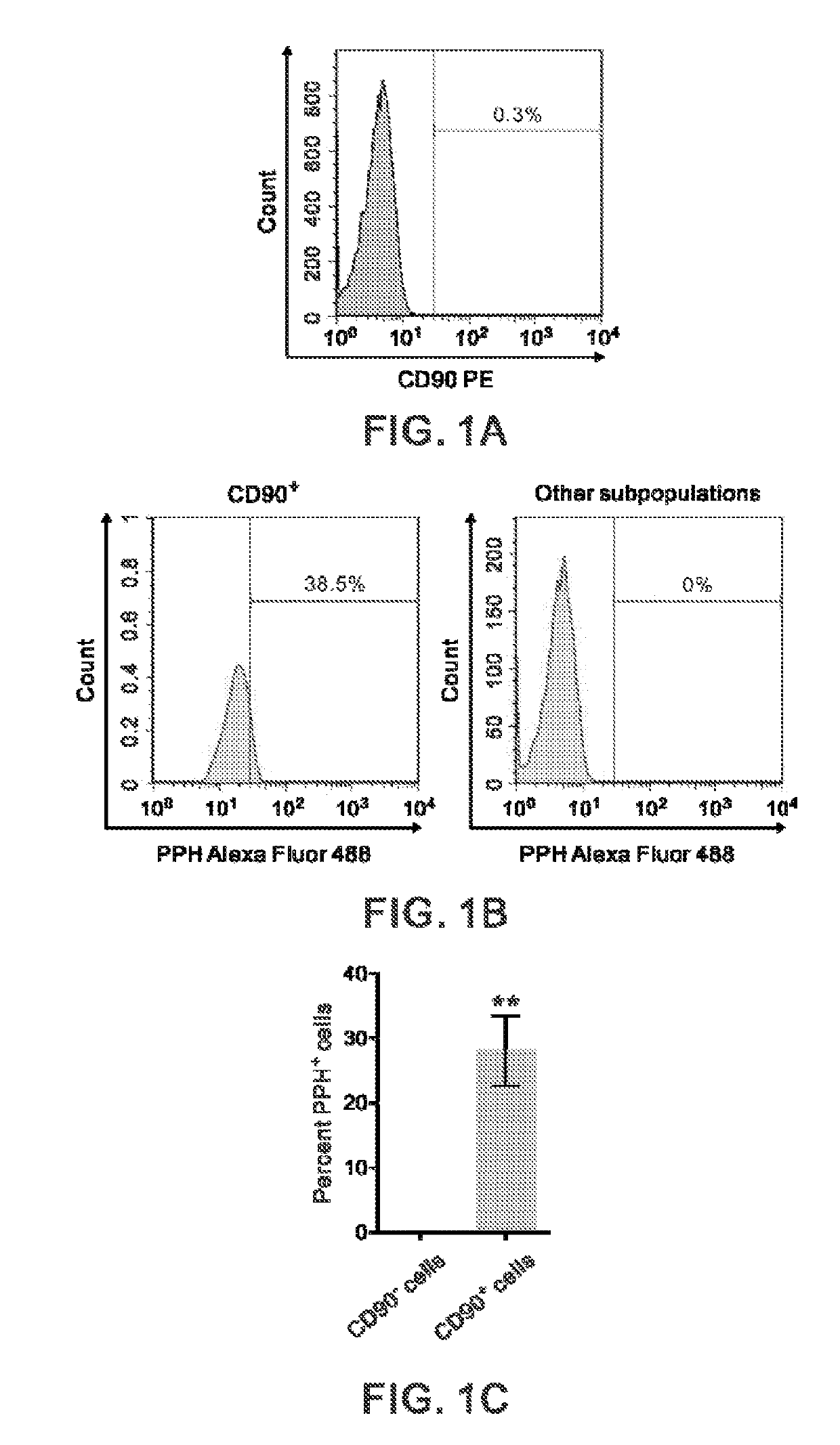
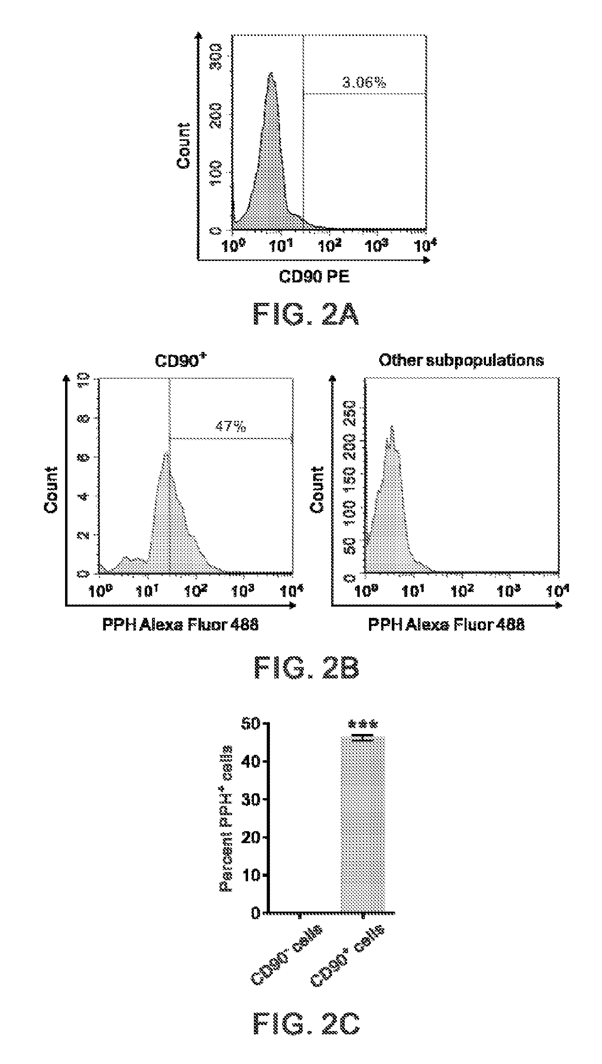
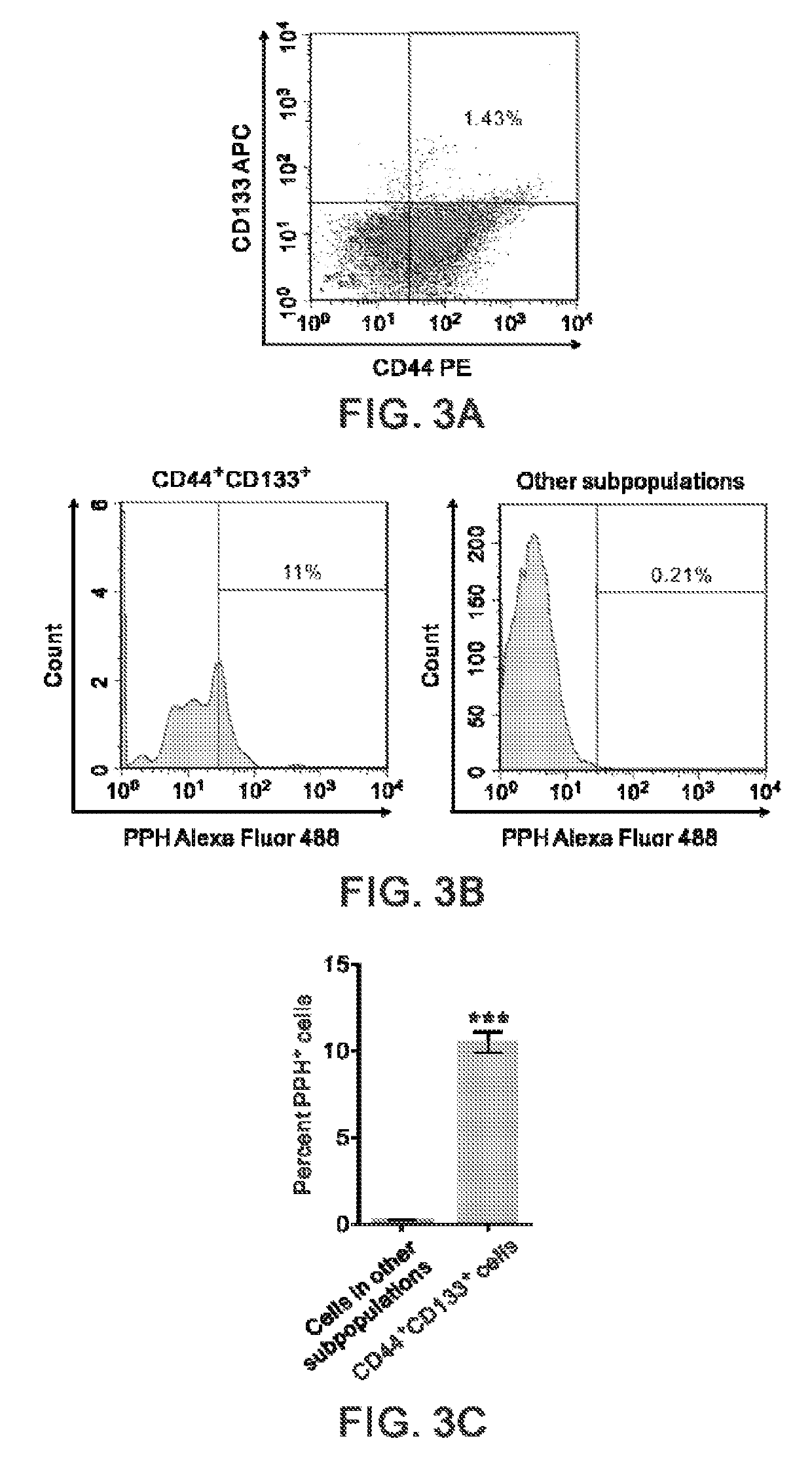
[0110]This example describes that cell-surface PPH marks a subpopulation of CSCs in gastric cancer and prostate cancers.

[0111]We analyzed the expression of cell surface PPH in gastric cancer and pancreatic cancer cells by the fluorescence-assisted cell sorting (FACS) analysis. We measured the proportion of primary gastric cancer-derived AGS cells (American Type Culture Collections) and metastatic gastric cancer SNU-16 cells (American Type Culture Collections) that expressed the surface marker CD90, which have been shown to contain the enriched CSCs in gastric cancer (Jiang et al., 2012). We also measured the proportion of primary prostate cancer-derived 22Rv-1 cells and metastatic prostate cancer-derived PC-3 cells (both from American Type Culture Collections) that expressed that surface markers CD133 and CD44, which have been shown to contain the enriched CSCs in prostate cancer (Dubrovska et al., 2009; Richardson et al., 2004). For the FACS analysis, cells were dissociated, antibo...

example 2

[0114]This example describes that cell-surface PPH marks a subpopulation of CSCs in malignant glioma and bladder cancer.

[0115]We analyzed the expression of cell surface PPH in malignant glioma and HCC cells by the FACS analysis. I measured the proportion of two malignant glioma (glioblastoma) cell lines, U-87MG and Hs-683 cells (both obtained from American Type Culture Collections) that expressed the surface marker CD133, which have been shown to contain the enriched glioma stem cells (Singh et al., 2004). We also measured the proportion of the bladder cancer line T24 (obtained from American Type Culture Collections) that expressed that surface marker CD44, which have been shown to contain the enriched CSCs in bladder cancer (Chan et al., 2009). For the FACS analysis, cells were dissociated, antibody-labeled (1-2 μg per 106 cells×1 hour) and resuspended in HBSS / 2% FBS as previously described (Hermann et al., 2007; Li et al., 2007). The antibodies used included APC-anti-CD133 (Milten...

example 3

[0117]This example describes that cell-surface PPH marks a subpopulation of LSCs in acute myeloid leukemia.

[0118]We analyzed the expression of cell surface PPH in AML cells by the FACS analysis. I measured the proportion of AML THP-1 cells and HL-60 cells (both obtained from American Type Culture Collections) that expressed CD34 but lacked the expression of CD38, which have been shown to contain the enriched LSCs in AML (van Rhenen et al., 2005). For the FACS analysis, cells were dissociated, antibody-labeled (1-2 μg per 10.sup.6 cells×1 hour) and resuspended in HBSS / 2% FBS as previously described (Hermann et al., 2007; Li et al., 2007). The antibodies used included FITC-CD34, APC-CD38 (both from BD Biosciences) and anti-PPH (PAS-12051; Thermo Scientific) in conjunction with Alexa Fluor 488-anti-mouse IgG (Invitrogen). Flow cytometry was done using a FACSAria III (BD Biosciences).

[0119]As shown in FIGS. 7A-7C, the majority (77.3% on average) of CD34.sup.+.CD38.sup.−. THP-1 cells wer...


PUM

PropertyMeasurementUnit
sizeaaaaaaaaaa
sizeaaaaaaaaaa
affinityaaaaaaaaaa

Abstract

The present invention provides an invasive cancer stem cell (iCSC) or a substantively homogeneous cell population including said iCSC based on a cell-surface biomarker specifically localizing to the invadopodia of said iCSC. The present invention further provides an invasive leukemia stem cell (iLSC) or a substantively homogeneous cell population including said iLSC based on a cell-surface biomarker specifically localizing to the invadopodia of said iLSC. The present invention also provides a method or a kit of determining a diagnosis of aggressive solid tumor or hematopoietic cancer.

Description

[0001]This application claims priority to U.S. Provisional Application Ser. No. 62 / 405,998 filed Oct. 10, 2016, which is incorporated herein by reference.[0002]This patent application incorporated by reference the material (i.e., Sequence Listing) in the ASCII text file named NP-2189-PCT-Sequence_Listing.txt, created on Sep. 14, 2017, having a file size of 11 kilobytes.BACKGROUNDField of Invention[0003]The present invention relates to an isolated invasive cancer stem cell and method for detecting and isolating and the use thereof. More particularly, the present invention relates to an isolated invasive cancer stem cell and method for detecting the same by employing cell-surface PPH as a biomarker.Description of Related Art[0004]A prevailing new paradigm of tumorigenesis proposes that only a subset of tumor cells with stem-like properties, termed “cancer stem cells (CSCs)”, or “tumor-initiating cells”, has the ability to self-renew and sustain tumorigenesis (Visvader and Lindeman, 20...

Claims


Application Information

Patent Timeline
Patent Type & Authority Applications(United States)
IPC IPC(8): G01N33/574C12N5/095
CPCG01N33/57492C12N5/0695C12Y402/01011G01N33/57426G01N2333/988C12Q1/68C12Q1/6886C12Q2600/158A61P35/00G01N33/57484
Inventor TSAI, KUN-CHIHYANG, PATRICK YUHMINCHAN, TZE-SIAN
Owner TSAI KUN CHIH
Why Eureka
Try Eureka