Method for monitoring metastasis of cancer cells using cells cultured in three dimensional collagen environment

a three-dimensional collagen and cancer cell technology, applied in the field of monitoring the metastasis of cancer cells using cells cultured in three-dimensional collagen environment, can solve the problems of difficult treatment of each individual, countless efforts and costs, and still not very successful in developing a satisfactory anticancer agent, and achieves high-efficiency

Inactive Publication Date: 2016-04-21
MEDICINAL BIOCONVERGENCE RES CENT
View PDF0 Cites 6 Cited by
  • Summary
  • Abstract
  • Description
  • Claims
  • Application Information

AI Technical Summary

Benefits of technology

[0023]The present invention can be used as a method for monitoring the effect of extracellular microenvironment on various cell functions by regulating the extracellular microenvironment in a three-dimensional culture that can copy in vivo environment, a method for monitoring cancer cell migration, invasion, metastasis, and the degree of metastasis by i

Problems solved by technology

The said primary tumor can be developed by various genetic reasons of a host, which makes the treatment of each individual difficult.
To develop an anticancer agent for chemotherapy, countless efforts and costs have been consumed.
Nevertheless, it is still not very successful to develop a satisfactory anticancer agent.
Animal models are genetically different systems from human, suggesting that the tests with animal models might bring inappropriate results for human in the aspects of cancer treatment and drug reaction and autoimmune disease, etc.
Besides, it takes

Method used


Image

  • Method for monitoring metastasis of cancer cells using cells cultured in three dimensional collagen environment
  • Method for monitoring metastasis of cancer cells using cells cultured in three dimensional collagen environment
  • Method for monitoring metastasis of cancer cells using cells cultured in three dimensional collagen environment

Examples


example 1

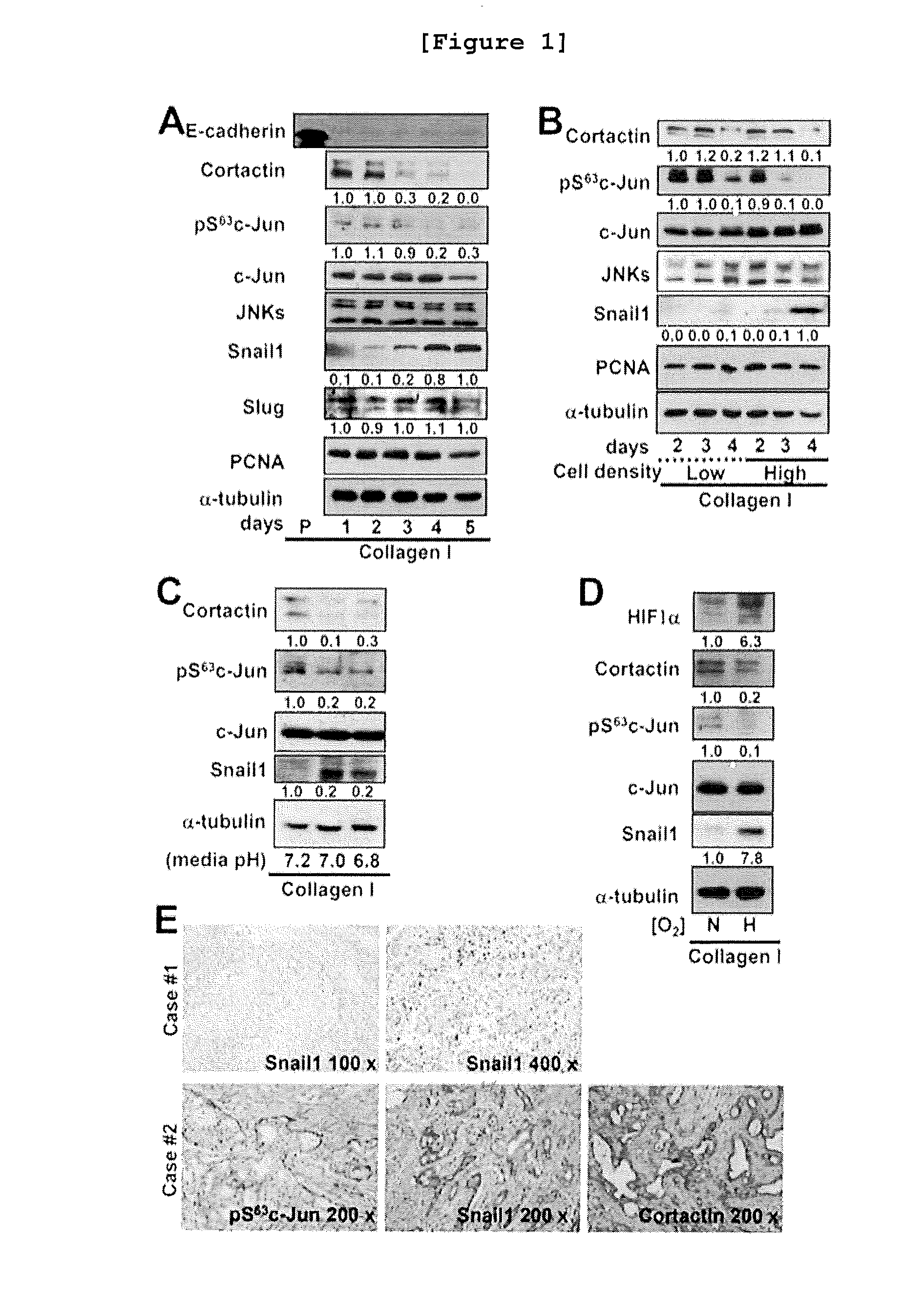
Changes of Intracellular Protein Caused by Microenvironment in the Breast Cancer Cell Line Cultured in a Three-Dimensional Collagen Gel Environment

[0144] Preparation of polydimethylsiloxane (PDMS) Culture Vessel for Three-Dimensional Cell Culture

[0145]To observe the cells growing in a three-dimensional environment under confocal microscope, the PDMS culture vessel equipped with a cover glass on one side was prepared.

[0146]Particularly, PDMS crude liquid was mixed with a hardener at the ratio of 10:1, which was hardened at 100° C. for 1 hour. The hardened PDMS was taken off from the mold and punched by using an 8 mm punch. A cover glass (24×60 mm, Marienfeld) was attached on the hole of the PDMS by treating oxygen plasma for 45 seconds, followed by drying in a 60° C. oven for 24 hours to recover hydrophobicity. The prepared PDMS was used after being irradiated with UV.

Culture of Breast Cancer Cell Lines in a Three-Dimensional Collagen Gel or Matrigel Environment

[0147]Various breast ...

example 2

Changes of Cell Shape and Cell Migration by JNK Inhibitor in the Breast Cancer Cell Line Cultured in a Three-Dimensional Collagen Gel Environment

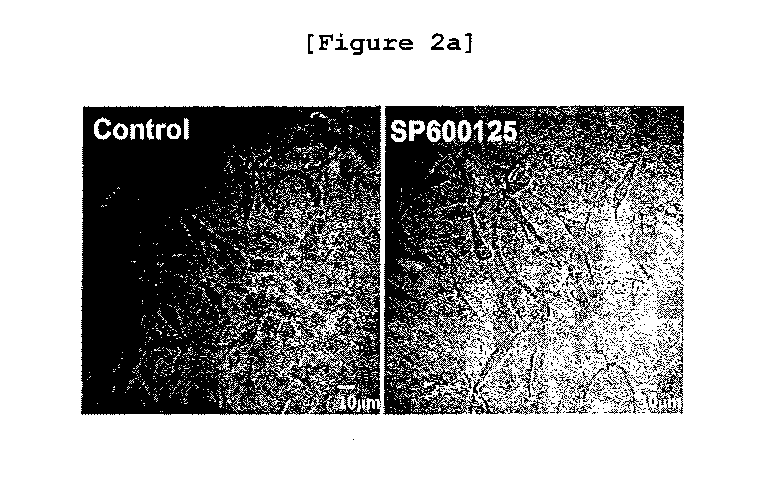
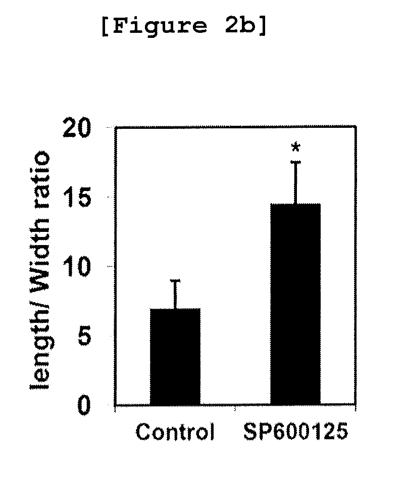
Changes of Cell Shape by JNK Inhibitor

[0156]When MDA-MB-231 cell line was cultured in a three-dimensional collagen gel environment for 3 days, the volume of cytoplasm was reduced and the cell shape became comparatively thinner and longer with very dynamic end part unlike when the cell line was cultured in a two-dimensional environment. After treating the JNK inhibitor SP600125 (LC Labs), the cell shape and the migration pattern were observed.

[0157]Particularly, when the gel mixture comprising the MDA-MB-231 cell line cultured by the method of Example and collagen was fully hardened, the culture medium supplemented with 10% FBS (control) and the culture medium supplemented with 50 μM of SP600125 (experimental group) were loaded on top of the gel, followed by culture for 3 days. Then, the shape of the cells and the migration pattern in the ...

example 3

Effect of JNK Inhibitor in the Breast Cancer Cell Line Cultured in a Three-Dimensional Matrigel Environment

[0169]In this example, it was investigated whether or not the above results obtained in Example 2 were consistent with those resulted from the culture in a matrigel (another ECM) environment.

[0170]Particularly, MDA-MB-231 cell line was cultured in a three-dimensional matrigel environment treated with SP600125 and then the shape of the cells was investigated by the method of Example . The changes in protein expression were also investigated by the method of Example .

[0171]As a result, as shown in FIGS. 3A and 3B, the shape of the cells was not changed by JNK inhibitor (FIG. 3A) and the expression of snail1 protein was not changed, either (FIG. 3B). It was also investigated if the treatment of another MAPK, p38 or ERK inhibitor could bring the consistent results with those obtained from the treatment of JNK inhibitor. As a result, ERK inhibition did not affect cortactin expressio...


PUM

PropertyMeasurementUnit
Concentrationaaaaaaaaaa
Densityaaaaaaaaaa
Protein activityaaaaaaaaaa

Abstract

The present invention relates to a method for monitoring migration, invasion, and metastasis of cancer cells by observing the shape of cancer cells cultured in a three-dimensional environment and measuring the activity, expression, and changes in expression sites of proteins associated with invadopodia formation and metastasis, and the degradation of an extracellular matrix; and to a method for screening a cancer metastasis inhibitor. More specifically, it was verified that the reduction in c-Jun phosphorylation induced the increase in snail1 and the decrease in cortactin expression in the cells cultured in a three-dimensional environment and the expression regulation relations between the proteins were identical to those in breast cancer tissues obtained from patients. In addition, it was verified that, when breast cancer cells in a three-dimensional collagen gel environment were treated with a JNK inhibitor, the shape of the cells became longer; the contact region of the cells and the extracellular matrix became flattened and thinner; the migration of cancer cells was decreased; and the changes in protein expression was observed, such as the increase in TGFβ1 expression, the increases in smad2 and smad3 expression and activity, the increase in snail1 expression, the decrease in cortactin expression, and the resulting decrease in invadopodia formation. In addition, in a three-dimensional collagen gel environment, MT1-MMP besides the cortactin can be used as a marker of invadopodia, and it was verified that the inhibition of JNK led to the decrease in cortactin expression and the increase in snail1 expression, badly influenced the site and role of MT1-MMP to inhibit the formation of invadopodia, and inhibited the degrading activity of a collagen gel substrate. Thus, the present invention can be used as a method for monitoring migration, invasion, metastasis, and the degree of metastasis of cancer cells and a method for screening a cancer metastasis inhibitor, and will be useful as one of screening methods capable of creating low-cost, high-efficient added value at the time of pre-clinical tests required for drug development.

Description

BACKGROUND OF THE INVENTION[0001]1. Field of the Invention[0002]The present invention relates to a method for monitoring migration, invasion, and metastasis of cancer cells by observing the shape of cancer cells cultured in a three-dimensional environment and measuring the activity, expression, and changes in the expression sites of proteins associated with metastasis, and the degradation of an extracellular matrix; and to a method for screening a cancer metastasis inhibitor.[0003]2. Description of the Related Art[0004]The reason that cancer is lethal to a patient is metastasis. Metastasis is the process of the dissemination of cells from the primary tumor, by which cancer cells can be spread wide. The said primary tumor can be developed by various genetic reasons of a host, which makes the treatment of each individual difficult. However, metastasis is the general phenomenon observed in every cancer, so that it is an important target of therapeutic intervention. The metastatic cance...

Claims


Application Information

Patent Timeline
IPC IPC(8): G01N33/574G01N33/50
CPCG01N33/574G01N33/5023G01N33/5011G01N2333/4704G01N2333/4706G01N2440/14G01N2333/4703C12N5/0693G01N33/5026G01N33/5029G01N33/57415C12N2501/727C12N2503/02C12N2533/54G01N2500/10C12Q1/00C12Q1/02C12Q1/04G01N33/15
Inventor LEE, JUNG WEONKIM, SUNGHOONLEE, MI-SOOK
Owner MEDICINAL BIOCONVERGENCE RES CENT
Why Eureka
Try Eureka