Compositions and methods for inhibition of cancer invasion and angiogenesis

a technology of angiogenesis and composition, applied in the field of medicine, can solve the problems that the membrane-bound proteases active at the site of angiogenesis have not yet been identified, and achieve the effect of increasing the blood supply to the affected tissue and increasing the blood supply

Inactive Publication Date: 2007-07-31
THE RES FOUND OF STATE UNIV OF NEW YORK
View PDF11 Cites 16 Cited by
  • Summary
  • Abstract
  • Description
  • Claims
  • Application Information

AI Technical Summary

Problems solved by technology

However, novel membrane-bound proteases active at sites of angiogenesis have not yet been identified.

Method used


Image

  • Compositions and methods for inhibition of cancer invasion and angiogenesis
  • Compositions and methods for inhibition of cancer invasion and angiogenesis
  • Compositions and methods for inhibition of cancer invasion and angiogenesis

Examples


example 1

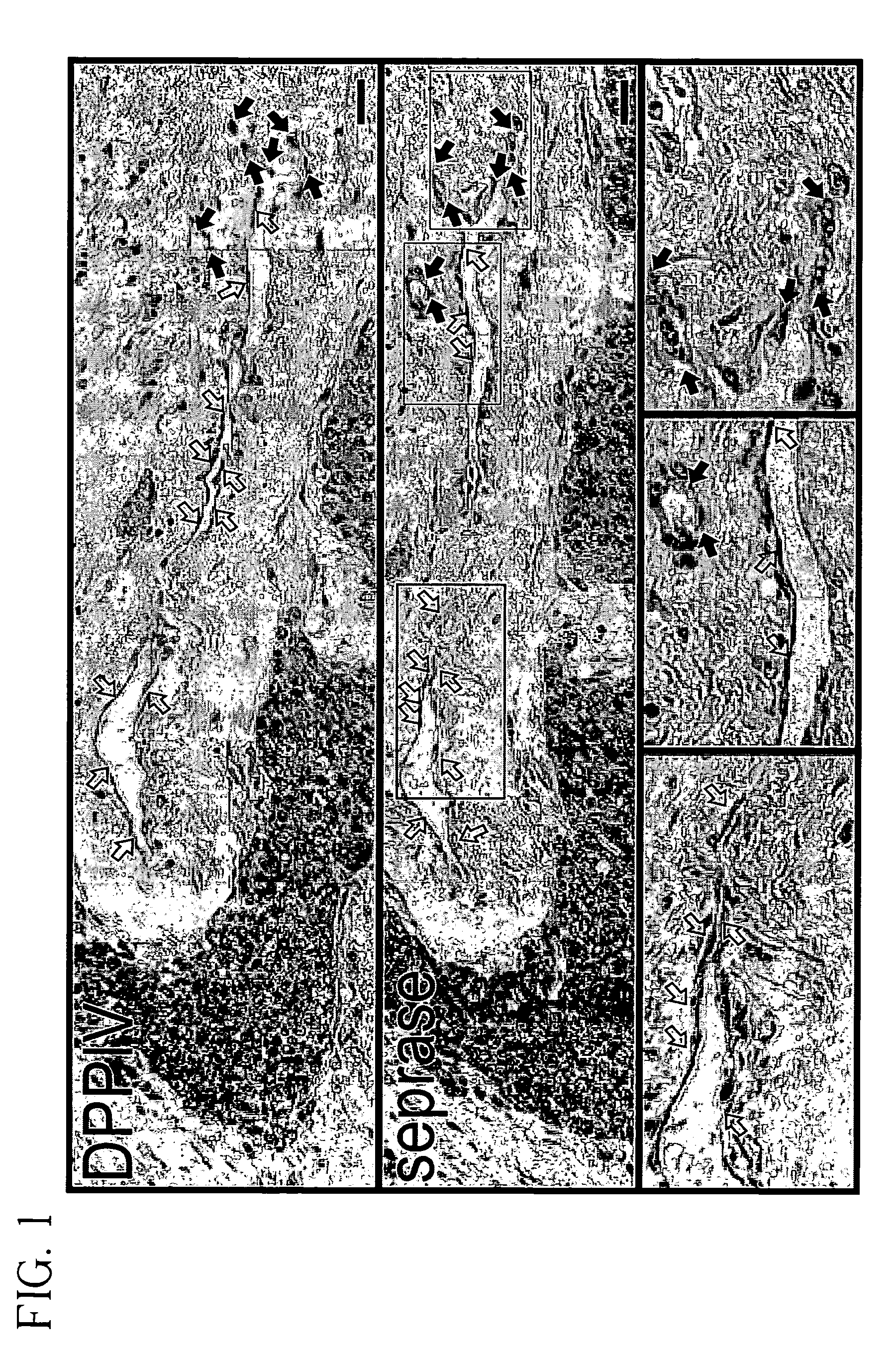
Cytoimmunohistochemical Staining for Seprase and DPPIV in Endothelial Cells Sprouting Vessels and in Normal Tissue.

[0081]To investigate the expression of seprase and DPPIV during angiogenesis, human malignant breast carcinoma tissue or adjacent normal skin were stained with antibodies specific for either seprase or DPPIV. Both seprase and DPPIV were abundantly expressed on the endothelial cells of sprouting vessels (FIG. 1, solid arrows) but were not detectable in other tumor vessels (FIG. 1, open arrows) or in adjacent normal skin from the same donor. These findings indicate that only sprouting sites of blood vessels involved in tumor angiogenesis have enhanced expression of seprase and DPPIV. Consistent with this result, expression of seprase and DPPIV on cultured endothelial cells can be induced by means or factors that enhance cell migration and vessel sprouting (see below).

example 2

Cytoimmunohistochemical Staining for Seprase and DPPIV in Human Primary Cell Culture Monolayers of Different Cell Densities.

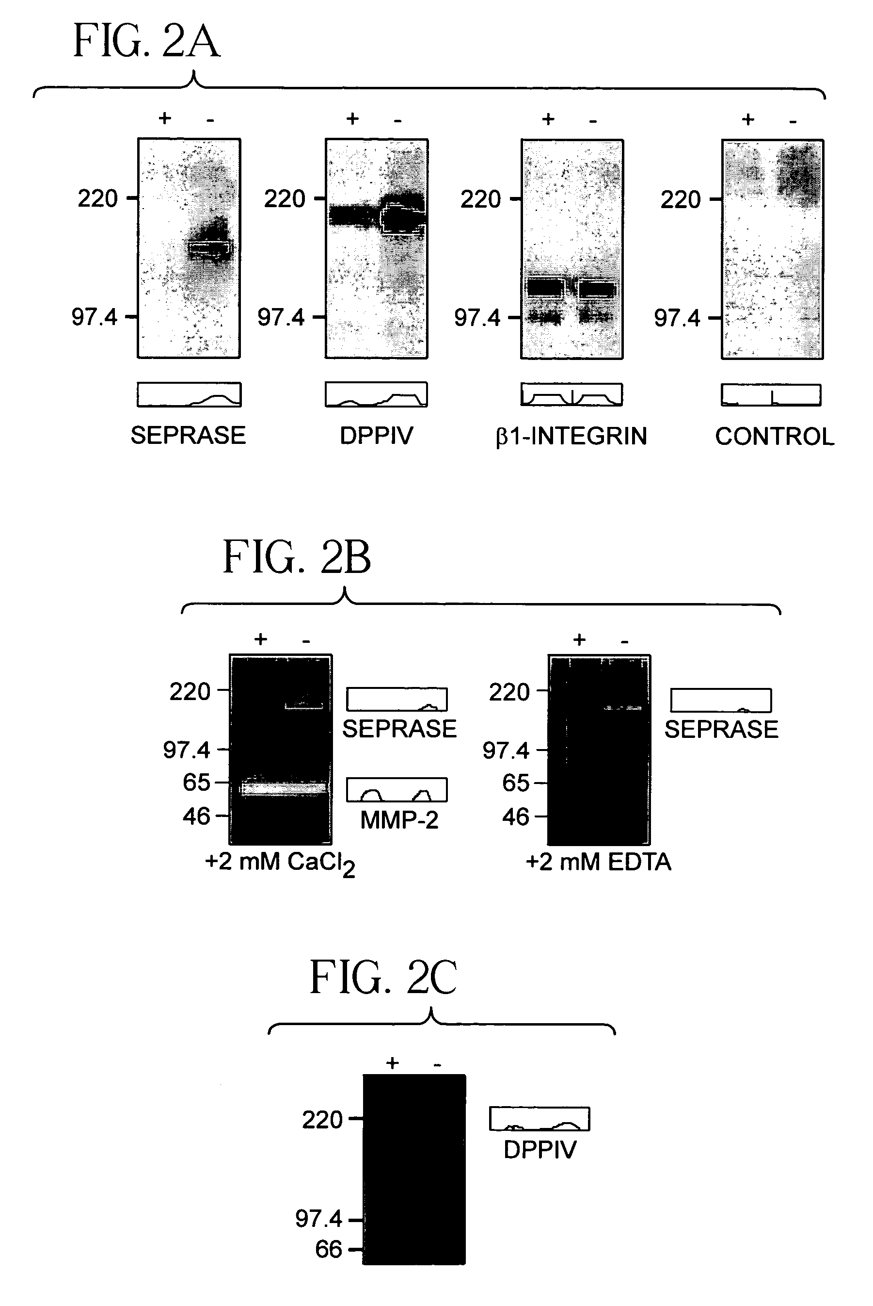
[0082]Endothelial cells of sprouting vessels are migratory and exhibit a lack contact inhibition (Pepper et al., 1993). Monolayer cultures of human umbilical vein endothelial cells (HUVEC) can be induced to migrate by wounding or passage to low cell density (Pepper et al., 1996). This assay was used to examine the expression of seprase and DPPIV in migratory endothelial cells. The confluent HUVEC monolayers were found to contain low levels of seprase and DPPIV and their proteolytic activities were also low (FIGS. 2a–c). Passage of monolayers into a sparse culture within 24 hours induced the expression of functional seprase protein; it also caused an increase in DPPIV protein and their proteolytic activities (FIGS. 2a–c).

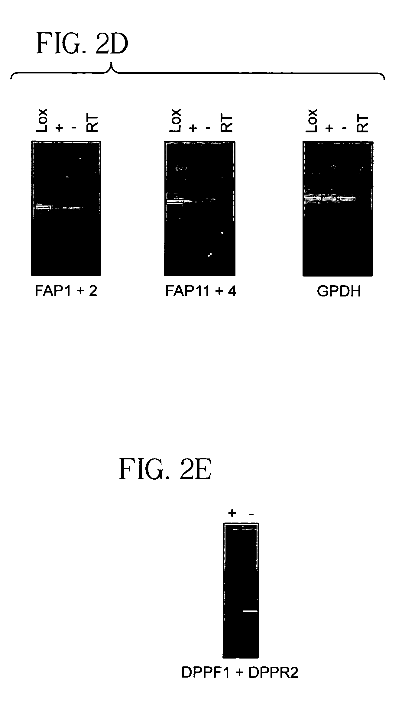
[0083]Both HUVEC cultures had detectable seprase and DPPIV mRNA (FIGS. 2d–e). As β1-integrins (Bloch et al., 1997) and membrane-bound MMP-2 (Hi...

example 3

Expression of Seprase / DPPIV and β1-Integrins During Wound-Induced Endothelial Migration.

[0084]The relative expression of seprase / DPPIV and β1-integrins during wound-induced endothelial migration was examined by double-labeled immunofluorescence. The monolayer wound model consists of a 300 μm width wound on the HUVEC monolayer; migratory activity of HUVEC was visible one hour after wounding (FIGS. 2f–g). Expression of β1-integrins was high in endothelial cells at both the wound edge (wounded) and in the monolayer (stationary); in contrast, seprase and DPPIV expression was restricted to migratory cells at the wound edge (FIGS. 2f–g, arrows).

[0085]In addition, seprase and DPPIV expression was found on invadopodia (FIGS. 2f–g, arrows) and on the perinuclear region (Golgi apparatus) of migratory cells but not in confluent cells. In phase contrast images (FIGS. 2f–g), β1-integrins were distributed widely on the surface of migratory cells (FIGS. 2f–g, solid arrows) and particularly concent...


PUM

PropertyMeasurementUnit
concentrationaaaaaaaaaa
widthaaaaaaaaaa
concentrationaaaaaaaaaa

Abstract

The invention provides antibodies that specifically bind a membrane protease complex, the complex consisting of two homodimers of seprase and dipeptidyl peptidase IV (DPPIV), obtained from mammalian, preferably human cell membranes. The antibodies specifically bind the DPPIV protease of the seprase-DPPIV complex. This membrane protease complex resides on cell surface invadopodia at the leading edge of angiogenic endothelia, migratory fibroblasts, and invading cancer cells. The antibodies and immunoconjugates of the invention specifically bind the membrane protease complex at the cell surface invadopodia, yet fail to react with resting cells in adjacent human tissues and blood vessels. These antibodies and immunoconjugates block interaction of collagen matrix with the seprase-DPPIV complex in the invasive cells during angiogenesis and cancer spreading but not that with other endothelia or tumor cells. The invention further provides methods for identifying and of using DPPIV antagonists to inhibit capillary sprouting, angiogenesis and cancer invasion in tumor tissues and metastases. Also provided are therapeutic compositions comprising DPPIV antagonists.

Description

RELATED APPLICATIONS[0001]This application is a continuation of U.S. application Ser. No. 09 / 823,277 filed on Mar. 30, 2001, now abandoned which is a continuation-in-part of U.S. application Ser. No. 09 / 541,785 filed on Apr. 3, 2000, now U.S. Pat. No. 6,573,096, which claims the benefit of U.S. Provisional Application No. 60 / 193,987 filed on Apr. 1, 2000. The specifications of U.S. application Ser. No. 09 / 823,277, U.S. application Ser. No. 09 / 541,785, and U.S. Provisional Application No. 60 / 193,987 are hereby incorporated by reference in their entirety.[0002]This work was supported by grants from one or more of the following: U.S. Public Health Service, National Cancer Institute and The National Institute of Aging. The government may have certain rights to this invention.FIELD OF THE INVENTION[0003]The present invention relates generally to the field of medicine, and relates specifically to angiogenesis and metastasis of cancer tissues, as well as treatment of cardiovascular disease...

Claims


Application Information

Patent Timeline
Patent Type & Authority Patents(United States)
IPC IPC(8): C12P21/08C12N15/02A61K39/395A61K45/00A61K47/48A61P3/10A61P9/00A61P9/10A61P9/14A61P17/02A61P35/04A61P43/00C07K14/705C07K16/28C07K16/40C07K19/00C12N5/10C12N9/48C12N9/64
CPCA61K47/48561C07K14/70596C07K16/2896C07K16/40C12N9/48C12N9/6424C12N9/6491C12Y304/14005A61K2039/505A61K47/6849A61P3/10A61P9/00A61P9/10A61P9/14A61P17/02A61P35/04A61P43/00
Inventor CHEN, WEN-TIEN
Owner THE RES FOUND OF STATE UNIV OF NEW YORK
Why Eureka
Try Eureka