
Radio frequency coil and medical imaging device including same

a radio frequency coil and medical imaging technology, applied in tomography, instruments, applications, etc., can solve the problems of severe image information distortion, and achieve the effect of minimizing image distortion, minimizing image distortion, and minimizing image distortion

Active Publication Date: 2019-11-07
IND ACADEMIC CORP FOUND YONSEI UNIV
View PDF0 Cites 0 Cited by
  • Summary
  • Abstract
  • Description
  • Claims
  • Application Information

AI Technical Summary

Benefits of technology

The patent text describes a new radiofrequency coil that can reduce image distortion caused by a beam-hardening artifact. This coil can be used in medical imaging devices such as MRI, CT, PET, and MRI / PET. The technical effect of this invention is to improve the quality and accuracy of medical imaging.

Problems solved by technology

Additionally, positrons can cause beam-hardening artifact due to hardening at the soldering site of the radiofrequency coil thereby severely distorting the image information.

Method used


Image

  • Radio frequency coil and medical imaging device including same
  • Radio frequency coil and medical imaging device including same
  • Radio frequency coil and medical imaging device including same

Examples


Embodiment Construction

[0032]Hereinafter, embodiments of the present inventive concept will be described in detail with reference to the accompanying drawings so that those skilled in the art can easily carry out the present inventive concept. However, the present inventive concept may be embodied in many different forms and is not limited to the embodiments set forth herein. In order to clearly illustrate the present inventive concept, parts not related to the description are omitted, and like reference numerals designate like elements throughout the specification.

[0033]In the present specification, duplicate descriptions are omitted for the same constituent elements.

[0034]Further, in the present specification, when it is described that a constituent element is “connected” or “electrically connected” to another constituent element, it should be understood that the element may be “directly connected” or “directly electrically connected” to the other constituent elements or may be “connected” or “electrica...


PUM


Abstract

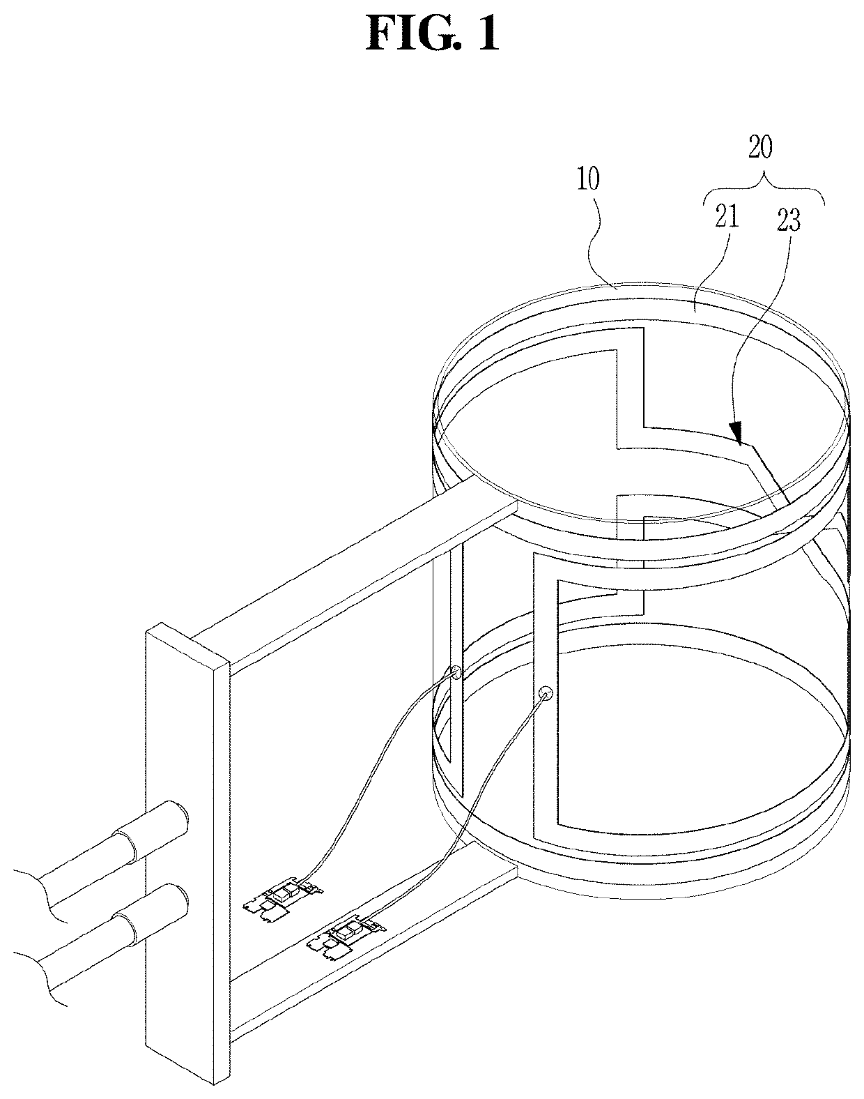
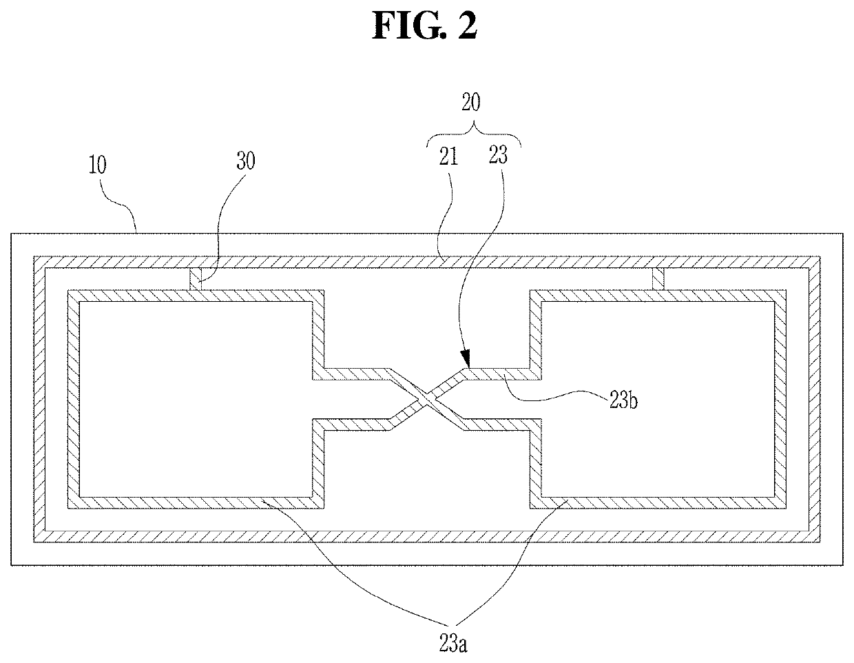
A medical radiofrequency coil may comprise: a base substrate; and a radio coil unit having a first coil element which has a rectangular shape and is formed along an edge of the inner peripheral surface of the base substrate, and a second coil element which is formed at the inner side of the first coil element and has a shape of paired paddles connected to each other. Therefore, the present inventive concept provides a radiofrequency coil, which can minimize an image distortion due to a beam-hardening artifact effect, and a medical imaging device including the same.

Description

TECHNICAL FIELD[0001]The present disclosure relates to a radiofrequency coil and a medical imaging device including the same, and more specifically, a radiofrequency coil which is used to photograph the human body using a magnetic field and a medical imaging device including the sameBACKGROUND[0002]Recently, various diagnostic imaging devices to diagnose the body for prevention and treatment of diseases, such as a magnetic resonance imaging (hereinafter, MRI) device using a magnetic field generated by a magnetic force, a positron emission tomography (hereinafter, PET) device that detects gamma rays derived from a radioactive tracer, a computed tomography (hereinafter, CT) device that uses an X-ray generator, e.g. are widely used.[0003]The MRI device used to determine the internal structure of a subject includes a transmit-and-receive radiofrequency coil (RF coil) that can receive and transmit radiofrequency (RF) signals so as to acquire images from an electromagnetic field. For opti...

Claims


Application Information

Patent Timeline
IPC IPC(8): G01R33/34A61B5/055A61B6/00G01R33/3415
CPCA61B5/055G01R33/34069G01R33/3415A61B6/502A61B6/032G01R33/481G01R33/34007G01R33/34061G01R33/34G01R33/341
Inventor LEE, YOUNG HANKIM, MIN JUNGYANG, JAEMOONSUH, JIN-SUCK
Owner IND ACADEMIC CORP FOUND YONSEI UNIV
Learn More