
Multimodal systems and methods for detecting and quantitating cell or other particle targets in a bloodstream of a living being

Inactive Publication Date: 2019-12-19
IVDIAGNOSTICS
View PDF0 Cites 0 Cited by
  • Summary
  • Abstract
  • Description
  • Claims
  • Application Information

AI Technical Summary

Benefits of technology

The patent describes a system and methods for detecting and measuring targets like cells, bacteria, and viruses in the blood of living subjects. This is done in a non-invasive way, without needing to collect blood from the subject and process it outside of the body. The system uses fluorescent tags that specifically bind to targets, allowing for real-time monitoring and analysis of the targets in real-time. This has applications in clinical medicine and biomedical research, providing a valuable tool for detecting and measuring targets in the blood of living subjects.

Problems solved by technology

These systems have drawbacks, as they are specifically adapted for, and limited to use with human clinical patients, are invasive, require lengthy sample processing times, can only detect cancer and blood cells, and are not configured for research use with laboratory animals.

Method used

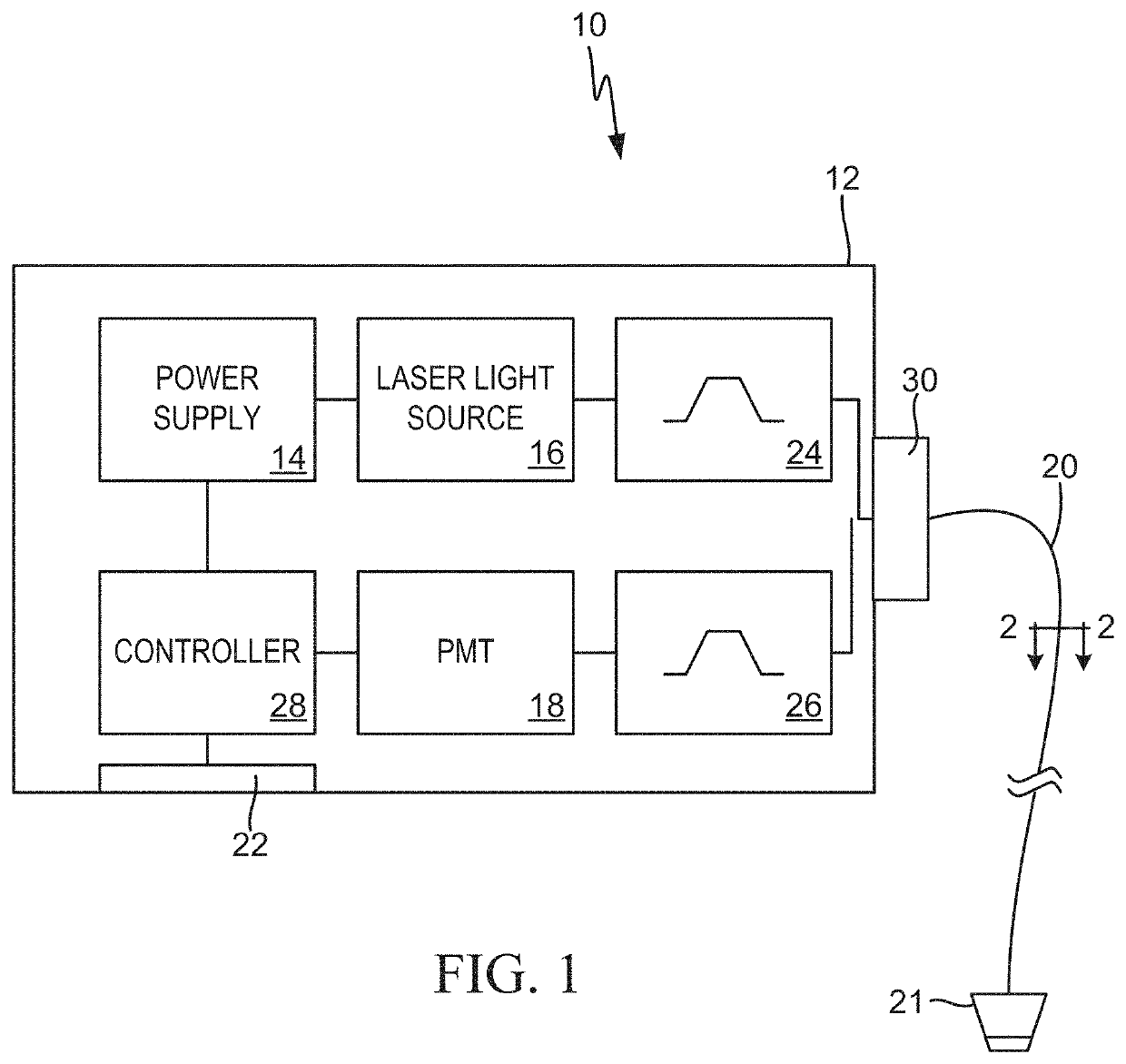
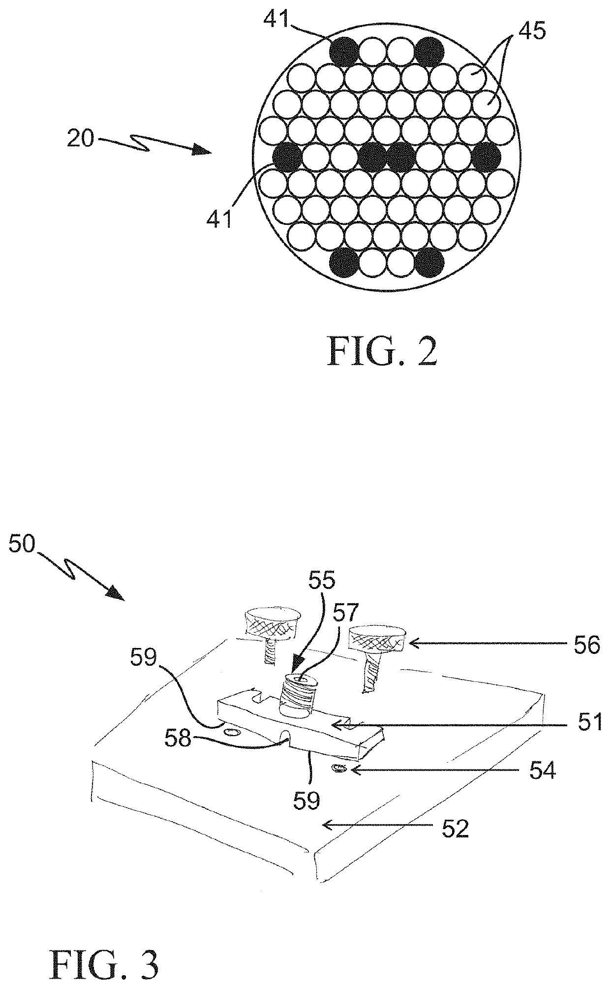
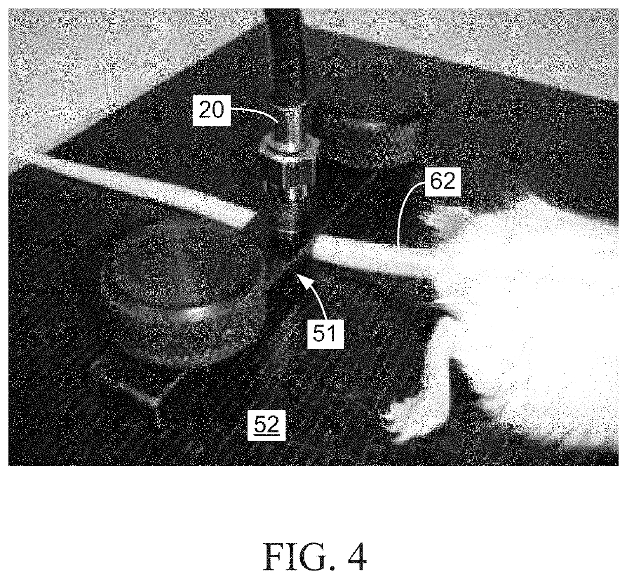

Image

  • Multimodal systems and methods for detecting and quantitating cell or other particle targets in a bloodstream of a living being
  • Multimodal systems and methods for detecting and quantitating cell or other particle targets in a bloodstream of a living being
  • Multimodal systems and methods for detecting and quantitating cell or other particle targets in a bloodstream of a living being

Examples


experimental / examples

EXPERIMENTAL / EXAMPLES

[0030]The following examples describe practical applications of the present invention in humans, laboratory animals and other research models. However, practical applications of the present invention are not limited to the examples described herein. It should be noted that various changes and modifications to the embodiments described herein will be apparent to those skilled in the art. Such changes and modifications may be made without departing from the spirit and scope of the present invention and without diminishing its attendant advantages. For example, various embodiments of the systems and methods may be provided based on various combinations of the features and functions from the subject matter provided herein.

example 1

In Vivo Detection and Quantitation of Circulating Tumor Cells (CTCs)

[0031]In neoplastic disease, malignant cells often express unique biomarkers not found on normal (non-malignant) cells, or that are present at levels that are significantly different than those found on normal cells. The present invention exploits this principle by providing a means by which to detect and quantitate cancerous cells circulating in the bloodstream, so called circulating tumor cells (CTCs).

[0032]For human clinical applications, a diagnostic reagent including a fluorescent dye conjugated to an antibody or other ligand which specifically binds to a unique cancer biomarker is intravenously (IV) injected into a cancer patient. The fluorescent conjugate could also be comprised of fluorescent microspheres, including magnetic or non-magnetic material derivatized with an antibody or other ligand. Once in circulation, the fluorescent conjugate would then bind to, and label, the CTCs in the patient's bloodstream...

example 2

In Vivo Detection and Quantitation of Metastatic CTCs

[0034]In a research application, tumor cells expressing a unique a cancer biomarker can be injected into a tissue site in the subject other than the blood to establish a focal tumor graft. The ability of the cells of the resulting tumor to extravasate into the bloodstream as CTCs can then be determined, over time, by detection and quantitation of the resulting CTCs using the system and methods of the present invention as described in example 1 above. In this example, use of the system and methods of the invention provides a convenient, in vivo, minimally invasive model for metastatic cancer in a laboratory animal.


PUM


Abstract

Provided is a device for excitation of a fluorescent component associated with a predetermined target circulating in blood flowing through a blood vessel of a living being. The device includes a clamping device, a coherent light source that emits light causing excitation of the fluorescent component forming a conjugate with the predetermined target in the blood stream. A fluorescence sensor senses and measures a fluorescent effect exhibited by the fluorescent component forming a portion of the conjugate in response to excitation of the fluorescent component by the light emitted by the coherent light source. Optical fibers transmit the emitted light between the light source and the clamping device, and the fluorescent effect exhibited by the fluorescent component of the conjugate between the clamping device and the sensor.

Description

CROSS-REFERENCE TI) RELATED APPLICATIONS[0001]This application claims the benefit of U.S. Provisional Application No. 62 / 206,502, filed Aug. 18, 2015, which is incorporated in its entirety herein by reference.BACKGROUND OF THE INVENTION1. Field of the Invention[0002]This application relates generally to a method and apparatus for the in vivo detection of particle targets in blood and, more specifically, to a minimally-invasive apparatus and method for detecting and quantitating fluorescently-labeled cells or other particles in the bloodstream of humans and animals in vivo.2. Description of Related Art[0003]Current systems available for detecting cells or other particle targets in the bloodstream, such as the Cell Search Circulating Tumor Cell Test (Janssen Diagnostics, LLC) and the MACS Whole Blood Analysis System (Miltenyi Biotec), require that a blood sample be invasively drawn from the subject prior to analysis, followed by sample processing, and final analysis of the results ext...

Claims


Application Information

Patent Timeline
IPC IPC(8): A61B5/1455A61B5/00G01N33/49
CPCA61B5/1455A61B5/0071A61B5/6838A61B5/4848G01N33/49A61B2503/40A61B2503/42A61B5/145A61B5/14546A61B2562/0233
Inventor SZCZEPANSKI, FRANKCLEMENS, CHRISTOPHERFIRCA, JOSEPH
Owner IVDIAGNOSTICS
Learn More