
System, method, and computer-accessible medium for generating magnetic resonance imaging-based anatomically guided positron emission tomography reconstruction images with a convolutional neural network

a convolutional neural network and anatomical imaging technology, applied in the field of magnetic resonance imaging, can solve the problems of requiring significant additional reconstruction time, requiring the use of pet raw materials, and contaminated clinical pet images

Active Publication Date: 2020-04-16
NEW YORK UNIV
View PDF0 Cites 2 Cited by
  • Summary
  • Abstract
  • Description
  • Claims
  • Application Information

AI Technical Summary

Benefits of technology

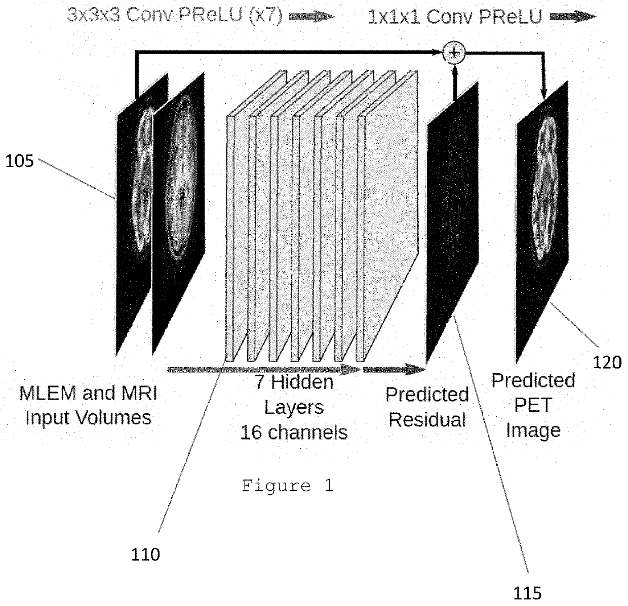
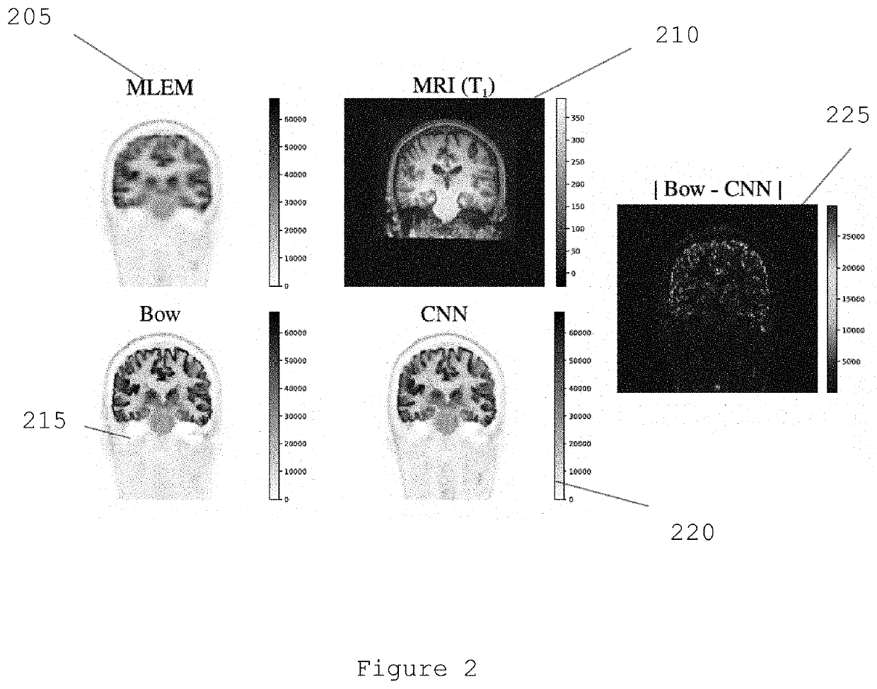
The patent describes a system, method, and computer-accessible medium for generating an image of a patient using information from positron emission tomography (PET) and magnetic resonance imaging (MRI). The system uses a convolutional neural network (CNN) to analyze the first information and generate second information, which is then used to generate the image. The PET information is reconstructed using a maximum likelihood estimation procedure. The system can include a plurality of hidden layers and filters, along with a plurality of parametric activation functions. The method can involve receiving the first information and using a residual image and a maximum likelihood estimation procedure to generate the second information, which is then combined with the MLE image to generate the image. The patent describes a technical effect of combining multiple imaging modalities to improve image quality and accuracy.

Problems solved by technology

Positron emission tomography (“PET”) is a clinical tool that provides valuable physiological information that may not be obtained from either computed tomography (“CT”) or MRI; however, compared with these imaging modalities, it suffers from several drawbacks.
Due to physical and technical constraints, clinical PET images are often contaminated by a high level of Poisson noise and general purpose clinical scanners are typically limited to a spatial resolution of 4-5 millimeters.
One limitation to the widespread clinical adoption of these Bayesian reconstruction procedures is that they require significant additional reconstruction time.
Another problem with MBIR approaches is that they require the use of the PET raw data, which typically has a proprietary format, and generally has a much larger size than a Digital Imaging and Communications in Medicine (“DICOM”)-based image.

Method used


Image

  • System, method, and computer-accessible medium for generating magnetic resonance imaging-based anatomically guided positron emission tomography reconstruction images with a convolutional neural network
  • System, method, and computer-accessible medium for generating magnetic resonance imaging-based anatomically guided positron emission tomography reconstruction images with a convolutional neural network
  • System, method, and computer-accessible medium for generating magnetic resonance imaging-based anatomically guided positron emission tomography reconstruction images with a convolutional neural network

Examples


Embodiment Construction

[0007]An exemplary system, method and computer-accessible medium for generating an image(s) of a portion(s) of a patient(s) can be provided, which can include, for example, receiving first information associated with a combination of positron emission tomography (PET) information and magnetic resonance imaging (MRI) information, generating second information by applying a convolutional neural network(s) (CNN) to the first information, and generating the image(s) based on the second information. The PET information can be fluorodeoxyglucose PET information. The CNN(s) can include a plurality of convolution layers and a plurality of parametric activation functions. The parametric activation functions can include a plurality of parametric rectified linear units. Each of the convolution layers can include a plurality of filter kernels. The PET information can be reconstructed using a maximum likelihood estimation procedure to generate a MLE image.

[0008]In some exemplary embodiments of t...


PUM


Abstract

An exemplary system, method and computer-accessible medium for generating an image(s) of a portion(s) of a patient(s) can be provided, which can include, for example, receiving first information associated with a combination of positron emission tomography (PET) information and magnetic resonance imaging (MRI) information, generating second information by applying a convolutional neural network(s) (CNN) to the first information, and generating the image(s) based on the second information. The PET information can be fluorodeoxyglucose PET information. The CNN(s) can include a plurality of convolution layers and a plurality of parametric activation functions. The parametric activation functions can include, e.g., a plurality of parametric rectified linear units. Each of the convolution layers can include, e.g., a plurality of filter kernels. The PET information can be reconstructed using a maximum likelihood estimation (MLE) procedure to generate a MLE image.

Description

CROSS-REFERENCE TO RELATED APPLICATION(S)[0001]This application relates to and claims priority from U.S. Patent Application No. 62 / 743,905, filed on Oct. 10, 2018, the entire disclosure of which is incorporated herein by reference.FIELD OF THE DISCLOSURE[0002]The present disclosure relates generally to magnetic resonance imaging (“MRI”), and more specifically, to exemplary embodiments of exemplary system, method, and computer-accessible medium for generating high-resolution, magnetic resonance imaging-based anatomically guided positron emission tomography reconstruction images with a convolutional neural network.BACKGROUND INFORMATION[0003]Positron emission tomography (“PET”) is a clinical tool that provides valuable physiological information that may not be obtained from either computed tomography (“CT”) or MRI; however, compared with these imaging modalities, it suffers from several drawbacks. Due to physical and technical constraints, clinical PET images are often contaminated by...

Claims


Application Information

Patent Timeline
Patent Type & Authority Applications(United States)
IPC IPC(8): G06T11/00
CPCG06T2211/408G06T11/003G06T11/006
Inventor BOADA, FERNANDORIGIE, DAVID
Owner NEW YORK UNIV
Learn More