Simulated eyeball, device for training in ophthalmic surgery, and method for training in ophthalmic surgery
a technology for simulated eyes and eyeballs, applied in the field of simulated eyeballs, can solve problems such as problems of conventional simulated eyeballs
- Summary
- Abstract
- Description
- Claims
- Application Information
AI Technical Summary
Benefits of technology
Problems solved by technology
Method used
Image
Examples
first embodiment
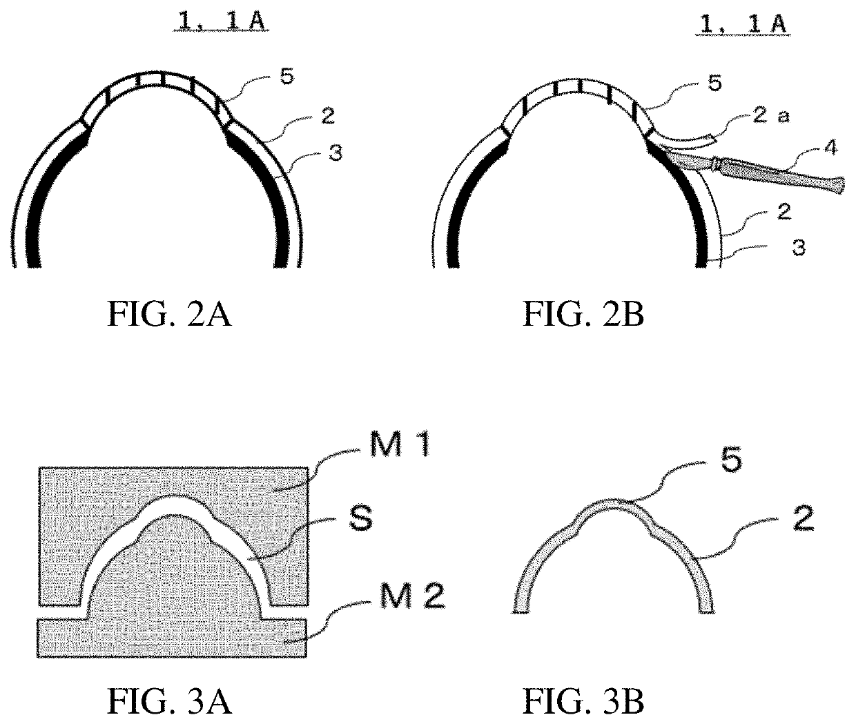
[0084]A simulated eyeball 1A in a first embodiment will be described with reference to FIGS. 2A and 2B. FIGS. 2A and 2B are schematic cross-sectional diagrams showing parts of the simulated eyeball in the first embodiment. Descriptions that are common to both FIGS. 2A and 2B sometimes refer to “FIG. 2” in the specification. The same applies for the other drawings.
[0085]The simulated eyeball 1A in the first embodiment has at least a simulated sclera region 2, and a conductor layer 3 formed on a side of the simulated sclera region 2 that is on an interior of the simulated eyeball. In the present description, the “simulated sclera region” refers to a “region” in which a “simulated sclera” is formed. Therefore, this element is described as the “simulated sclera” in cases illustrating the characteristics or the like of the “simulated sclera” and as the “simulated sclera region” in cases illustrating the region in which the “simulated sclera” is provided. The same reference symbol is appl...
second embodiment
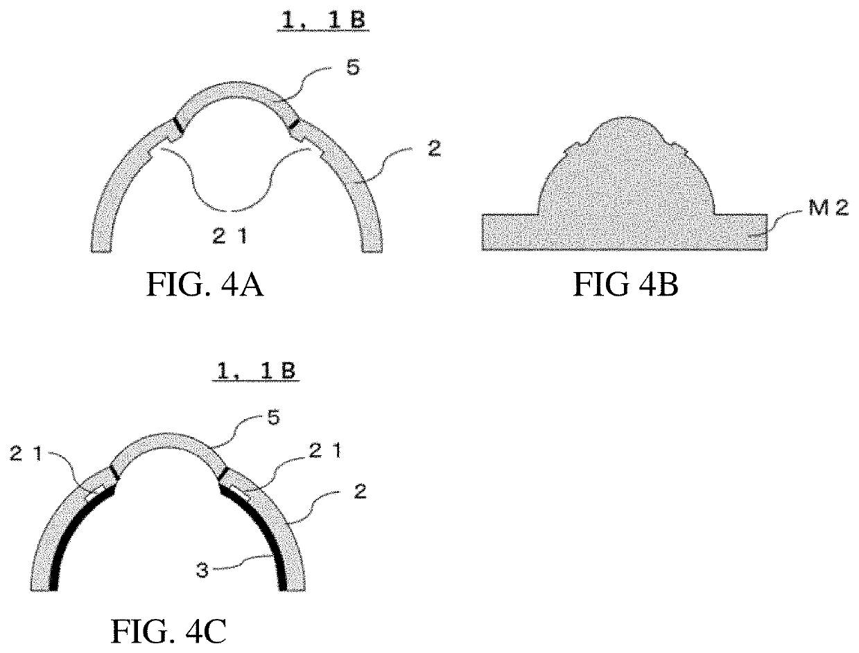
[0100]FIG. 4A is a cross-sectional diagram showing a schematic of a simulated eyeball 1B of a second embodiment. In the simulated eyeball 1B of the second embodiment, a recess 21 is formed on the simulated sclera 2 near the simulated cornea region 5, the recess 21 being formed on the inner side of the simulated sclera 2 (the side on which the conductor layer 3 is layered). The recess 21 can be used as a simulated Schlemm's canal.
[0101]Within a human eyeball, there is a venous system having a lumenal structure that is referred to as “Schlemm's canal” and that functions to discharge the fluid from inside the eye. In the trabeculotomy type of glaucoma surgery, after the sclera is thinly sliced, it is necessary to insert a narrow metal rod having a diameter of about 0.5 mm into Schlemm's canal and cut out a fiber cylinder. However, in conventional simulated eyeballs, no lumenal structure simulating Schlemm's canal is provided whatsoever, and it is impossible to conduct training in makin...
third embodiment
[0104]FIG. 6A is a cross-sectional diagram showing a schematic of a simulated eyeball 1C of a third embodiment. The simulated eyeball 1C of the third embodiment includes an extension region 31 by which the conductor layer 3 extends away from the simulated sclera 2 into the simulated eyeball 1C, from a position near a boundary between the simulated cornea region 5 and the simulated sclera region 2. The extension region 31 forms a simulated iris region 31. When the simulated eyeball 1C of the third embodiment is used, an iris region of the eyeball is formed by the conductor layer 3. Therefore, in addition to training in glaucoma surgery, in procedures such as making an incision in a fiber cylinder, it is possible to perform surgery so that the metal rod does not mistakenly touch the iris during making of an incision in the fiber cylinder. Alternative uses involve performing training for micro-glaucoma surgery that involves inserting an instrument such as an iStent, a trabectome, a hyd...
PUM
 Abstract
Description
Claims
Application Information
 - R&D
- Intellectual Property
- Life Sciences
- Materials
- Tech Scout
- Unparalleled Data Quality
- Higher Quality Content
- 60% Fewer Hallucinations
Browse by: Latest US Patents, China's latest patents, Technical Efficacy Thesaurus, Application Domain, Technology Topic, Popular Technical Reports.
© 2025 PatSnap. All rights reserved.Legal|Privacy policy|Modern Slavery Act Transparency Statement|Sitemap|About US| Contact US: help@patsnap.com


