Method for removal of yolk

Inactive Publication Date: 2020-07-16
CRYOGENETICS
View PDF0 Cites 0 Cited by
  • Summary
  • Abstract
  • Description
  • Claims
  • Application Information

AI Technical Summary

Benefits of technology

[0088]The yolk may also be removed at later embryonic stages according to the present invention, such as e.g. the somite stage or also later stages (e.g. about prim 15 stage, such as prim 16 stage). The present method have inter alia been used to remove the yolk of zebrafish embryos in the somite 18 stage, somite 21 stage and prim16 stage (cf. FIG. 4-6), as well as in the blastula stage (FIG. 3).
[0089]The term non-specific protease is to be understood to mean a protease or a mixture of protease that is non-specific in respect of substrate protein. A non-specific protease or a mixture thereof do not only degrade or cleave one specific type protein or a specific group of proteins but cleave various types of protein irrespective of the type of protein, its function and where and when in an organism a protein is expressed. In particular, it is to be understood that proteases that are not to be considered comprised by the definition of non-specific proteases given herein is e.g. Collagenase type IV and dispase.
[0090]A non-specific protease that is particularly useful according to the present invention is pronase. Pronase is commonly known as a mixture of proteases isolated from the extracellular fluid of Streptomyces griseus. Various manufacturers provide pronases that may be used to remove egg yolk according to the present invention. As a non-limiting example of a pronase useful according to the present invention is Pronase E (CAS 9036-06-0) provided e.g. by SERVA Electrophoresis, Heidelberg, Germany.
[0091]The concentration of the non-specific protease to be used according to the present invention may vary dependent upon the species of the embryo and the stages of development

Problems solved by technology

Successful preservation of whole fish embryos has so fare shown to be difficult (cf.
However, the

Method used


Image

  • Method for removal of yolk
  • Method for removal of yolk
  • Method for removal of yolk

Examples


example 1

val and Isolation of Blastoderms

[0111]Embryos used in this experiment are the results of natural breeding. Embryos were collected from fish kept in tanks at the zebrafish facility at the Norwegian Veterinary School, at a temperature of 27.5° C., light:dark cycle 14:10, and fed three times daily with artemia / SDS 400.

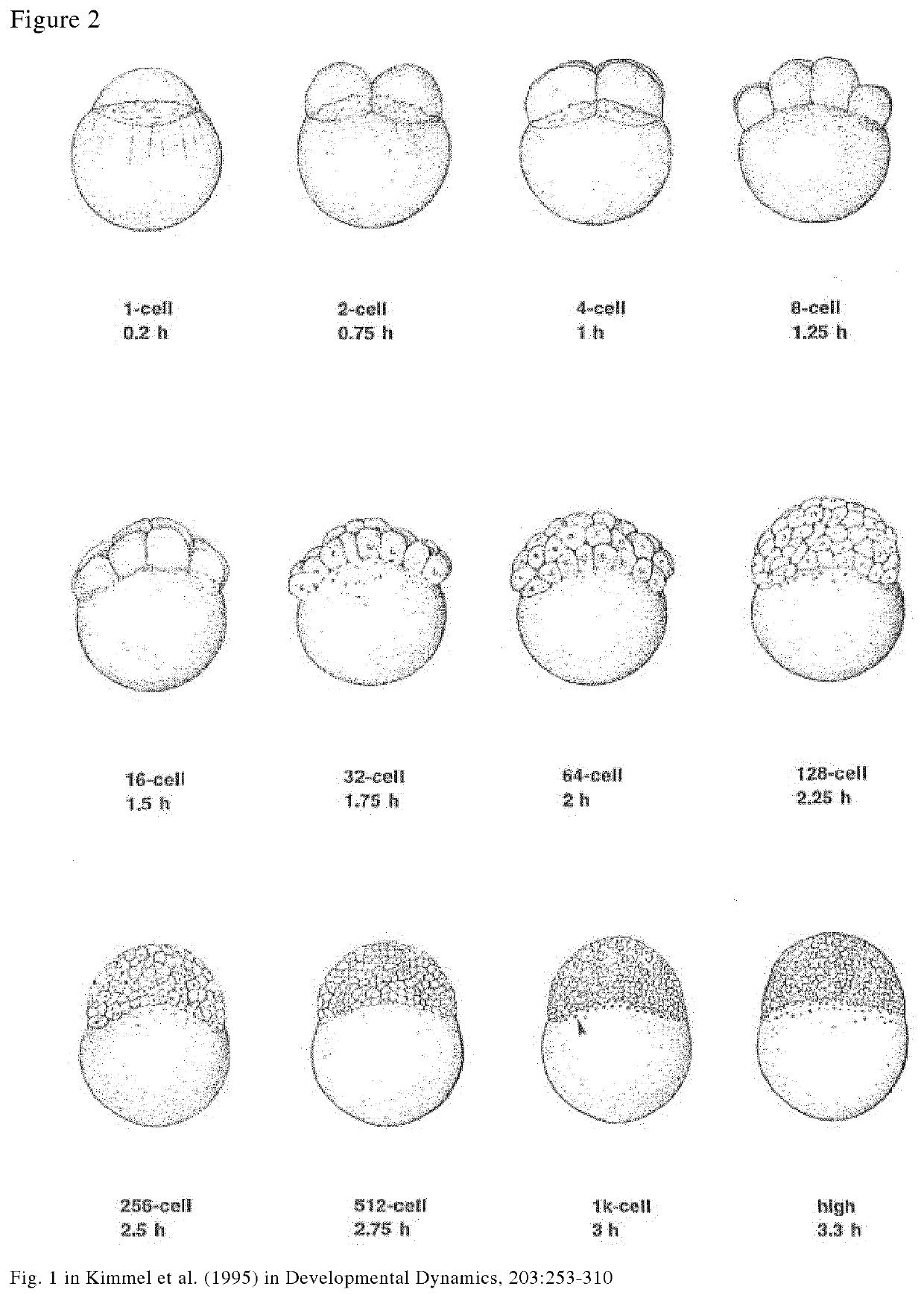
[0112]The pronase solution was pre-warmed to 25° C. and the further treatment is performed at RT. A number from −10 to −100 of embryos between the 512-cell to 1K cell stage was treated with 1 mg / ml pronase for 30 minutes in plastic petri dishes at room temperature. The treatment may also be performed at 25° C. in order for the removal of chorion and yolk to be performed more quickly.

[0113]The pronase treated embryos was then washed three times in Hank's Balanced Salt Solution (HBSS) with fetal bovine serum (FBS) to remove and quench the pronase. The isolated blastoderms are kept in HBSS comprising FBS until further use (e.g. transplantation, cryoconservation).
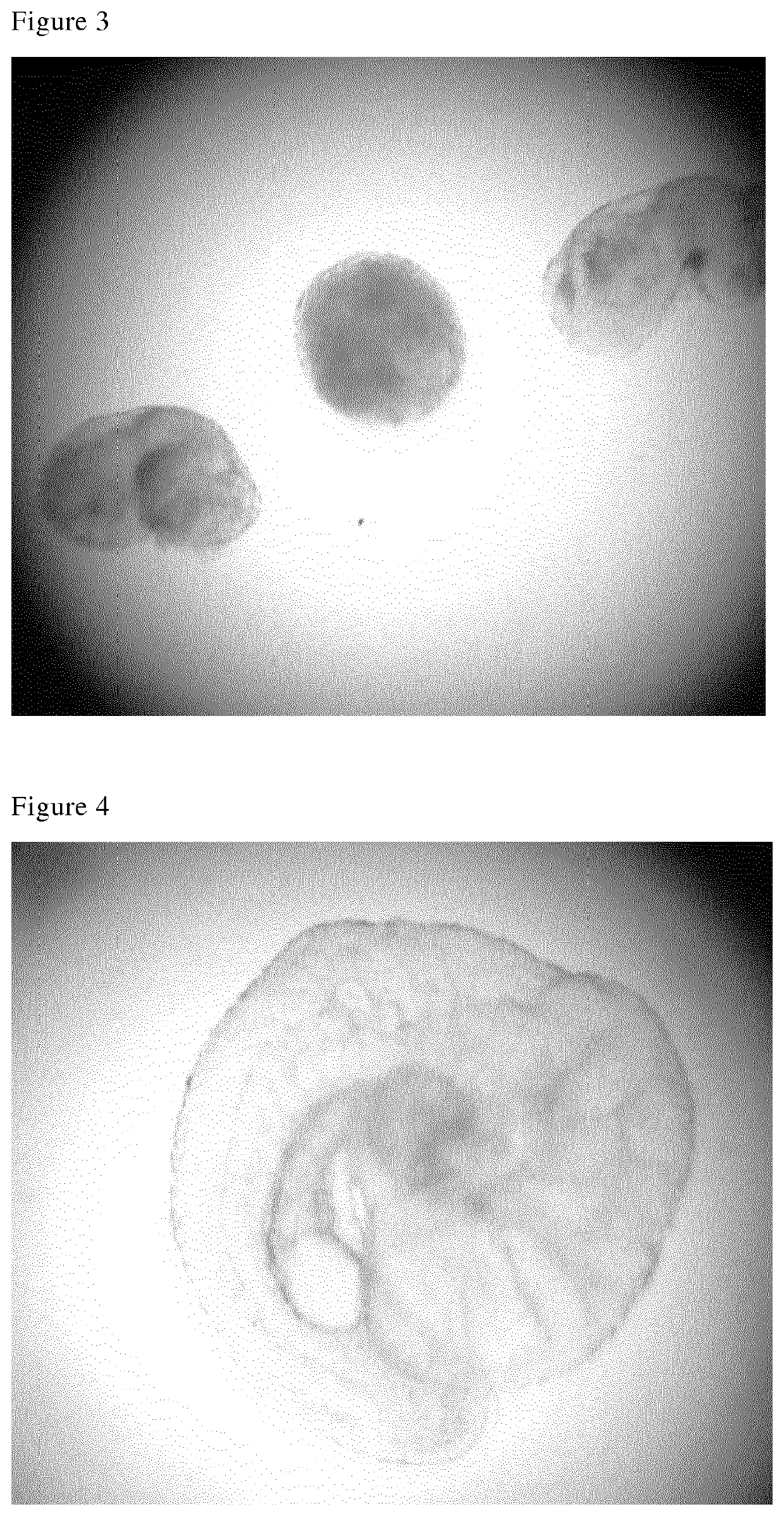
[0114]In FIG...

example 2

tation of Embryo Blastoderm on Donor Yolk

[0116]Zebrafish embryos was exposed to pronase and washed according to the method of example 1. The isolated blastoderm where then transplanted onto a donor yolk according to the method disclosed in Mizuno et al, From Methods in Molecular Biology, Vol 127, Cell and Tissue Transplantation in Zebrafish Embryos (1999) and Yamaha et al, (2001), Germ-line chimera by low-part blastoderm transplantation between diploid goldfish and triploid crucian carp.

[0117]FIG. 7 is a picture of an intact donor embryo (left) and a blastoderm (right) isolated according to the present invention.

[0118]The blastoderm treated according to the present invention attached to the donor yolk short time after transplantation (FIG. 8), and developed normally thereafter, see FIG. 9 showing the transplanted embryo 1-2 hours after transplantation of the blastoderm to the donor yolk.

example 3

val and Isolation of Blastoderms from Carp

[0119]Embryos being the result of natural breeding of carp belonging to the species Puntius conchonius were treated in the same manner as disclosed in example 1.

[0120]As shown in FIG. 10-13, yolk were successfully removed resulting in isolation of blastoderms from Puntius conhonius embryos in the blastula stage.

[0121]The method of example 1 was also applied on naturally breeded embryos of goldfinned barbs (Puntius semifasciolatus) with similar results (data no shown).


PUM


Abstract

A method for isolating the blastoderm of a non-human vertebrate embryo by enzymatic removal of the yolk.

Description

FIELD OF THE INVENTION[0001]The present invention relates to a method for yolk removal from embryos and isolation of blastoderms, in particular from fertilized embryos of aquatic origin.BACKGROUND OF THE INVENTION[0002]Embryogenesis is the process where an embryo develops from the fertilization of the ovum. In normal development, the newly fertilized egg (zygote) undergoes a rapid series of mitotic divisions resulting in a larger and larger number of smaller and smaller cells producing a cluster of cells. This first period of division is not accompanied by an increase in the overall size of the embryo, but resulting in tightly packed mass of cells (blastomeres) forming a blastoderm. As the cleavage continues, the embryo enters the blastula stage, and the blastula cells begin to migrate into the embryo, marking the beginning of the next stage, the gastrula stage, where the gastrulation process begins.[0003]All vertebrate embryos undergo a similar pattern of development after fertiliz...

Claims


Application Information

Patent Timeline
IPC IPC(8): C12N5/073
CPCC12N2506/02C12N5/0605C12N5/0606
Inventor GRONENG, LINE CECILIEKJOS, MARI
Owner CRYOGENETICS
Why Eureka
Try Eureka