

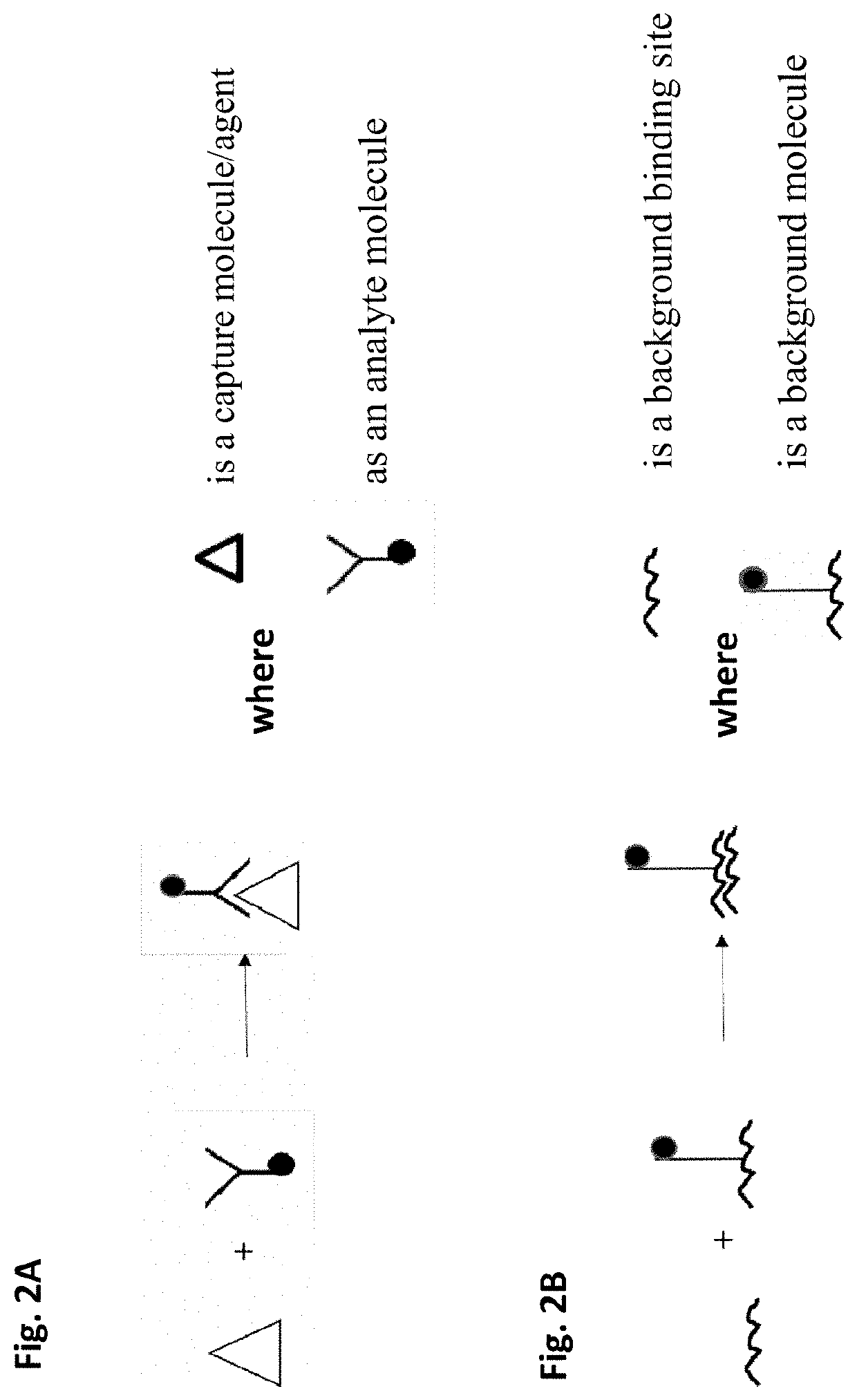
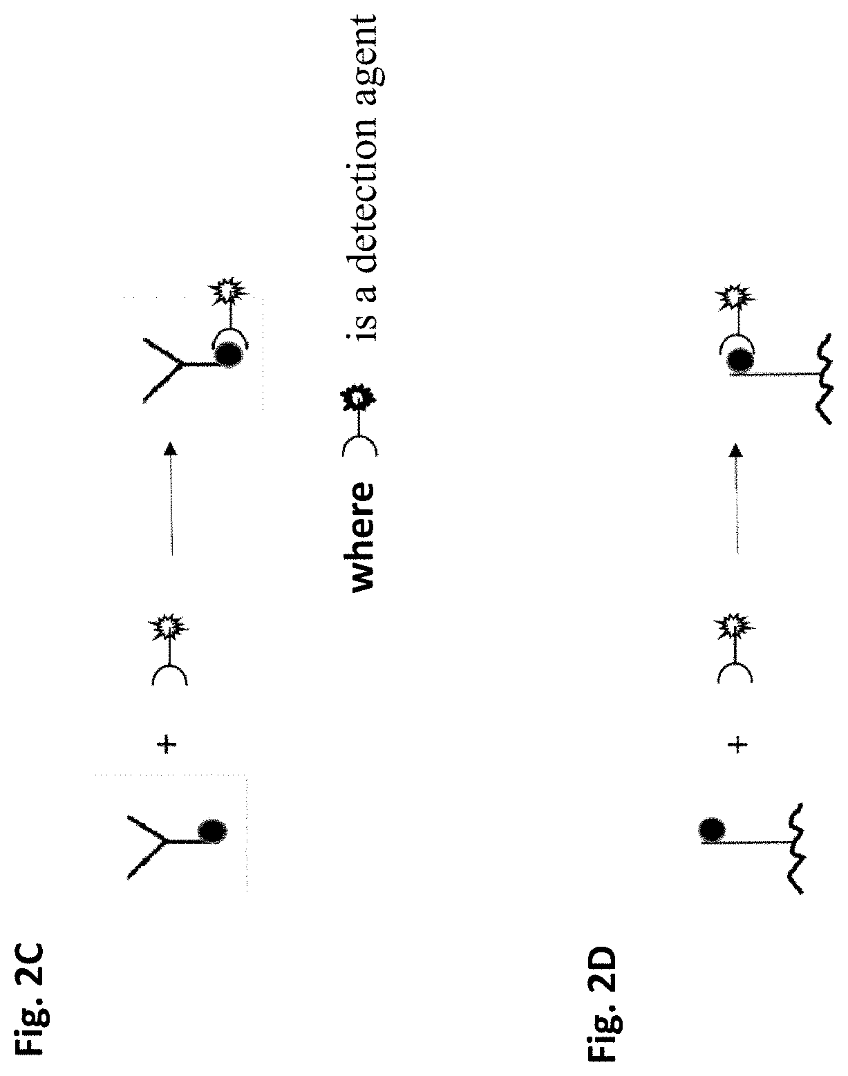
Method of removing non-specific binding signals using microparticle assay
- Summary
- Abstract
- Description
- Claims
- Application Information
AI Technical Summary
Benefits of technology
Problems solved by technology
Method used
Image

Examples
example 1
of an Anti-PEG Antibody in Serum
[0160]Mouse immunized serum: Leading Life Technology, obtained through CRO
[0161]Capture bead: CBA B4-PEG (prepared by LLT)
[0162]Associated control bead: CBA B8-BSA (LLT)
[0163]Samples: PEG-immunized mouse serum sample (CRO of LLT)
[0164]Analytes: anti-PEG Igs in mouse serum
[0165]Detection agent: PE-labeled goat anti-mouse Igs, IgG polyclonal (BD Biosciences)
[0166]Polyethylene glycol (PEG) immunized mouse serum was collected and tested for mouse anti-PEG antibody titer using a pair of capture microparticles and corresponding control microparticles. Capture microparticles were made by conjugating 20KD PEG with CBA B4 beads (purchased from BD Biosciences, San Jose, Calif.) as illustrated in FIG. 2E; the corresponding control microparticles were made by conjugating BSA with CBA B8 beads (BD Biosciences) as illustrated in FIG. 2E. B4 and B8 beads are the same size, indistinguishable by FSC and SSC by flow cytometry, i.e., showed as the same region in the FSC...
example 2
of Human Anti-PEG IgG, IgM and IgE Isotype (Chimeric Anti-Human IgG, IgM and IgE Detection)
[0169]Microparticles (CBA beads and conjugation buffer were purchased from BD Biosciences): Capture microparticle 1, E4-IgE, CBA bead E4 conjugated with anti-human IgE; associated control microparticle 1, B4-IgE, CBA bead B4 conjugated with anti-human IgE. The conjugation of capture bead 1 and the associated control bead 1 with IgE are processed with the same procedure including anti-IgE dosage. The FSC and SSC of the two microparticles are indistinguishable (FIG. 9A).
[0170]Capture microparticle 2, E6-IgM, CBA bead E6 conjugated with anti-human IgM;
[0171]associate control microparticle 2, B6-IgM, CBA bead B6 conjugated with anti-human IgM. The conjugation of capture bead 2 and the associated control bead 2 with IgM are processed with the same procedure including anti-IgM dosage, the FSC and SSC of the two beads are indistinguishable (FIG. 9A).
[0172]Capture microparticle 3, E8-IgG, CBA bead E8 ...
example 3
of Anti-PEG Antibody in Serum by Mass Cytometry
[0181]Chimeric human anti-PEG IgG (Leading Life Technology, purchased from Institute of Biomedical Sciences Academia Sinica, Taiwen)
[0182]Capture microparticle: 174Yb-tagged anti-PEG microparticle named “CBA B4-Anti-PEG-174Yb”, made by CBA B4 beads coupled with Anti-Mouse I-A / I-E (M5 / 114.15.2)-174Yb (Fluidigm, CA) and a mouse anti-PEG monoclonal antibody (prepared by Life Diagnostics, Inc., West Chester, Pa.).
[0183]Corresponding control microparticle: 209Bi-tagged anti-PEG microparticle named “CBA B8-Anti-PEG-209Bi”, made by CBA B8 beads coupled with Anti-Mouse I-A / I-E (M5 / 114.15.2)-209Bi (Fluidigm, CA) and a mouse anti-PEG monoclonal antibody (Life Diagnostics, Inc., West Chester, Pa.).
[0184]The particle size, amount of surface-coupled anti-mouse I-A / I-E and anti-PEG antibody were validated to be the same for capture microparticle and associated control microparticle.
[0185]Samples: 1 μg and 0.5 μg of chimeric human anti-PEG IgG were di...
PUM
 Abstract
Description
Claims
Application Information