Three dimensional porous cartilage template

a cartilage template and three-dimensional technology, applied in the field of three-dimensional porous cartilage templates, can solve the problems of severe bone defects and fractures, generating an annual cost of $2.5 billion, and bone tissue is also susceptible to malignant growths and metastases from surrounding organs, so as to promote the repair of severe bone defects and promote the repair of bone defects

Pending Publication Date: 2021-07-15
DREXEL UNIV
View PDF0 Cites 2 Cited by
  • Summary
  • Abstract
  • Description
  • Claims
  • Application Information

AI Technical Summary

Benefits of technology

[0030]In another aspect, the invention provides a method of promoting the repair of a bone defect in a patient, the method comprising preparing a porous cartilage template having a bone-mimicking internal structure, embedding a plurality of cells into the porous cartilage template, and implanting the porous cartilage template into the bone defect in the patient, thereby promoting the repair of the bone defect.

Problems solved by technology

Although bone has an exceptional capacity for regeneration, repairing severe bone defects and fractures remains a critical challenge.
Every year, over 600,000 cases linked to cancer or traumatic injury require the use of bone grafting, generating an annual cost of $2.5 billion.
These pre-formed grafts, which are either autogeneic or allogeneic, are associated with a number of complications including donor site morbidity for autografts and immune rejection for allografts.
Bone tissue is also susceptible to malignant growths and metastases from surrounding organs.

Method used


Image

  • Three dimensional porous cartilage template
  • Three dimensional porous cartilage template
  • Three dimensional porous cartilage template

Examples


example 1

ion of 3D Porous Cartilage Template Printing Using Human MSCs

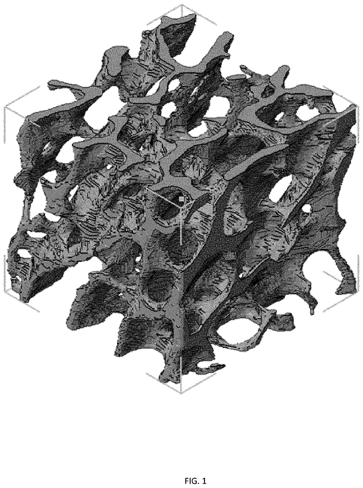
[0139]Rationale. The first step in developing a model of endochondral ossification is the generation of a porous cartilage template. While cartilage has been engineered for decades using human MSCs cultured on porous scaffolds, chondrocytes do not reside on porous structures in the body. Rather, they are encapsulated within dense ECM, even when this structure constitutes a macroscopically porous template like it does during endochondral ossification. For this reason, cartilage engineering is typically conducted by encapsulating the cells within a matrix that closely resembles the native ECM, such as a hydrogel. Hydrogels, 3D crosslinked polymer networks swollen with water, can be prepared from synthetic or naturally derived polymers. The stiffness and crosslinking density of the hydrogel matrix affects the development of cartilage tissue. However, the generation of a porous hydrogel structure in which chondrocytes are enca...

example 2

f Dynamic Matrix Composition on Chondrocyte Hypertrophy

[0143]Rationale. The composition of the ECM is extremely important in cartilage development. Many investigators have explored the effects of incorporating various ECM components into hydrogels, including glycosaminoglycans (GAGs) and different types of collagen. However, in normal cartilage development, the content of the ECM varies dramatically over time. For example, MSCs undergoing chondrogenic differentiation produce the ECM component fibronectin for about 10 days, and then it is downregulated. The importance of temporal control over this biochemical cue in MSC chondrogenesis was demonstrated when fibronectin fragments were released from synthetic hydrogels via a light-activated degradation strategy according to the temporal profile observed in development. Chondrogenic differentiation of encapsulated MSCs was enhanced compared to hydrogels containing persistent levels of fibronectin. In order to examine the effects of signa...

example 3

es Between Bone-Like Structure (Described Embodiments), a Lattice Structure (Control 1) and a Non-Porous Structure (Control 2)

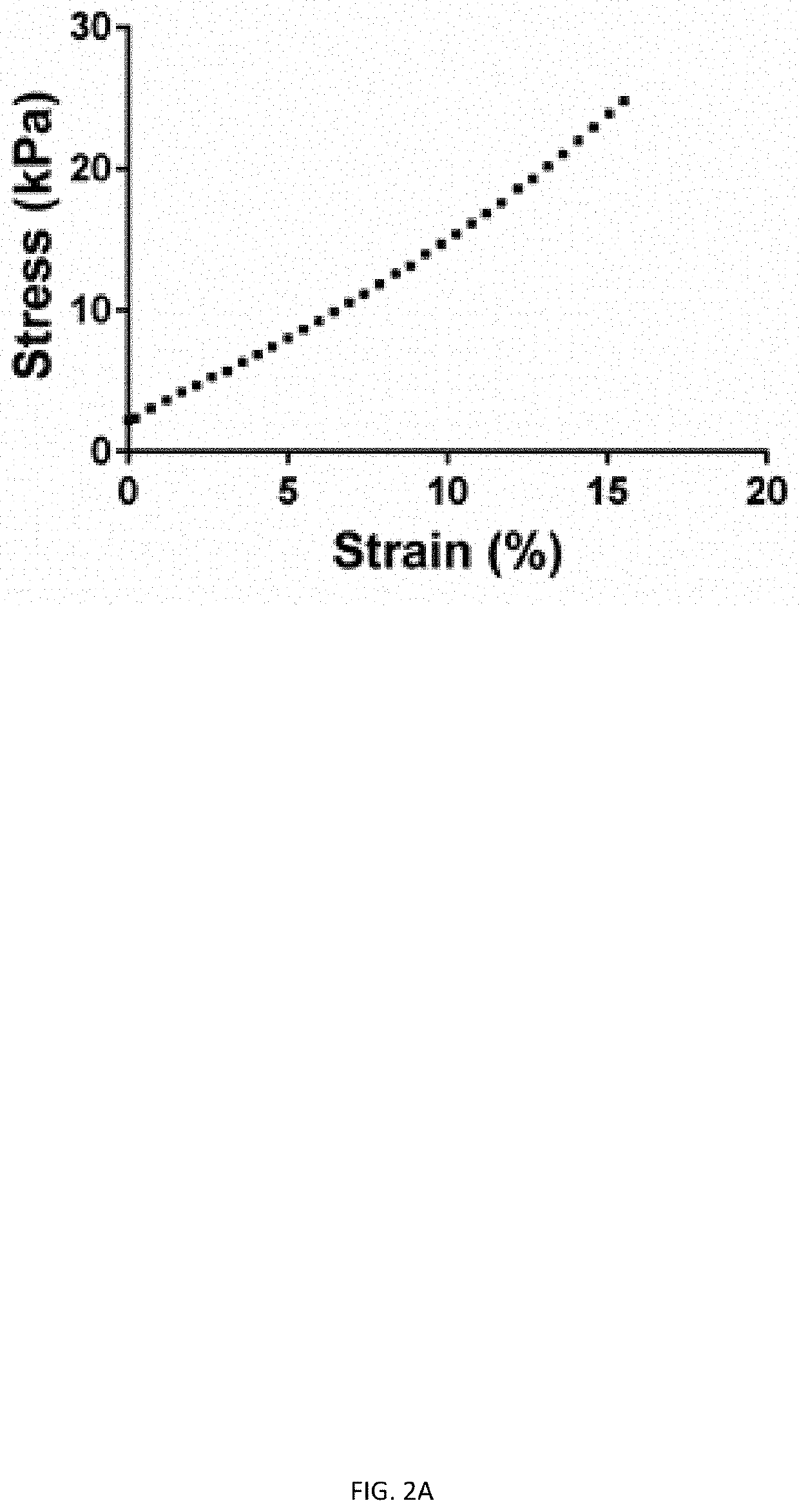
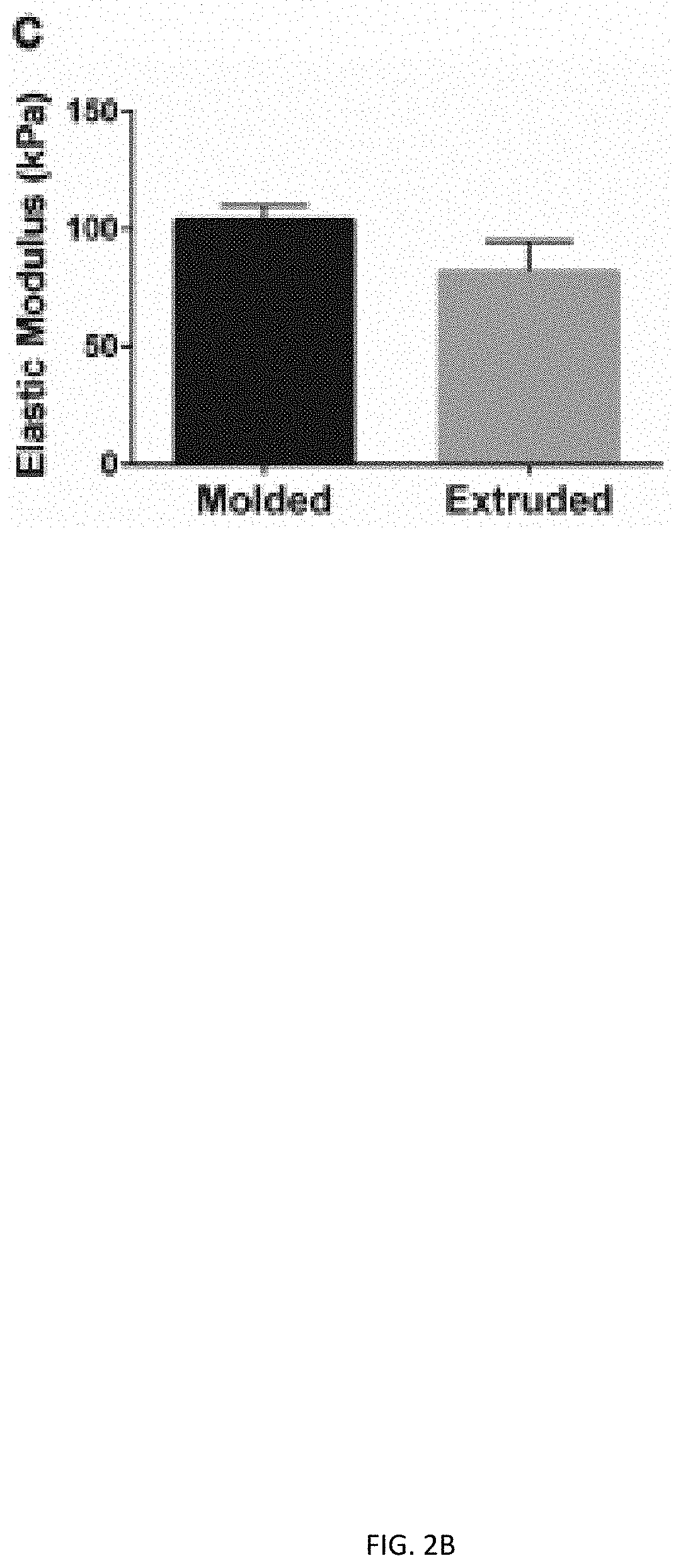
[0146]A. Percent porosity and pose size of bone-like and lattice structures is calculated from CAD designs. Similar measurement values between the two structures removes porosity as a variable in experiments comparing structure.[0147]B. The stress distribution of all three structures is evaluated using finite element analysis. The described embodiments have preferred stress distribution characteristics.[0148]C. Chondrocyte pellet-laden gels are printed into all three structures and cultured for three days. Live / dead staining of all constructs is performed to assess cell viability. Glycosaminoglycans (GAGs) staining of all constructs after three days of culture is performed to evaluate cartilage tissue formation.[0149]D. The culture described in (C) is extended to three weeks after extrusion of all structures. The constructs are stained for collagen and GAGs t...


PUM

PropertyMeasurementUnit
volumeaaaaaaaaaa
volumeaaaaaaaaaa
volumeaaaaaaaaaa

Abstract

This application relates to biologically compatible porous cartilage templates for in vitro and in vivo generation of bone with enhanced structural characteristics. Provided herein are compositions having an internal structure desirable for the generation and regeneration of bone, along with methods of preparation and use.

Description

CROSS-REFERENCE TO RELATED APPLICATIONS[0001]This application is a continuation under 35 U.S.C. § 120 of U.S. patent application Ser. No. 16 / 310,576, filed Dec. 17, 2018, which is a national phase application under 35 U.S.C. § 371 of International Application No. PCT / US2017 / 038718, filed Jun. 22, 2017, which claims the benefit of priority under 35 U.S.C. § 119(e) to U.S. Provisional Patent Application Ser. No. 62 / 353,799, filed Jun. 23, 2016. The entire content of each application is hereby incorporated by reference herein.STATEMENT REGARDING FEDERALLY SPONSORED RESEARCH OR DEVELOPMENT[0002]This invention was made with government support under Grant No. R01 HL130037 awarded by the National Institutes of Health. The U.S. Government has certain rights in the invention.BACKGROUND OF THE INVENTION[0003]Although bone has an exceptional capacity for regeneration, repairing severe bone defects and fractures remains a critical challenge. Every year, over 600,000 cases linked to cancer or tr...

Claims


Application Information

Patent Timeline
Patent Type & Authority Applications(United States)
IPC IPC(8): A61L27/56C09D11/04A61F2/44A61F2/46A61L27/22A61L27/38A61L27/52A61L27/54C09D11/101
CPCA61L27/56C09D11/04A61F2/44A61F2/46B33Y70/00A61L27/3834A61L27/52A61L27/54C09D11/101A61L27/222B33Y80/00A61L2300/25A61L2300/62A61L2300/64A61L2430/02A61L2430/06
Inventor SPILLER, KARA LORRAINEERSUMO, NATHAN TESSEMA
Owner DREXEL UNIV
Why Eureka
Try Eureka