Electronic endoscope system

a technology of endoscope and endoscope, which is applied in the field of electronic endoscope system, can solve the problem of inability to specify the place of the lesion in the organ, and achieve the effect of efficient acquisition of information

Pending Publication Date: 2022-07-07
HOYA CORP
View PDF0 Cites 0 Cited by
  • Summary
  • Abstract
  • Description
  • Claims
  • Application Information

AI Technical Summary

Benefits of technology

[0030]According to the electronic endoscope system described above, it is possible to efficiently acquire the information of the imaging place of the still image without providing a dedicated sensor in the electronic endoscope and associate the information with the still image.

Problems solved by technology

An endoscope system images a moving image, so that a still image is acquired from the moving image according to an instruction of an operator and recorded to be stored in a memory and the like; however, in a case where it is not clear where in an organ the still image is imaged, it is often impossible to specify a place of the lesion in the organ.

Method used


Image

  • Electronic endoscope system
  • Electronic endoscope system
  • Electronic endoscope system

Examples


Embodiment Construction

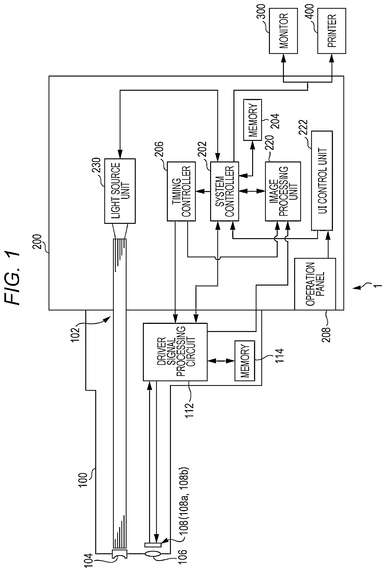
[0041]Hereinafter, an electronic endoscope system of an embodiment is described with reference to the drawings.

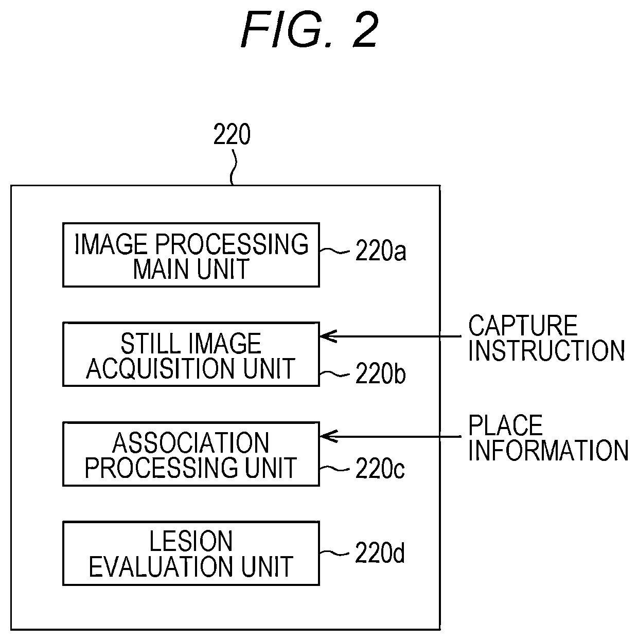
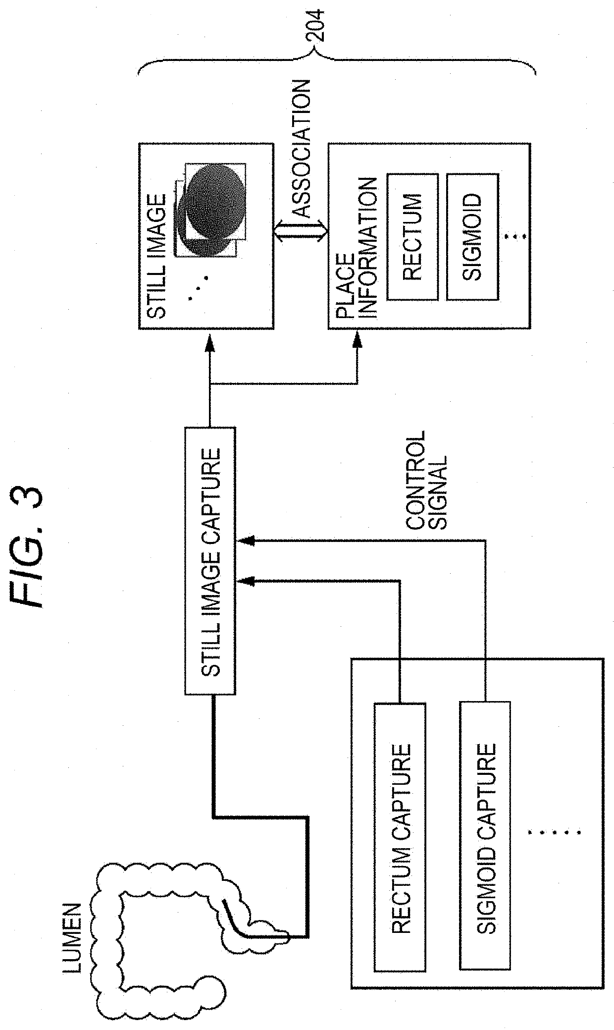
[0042](Place Information Regarding Imaging Place of Still Image and Capture Instruction)

[0043]A processor of an electronic endoscope system of the embodiment described below acquires a still image from a moving image of living tissue in an organ imaged by an electronic endoscope. This still image is used, for example, for evaluating a lesion degree in a lumen. In a case of imaging the living tissue in the organ as the moving image, for example, the electronic endoscope (hereinafter, referred to as an electronic scope) is inserted from an open end of a tubular organ to a deepest position in the organ in a depth direction to be imaged, and this images the living tissue in the organ while moving from there substantially continuously toward the open end of the organ. The depth direction includes both a direction from the open end toward the deepest position and a direction from...


PUM


Abstract

An electronic endoscope system is provided with an electronic endoscope, a processor provided with a still image acquisition unit that acquires a part of a moving image imaged by the electronic endoscope as a still image, an association processing unit that associates place information regarding an imaging place of living tissue in a the lumen with the still image, and a recording unit, a monitor, and an input operation device that transmits a capture instruction for acquiring the still image from the moving image to the still image acquisition unit in response to an operation of determining one place out of a plurality of places in the lumen set in advance as a selected place as a trigger, and further transmits information of the selected place to the association processing unit as the place information regarding the imaging place during an imaging period of the moving image.

Description

TECHNICAL FIELD[0001]The present invention relates to an electronic endoscope system that acquires a still image from a moving image of living tissue in an organ.BACKGROUND ART[0002]In a lesion area in living tissue, there are various levels of severities from an inflammation with a thinned mucosal layer of the living tissue that exhibits redness to an ulcer in which the mucosal layer and a lower layer thereof are partially lost. For example, an ulcer part of a lesion of ulcerative colitis (UC) includes white moss and purulent mucus to be white, and an inflammation part causes edema and exhibits redness due to easiness in bleeding. Such lesion area may be imaged to be observed and inspected with an endoscope system.[0003]An endoscope system images a moving image, so that a still image is acquired from the moving image according to an instruction of an operator and recorded to be stored in a memory and the like; however, in a case where it is not clear where in an organ the still ima...

Claims


Application Information

Patent Timeline
Patent Type & Authority Applications(United States)
IPC IPC(8): A61B1/00A61B1/045A61B1/06G06T1/00
CPCA61B1/000094A61B1/045A61B1/000095A61B1/00006G06T1/00A61B1/0684A61B1/04A61B1/00009G06T7/0012G06T2207/10068G06T2207/10024G06T2207/30028G06T2207/30096A61B1/05A61B1/07A61B1/0005A61B1/0004A61B1/00186A61B1/0661
Inventor MAKINO, TAKAO
Owner HOYA CORP
Why Eureka
Try Eureka