Analytical method for controlled and measured internal fluid after surgery

a technology of internal fluid and analytic method, which is applied in the field of medical devices, can solve the problems of high pressure, possible loss of vacuum, large volume, etc., and achieve the effects of preventing the loss of vacuum, facilitating handling, and maintaining sterility and a seal

Active Publication Date: 2022-08-18
SOMAVAC MEDICAL SOLUTIONS INC
View PDF0 Cites 0 Cited by
  • Summary
  • Abstract
  • Description
  • Claims
  • Application Information

AI Technical Summary

Benefits of technology

[0015]In several embodiments, the present invention makes use of a powered source of negative pressure which helps overcome clogging observed in prior art devices and one or more reservoirs which allow excess air to be removed. The invention comprises disposable reservoirs with one-way valves that are easy to handle while maintaining sterility and a seal to prevent the loss of vacuum. The present invention further provides continuous negative pressure suction which assists in providing constant drainage. Prior art devices do not provide a means of applying continuous negative pressure to the percutaneous wound site.
[0016]In addition, the measurements of the output can be performed automatically, relieving the need for the patient to perform measurements directly (and thus resolving the potential conflict of interest in self-measuring so that the best clinical decisions can be made). The measurements of output can be relayed to the caregiver, doctor, or the nurse via wired or wireless communications, and enables patients who do not have companions to manage their drain care. There is a potential diagnostic value in taking various measurements associated with the collected fluid. Measurements can include and are not limited to collected fluid amount, pH, certain known harmful mediators (cytokines, chemokines, reactive oxygen species), protein levels, blood content, etc. For example, amount of fluid collected can be an indicator of possible seroma development in some hernia surgeries. Additionally, pH has also been shown to act as an indicator of possible seroma formation. The present invention thus allows for the detection of infectious materials, and any other chemicals or substances which may indicate infection, or the presence of some medical condition which may naturally arise in response to the surgical procedure, initial pathology, or additional complications (of either the surgical procedure or the initial pathology) in the fluid collected from percutaneous (internal) wounds.

Problems solved by technology

There are large amounts of fluid that collect in patients who undergo large void-forming surgeries.
This results in large volumes to be collected, measured, and emptied.
Otherwise, either the reservoir is filled quickly with air / liquid mixture and emptying must take place to remove fluid often, or the reservoir overfills leading to high pressure levels and possibly backflow.
Prior art devices do not provide a means of applying continuous negative pressure to the percutaneous wound site.

Method used


Image

  • Analytical method for controlled and measured internal fluid after surgery
  • Analytical method for controlled and measured internal fluid after surgery
  • Analytical method for controlled and measured internal fluid after surgery

Examples


Embodiment Construction

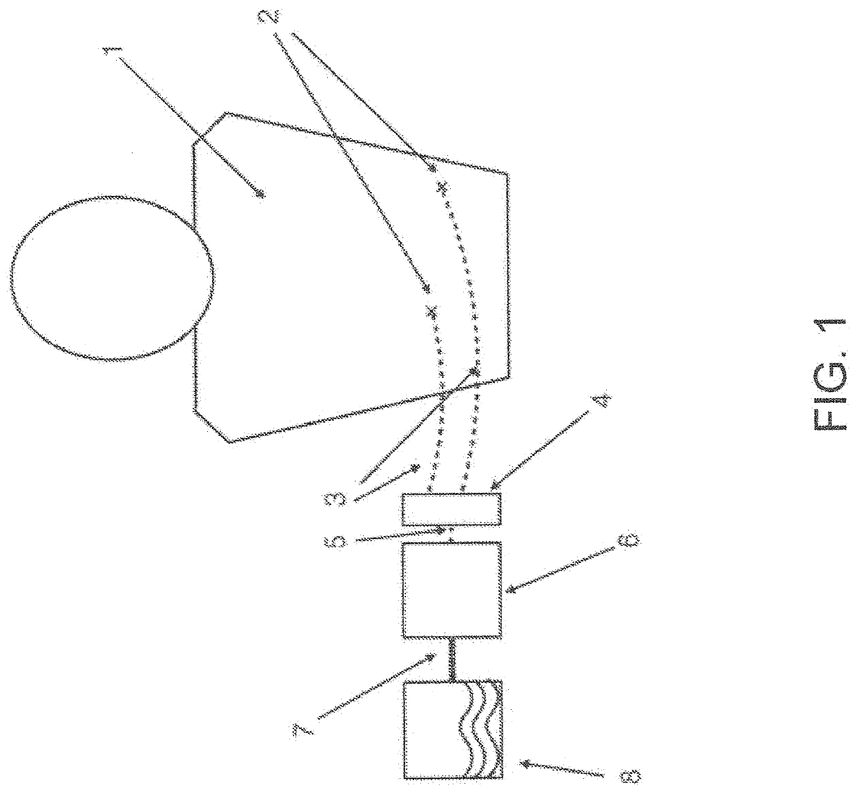
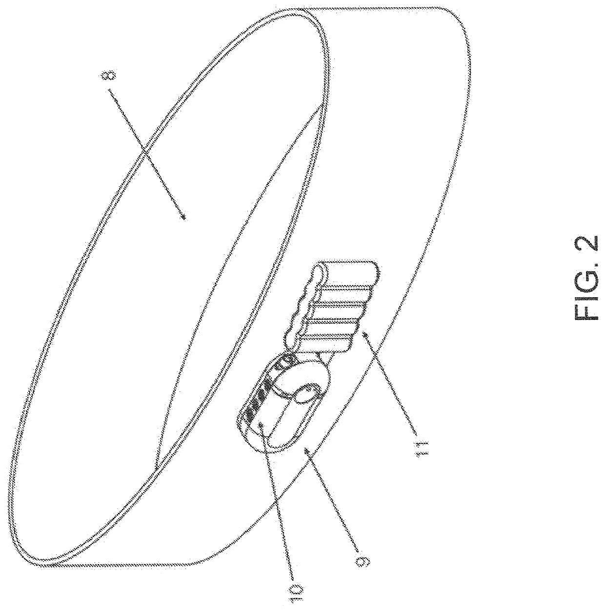
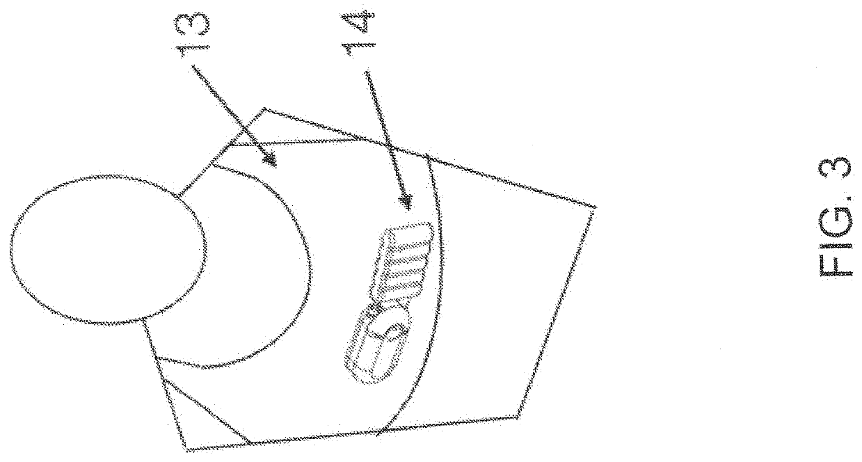
[0060]In various exemplary embodiments, the present invention comprises a system and apparatus for the collection of serous or serosanguinous fluid from the percutaneous site after surgery. There are large amounts of fluid that collect in patients who undergo large void-forming surgeries. This results in large volumes to be collected, measured, and emptied. In order to effectively remove the fluid in a continuous manner, air must be removed from the collection reservoir. Otherwise, either the reservoir is filled quickly with air / liquid mixture and emptying must take place to remove fluid often, or the reservoir overfills leading to high pressure levels and possibly backflow.

[0061]In several embodiments, the present invention makes use of a powered source of negative pressure which helps overcome clogging observed in prior art devices, and one or more reservoirs which allow excess air to be removed. The invention comprises disposable reservoirs with one-way valves that are easy to ha...


PUM


Abstract

A system and apparatus for the collection of serous or serosanguinous fluid from a percutaneous site after surgery. A pump unit with one or more pumps or powered sources provide continuous negative pressure suction to draw fluid from the percutaneous site and pumps the fluid into disposable reservoirs with one-way valves that are easy to handle while maintaining sterility and a seal to prevent the loss of vacuum. Air is continuously removed from the reservoirs. Measurement and analysis of the output is performed automatically. Additionally, through such a peristaltic pump device, controlled collection of measured surgical and / or bodily fluid from a subject patient may be undertaken allowing for more effective and closer analysis, as well.

Description

[0001]This application is a continuation-in-part of co-pending U.S. Non-Provisional patent application Ser. No. 15 / 604,254, filed on May 24, 2017, which claims benefit of and priority to U.S. Provisional Applications Nos. 62 / 340,853, filed May 24, 2016, now expired, and 62 / 409,400, filed Oct. 18, 2016, expired, and is entitled to priority to those filing dates. The specifications, drawings, appendices, and complete disclosures of U.S. Provisional Applications Nos. 62 / 340,853 and 62 / 409,400 as well as U.S. Non-Provisional patent application Ser. No. 15 / 604,254, are incorporated herein by specific reference for all purposes.FIELD OF INVENTION[0002]This invention relates to medical devices, particularly those used to drain serous or serosanguinous fluid from the percutaneous site after surgery. Additionally, this disclosure relates to the utilization of a disclosed peristaltic device for uniform and controlled collection of seroma fluid for analysis thereof.BACKGROUND OF THE INVENTION[...

Claims


Application Information

Patent Timeline
Patent Type & Authority Applications(United States)
IPC IPC(8): A61M1/00A61B5/145
CPCA61M1/962A61B5/14507A61M1/98A61B5/14539A61M1/631A61M1/73A61M1/78A61M1/79A61M1/83A61M1/87A61M1/90A61M1/918A61M1/984A61M2205/14A61M2205/3324A61M2205/3389A61M2205/3576A61M2209/088F04B23/04F04B43/1253A61M1/74
Inventor HERWIG, JOSHUA D.ROAN, ESRA
Owner SOMAVAC MEDICAL SOLUTIONS INC
Why Eureka
Try Eureka