Imaging gore loading tool

a technology of imaging gore and loading tool, which is applied in the field of medical devices, can solve the problems of axial load reaching its critical value, imaging core prone to lose column strength, and imaging core with decreased column strength inclined to collapse or buckl

Inactive Publication Date: 2001-01-09
SCI MED LIFE SYST
View PDF9 Cites 297 Cited by
  • Summary
  • Abstract
  • Description
  • Claims
  • Application Information

AI Technical Summary

Problems solved by technology

However, as the diameter of the imaging core is reduced, the imaging core tends to lose column strength.
Unfortunately, an imaging core with decreased column strength is inclined to collapse or buckle as the imaging core is advanced into the catheter lumen and encounters any type of increased friction due to sharp bends or constrictions, causing the axial load to reach its critical value.
This occurrence is problematic since imaging cores are typically loaded into cath

Method used


Image

  • Imaging gore loading tool
  • Imaging gore loading tool
  • Imaging gore loading tool

Examples


Embodiment Construction

The present invention provides for coaxially loading a flexible member into a lumen of a catheter. The present invention can work with any imaging core that incorporates a flexible drive shaft that can be delivered into a catheter body. Specifically, the imaging core will be a small diameter imaging core that has relatively little column strength. The catheter body can include, but is not limited to, ultrasound catheters, angioplasty balloon catheters, radiation catheters, stent delivery catheters, and aneurysm coil delivery catheters.

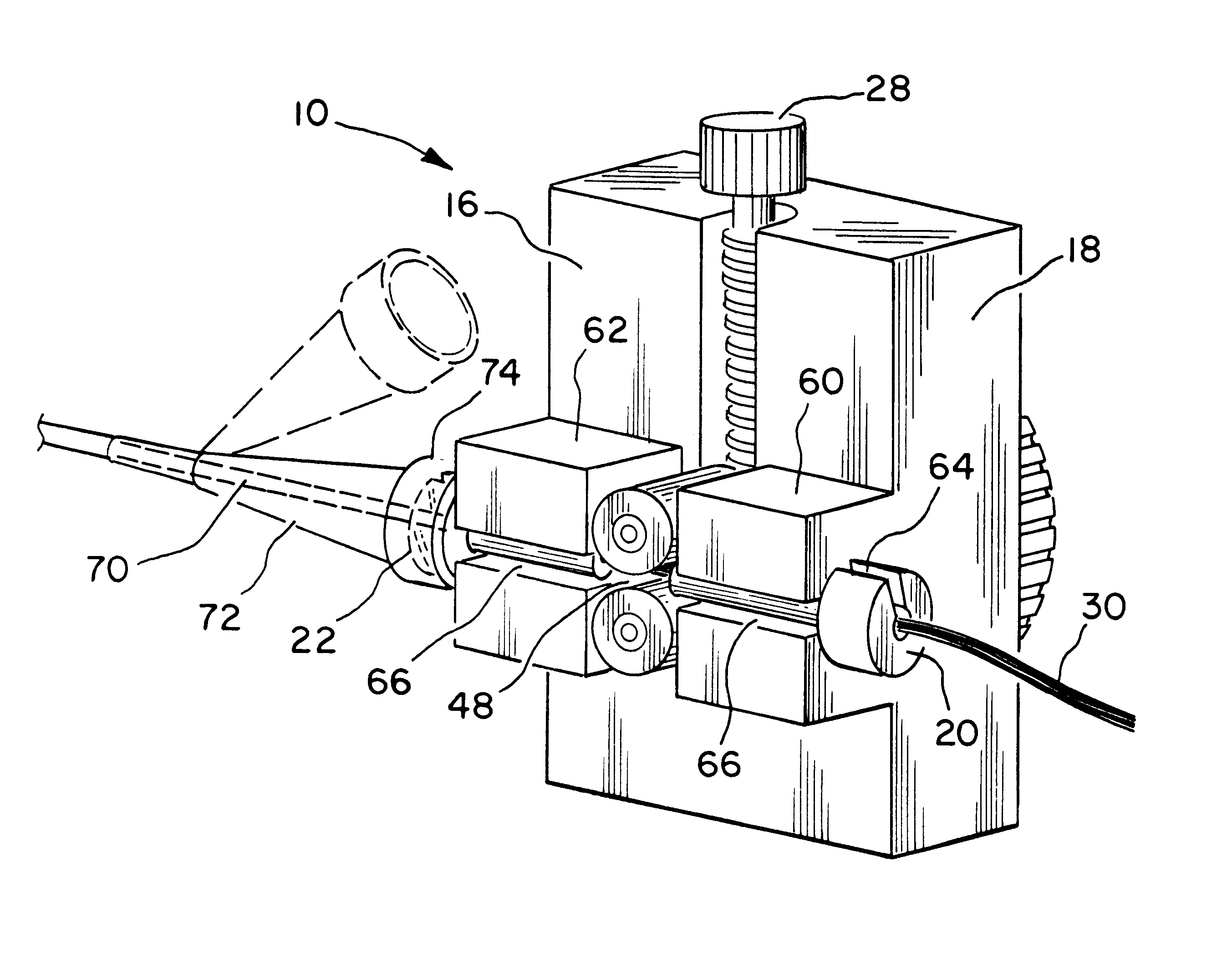
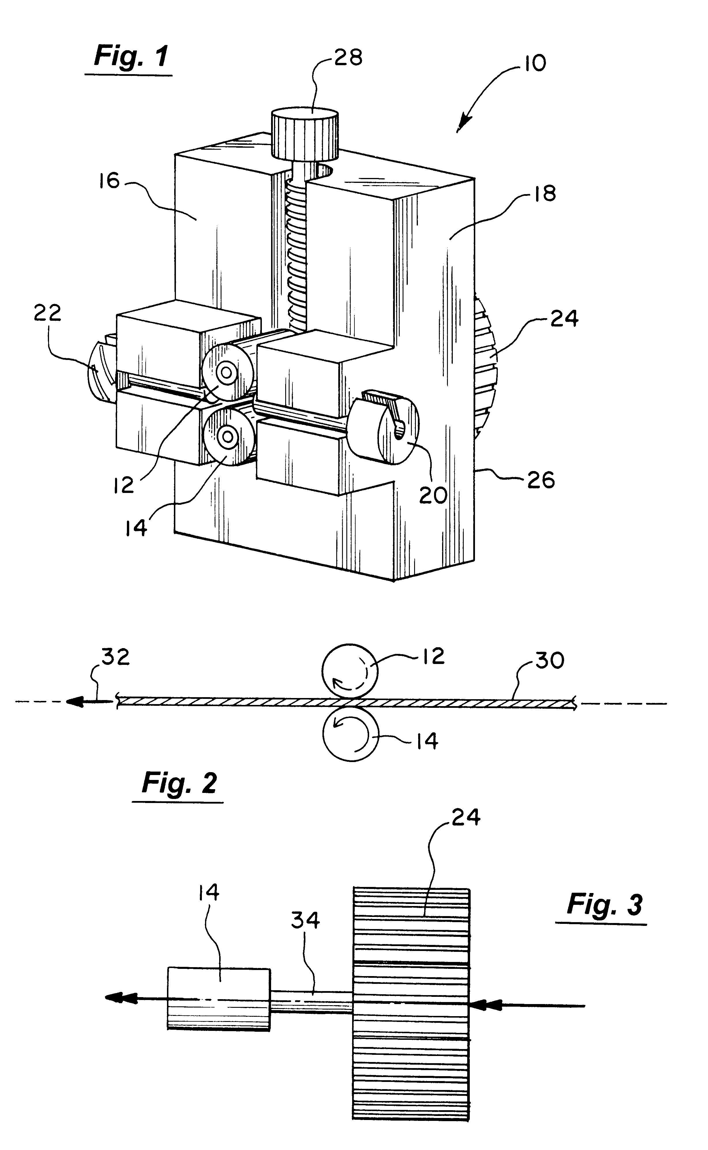
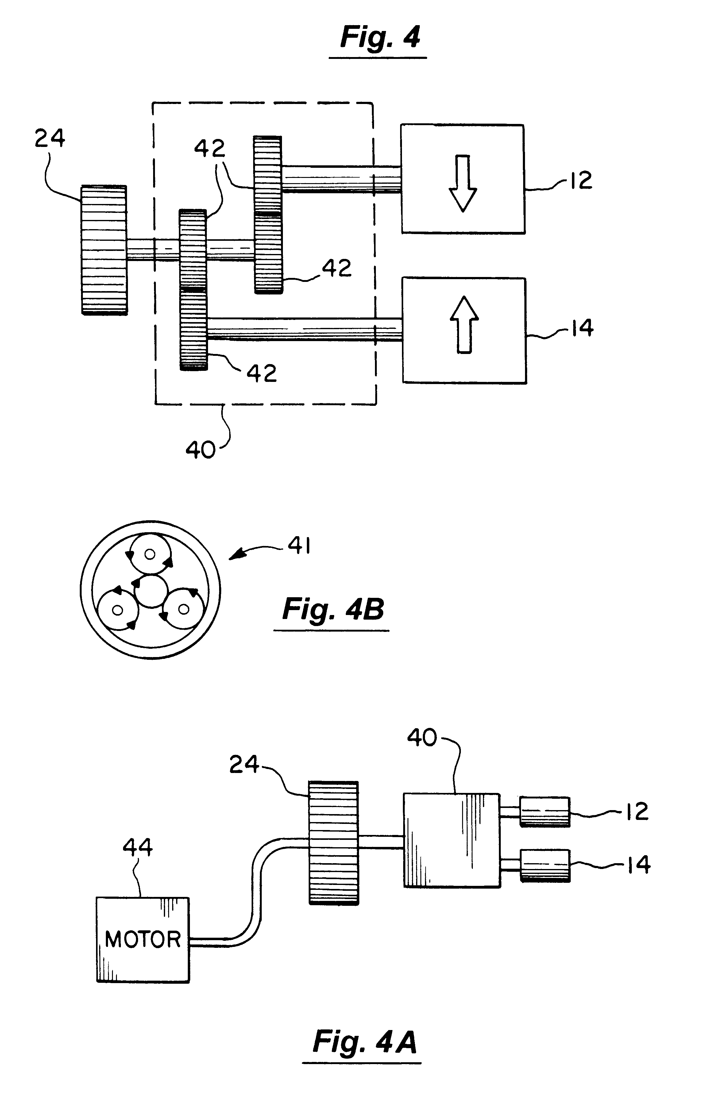
Referring to FIG. 1, a tool 10 for coaxially loading a flexible member into a lumen of a catheter, is shown according to the present invention. The tool 10 includes a pair of opposed rollers 12 and 14 mounted to a front face 16 of tool body 18, which also has an alignment mechanism 20 and a manifold coupling device 22, which removably receive the inner catheter, and a regulator assembly 28. A turning device 24 is mounted on an opposing face 24 of tool ...


PUM


Abstract

A tool for coaxially loading a flexible member into a lumen of a catheter. Typically, the flexible member is a flexible imaging core or similar device. The tool includes a body which has a connector, which connects the body to a hub located on the catheter. A pair of opposed rollers, disposed proximate to the connector, are used to advance the flexible member, through the connector, and into the catheter, which is connected to the connector. The flexible member is advanced by counter rotating the rollers.

Description

1. Field of the InventionThe present invention relates generally to medical devices and methods for their fabrication. In particular, the invention relates to a tool for loading a small diameter flexible imaging core into a catheter body.2. Description of the Relevant ArtIntravascular imaging of blood vessel lesions prior to percutaneous transluminal angioplasty, arthrectomy, and other interventional procedures, is of great benefit. A particularly successful design for an intravascular imaging apparatus employs a rotatable ultrasonic transducer, where the transducer is attached to the distal end of a flexible drive cable or imaging core. The transducer may be disposed within a catheter body or sheath and rotated in order to transmit an ultrasonic signal and produce a video image by well-known techniques.To be compatible with introduction of the catheter into very small coronary arteries, the imaging core is reduced to correspondingly small diameters. In addition to the small diamete...

Claims


Application Information

Patent Timeline
IPC IPC(8): A61B1/012A61B1/018A61M25/01A61B8/12
CPCA61B1/018A61M25/0113A61M25/09041
Inventor WHITE, DAVID A.LAMMERTS VAN BUEREN, LINDA H.
Owner SCI MED LIFE SYST
Why Eureka
Try Eureka