Targeted molecular bar codes and methods for using the same
Inactive Publication Date: 2002-10-15
RGT UNIV OF CALIFORNIA
12 Cites 66 Cited by
AI-Extracted Technical Summary
Problems solved by technology
The only limitation with respect to the physical dimensions of the molecular bar code is that it be able to assume a configuration that allows it to pass or translocate lengthwise through the nanopore from the cis sid...
Method used
6-Bromohexanoic acid may be protected, where desired, prior to coupling with the phenol. If it is protected with TMS, it yields 7 the silyl ester of 6-bromohexanoic acid. 7 then alkylates p-hydroxy methylbenzoate to yield 8. 7 behaves like the methyl ester during Grignard reaction. If alkylation at the silyl ester occurs, it is necessary to reverse the addition order and form the tertiary alcohol before alkylating with the 6-Bromohexanoic acid. The next step involves a double Grignard reaction at the ester, yielding alcohol 9. If the TMS protection of the caboxylate is not necessary, three equivalents of the Grignard will be necessary. The tertiary alcohol is then substituted with Cl using acetyl-Cl, converting the compound to the linker/protecting group 10. This halide is then coupled to a primary alcohol using the same procedure as DMT protection. Once coupled to an oligo, the DMT linker is then attached to an amine functionality using standard peptide coupling techniques. Treatment with trichloroacetic acid is the standard deprotection method for DMT. The linker is more labile than the DMT because it has three electron donating groups vs. the two on DMT, so a milder acid treatment cleaves it readily. The general scheme is listed below. ##STR6## ##STR7##
A targeted molecular bar code was synthesized at 1 .mu.mol scale on an ABI 392 DNA Synthesizer using standard conditions. The targeted bar code (top of FIG. 9) was composed of a poly dT.sub.18 mer at its 5' end, linked by a disulfide bond (Glen Research, Sterling, Va.) to a 50 base-long, anti-sense segment of N-ras Exon 1. This targeted bar code (10 .mu.M final concentration) was hybridized at 40 degrees C. for four hours in 6.times. SSC to either a 40 base segment of N-ras Exon 1 (sense strand) or to a 70 base segment of the p53 open reading frame. Each of the target oligonucleotides was attached to polystyrene beds to facilitate recovery and washing of the bound bar code. Following hybridization, the beads were washed twice in 0.2.times. SSC at room temperature. The beads were then suspended in 20 .mu.l of 0.1M DTT (pH 8.3/Tris) for 30 minutes to cleave the disulfide linkage between the dT.sub.18 code and the N-ras targeting sequence. The beads were subsequently spun down and the dT.sub.18 bar code was recovered in the supernatant. One half the supernatant was then added to ca. 100 .mu.l of 1M KCl buffer bathing a single alpha hemolysin nanopore. The recovered bar codes were then counted by translocating them through the pore at 150 mV applied potential and identifying dT.sub.18 -specific channel blockades. A diagram of the above protocol is provided in FIG. 9.
FIG. 12 shows that including hairpins with 2-to-10 base pair stems in DNA oligomers increases the blockade duration by 15-18 .mu.s per base pair relative to randomized controls. A two-base pair difference is easily distinguishable. Furthermore, oligomers containing different number of base pairs but the same number of monomer units can be distinguished based on blockade duration alone given a sample size of one zeptomole (1000 molec...
Abstract
Targeted molecular bar codes and methods for using the same are provided. The subject targeted molecular bar codes include a molecular bar code and a member of a specific binding pair, where the specific binding pair member is generally bonded to the bar code through a linking group. The subject molecular bar code may be read during translocation through a single nano-meter scale pore. The subject targeted molecular bar codes find use in a variety of different applications involving analyte detection, such as screening and diagnostic applications.
Application Domain
Sugar derivativesMicrobiological testing/measurement +2
Technology Topic
Subject specificBarcode +3
Image
Examples
- Experimental program(1)
Example
I. Preparation of a Horizontal Bilayer Containing a Single Nanopore Using a Miniature Horizontal Support
A single channel was inserted into a bilayer on the horizontal aperture as follows:
A. Formation of Diphytanoyl PC/hexadecane Bilayers on a Horizontal Aperture
A miniature support was manufactured as described under `Preparation of Subject Devices` as disclosed in copending application Ser. No. 60/107,307 filed Nov. 8, 1998, the disclosure of which is herein incorporated by reference. A lipid bilayer was then formed as follows. The aperture and the Teflon bath holding the aperture were first cleaned for 10 min in boiling 5% nitric acid, then rinsed in nanopure water. Just before use, the aperture and bath were rinsed with ethanol followed by hexane, and then air dried. The aperture was then coated with a thin film of diphytanoyl PC (obtained from Avanti Polar Lipids, Birmingham, Ala.) by applying 5 .mu.l of a 200 .mu.g per mL solution in spectroscopy grade hexane which was then evaporated with a light stream of air injected through the U-tube from the trans side. The chambers on both sides of the aperture were then filled with 65 .mu.l of buffer composed of 1.0 M KCl, 5 mM HEPES/KOH at pH 7.5. Silver chloride electrodes manufactured using standard methods were placed directly into each bath and were attached to an Axopatch 200B amplifier. To paint a bilayer, a single one-centimeter-long bristle on a 000 brush was dipped into a 3 mg per mL diphytanoyl PC solution in spectroscopy grade hexadecane. The bristle was then gently brushed across the aperture as viewed by a standard dissecting microscope. A 5 mV, 60 cycle square wave was applied across the aperture as a seal test. Once a seal was achieved the aperture was brushed repeatedly with a clean bristle until a capacitance of about 0.6 .mu.F cm.sup.-2 was achieved.
B. Insertion of Individual .alpha.-hemolysin Channels into the DiphytanoylPC/hexadecane Bilayer
.alpha.-hemolysin lyophilized in phosphate buffer (Calbiochem, La Jolla, Calif.) was dissolved in nanopure water at 2 .mu.g per .mu.l and dispensed as 2 .mu.l aliquots into 0.2 mL polypropylene tubes. These aliquots were frozen at -20.degree. C. On the day of an experiment, a single tube of toxin was placed on ice and diluted in 1.0 M KCl/HEPES buffer to a final concentration of 0.04 .mu.g per .mu.l. One .mu.l of this diluted stock was added to the cis side of the bilayer and mixed gently. Voltage (120 mV trans positive) was then applied across the bilayer. A single channel typically inserted into the bilayer within 10-60 minutes as indicated by an abrupt increase in current. In the event that no channel insertion was observed in one hour, a second 0.04 .mu.g aliquot of toxin was added. This generally resulted in a channel within an additional 15 minutes. Upon channel insertion, the cis chamber was immediately perfused with 2 mL of buffer, i.e. about 30 times the bath volume. Single rectifying channels (ca. 120 pA current in 1 M KCl at 120 mV potential trans positive) were used immediately for molecular bar code analysis.
II. Use of the Device
The nanopore device described above was used to characterize molecular bar codes as follows:
A. Preparation of Molecular Bar Codes
1. Preparation of RNA Homopolymer Molecular Bar Codes
Homopolymers of polycytidylic acid and polyadenylic acid (2000+nt) were purchased from Fluka (Ronkonkoma, N.Y.). To generate shorter fragments, 5 mg of full length RNA homopolymers were weighed into a 12 mL polypropylene tube. To this was added 1 mL of alkaline buffer (pH 10.2, 40 mM NaHCO.sub.3, 60 mM Na.sub.2 CO.sub.3) pre-warmed to 60.degree. C. For a product ranging in size from 100 to 500 nt in length, the solution was incubated at 60.degree. C. for 23.5 minutes and the reaction stopped by adding 100 .mu.l of 3 M sodium acetate, pH 5.2, and 50 .mu.l of 10% glacial acetic acid. The RNA was precipitated in 2.5 volumes of ethanol at -20.degree. C. The pellet was rinsed in 80% ethanol, then redissolved in 1 volume water and 1 volume 2.times. formamide loading buffer (90% formamide, 10% 10.times. MOPS RNA buffer). The product was loaded on an 8% polyacrylamide/MOPS gel and run at 4 volts per cm alongside RNA markers (Century Markers, Ambion Inc., Austin, Tex.). The gel was then examined by UV shadowing and RNA fragments of varying length were excised and eluted from the gel by electrophoresis. The sized RNA was then ethanol-precipitated and redissolved in water or pH 7.5 TE buffer at 2-to-5 .mu.g per .mu.l.
2. Synthesis of a Molecular Bar Code Comprised of an RNA Block Copolymer
A 134 base DNA oligo-nucleotide composed of the sequence
TAATACGACTCACTATAGGGA(A.sub.29)/C(.sub.70)GGTACCACACAC (SEQ ID NO:01)
was purchased from Midland Certified Reagents (Midland, Tex.). Full-length 134 nt strands were separated from incomplete strands by electrophoresis on an 8% preparative PAGE/TBE gel at 100 V for 4 hours. The desired band was excised, the full length material was electroeluted from the gel slice, precipitated in ethanol, rinsed twice with 80% ethanol, air-dried, then dissolved in water to give a final concentration of 1 .mu.g per .mu.l.
Double-stranded template was synthesized from the purified single-stranded 134mer using Sequenase (Amersham/U.S. Biochemical, Cleveland, Ohio). Briefly, 1 .mu.g of the 134 mer (25 pmol final) were combined with 0.2 .mu.g of a 14 base reverse complement to the 3' end of the 134 mer (50 pmol final), 4 .mu.l of Sequenase 5.times. buffer, and 3 .mu.l nanopure water. This mixture was heated to 65.degree. C. for 2 minutes and gradually cooled to 4.degree. over 30 minutes to permit annealing of the reverse complement to the 134 nt strand. This solution was then heated to 37.degree. for two minutes and combined with 1 .mu.l 0.1 M DTT, 2.4 .mu.l of a 2.5 mM dNTP mixture at room temperature, and 6 .mu.l of pure water. This solution was brought to 37.degree. C. for 1 minute, combined with 1 .mu.l of 13 U per .mu.l Sequenase and then incubated at 37.degree. C. for 45 minutes. The resulting double-stranded DNA product was stored at -20.degree. C.
RNA was synthesized using the 134nt double-stranded DNA template and a T7 RNA polymerase-based kit designed to give very high yields of short transcripts (Megashortscript, Ambion Inc., Austin, Tex.). Briefly, we combined, in order, at room temperature, 4 .mu.l nanopure water, 2 .mu.l 10.times. transcription buffer, 2 .mu.l each of 75 mM ATP, CTP, UTP, GTP, 4 .mu.l of dsDNA template from the previous step, and 2 .mu.l Megashortscript(T7 RNA polymerase) enzyme stock. This mixture was incubated at 37.degree. C. for 2 hours. At the end of the incubation, 1 .mu.l of 2 U/.mu.l DNAse 1 was added along with 0.25 .mu.g of RNAse T1 (Life Technologies) to cleave undesired ends of the RNA product at G residues. This digestion was incubated at 37.degree. for 15 minutes. The product was then run on an 8% PAGE gel in 1.times. MOPS RNA buffer at 80 V. The desired 101 nt band was excised and eluted by electrophoresis. The elution buffer was then exchanged for pH 7.5 TE buffer using a Bio-Rad 30 spin column (Hercules, Calif.). The final product was stored at 2 .mu.g per .mu.l in a -20.degree. C. freezer.
3. Synthesis of Molecular Bar Codes Using a DNA Synthesizer
Molecular bar codes were synthesized by conventional phosphoramidite chemistry using an automated device. The coupling and deprotection steps were in accordance with standard methods recommended by the manufacturer of the DNA synthesizer. The synthesis scale ranged from 40 nmol to 1 .mu.mol, and the column support was typically dT-CPG with a pore size of 500 angstroms to 1000 angstroms. These syntheses were typically performed `trityl on` after the last coupling step, permitting purification of full length molecular bar codes using a widely practiced column chromatography procedure. An example molecular bar code synthesized using 69 coupling reactions at 0.2 umol scale was:
PolydT.sub.(33) /PEG.sub.(18) /dT.sub.(1) /PEG.sub.(18) /polydT.sub.(34)
A current blockade profile for this molecular bar code is illustrated in FIG. 8.
In one embodiment, an automated DNA synthesizer is used to directly couple a bar code to a DNA oligomer that is a reverse complement to a targeted gene (e.g. Xenopus elongation factor). A cleave-able site (the Xba I recognition sequence) is built into the synthetic strand between the bar code and the specific binding pair member using a hairpin and a C18 spacer. The complete targeted molecular bar code is then
5nitroindole(20)/Spacer9/dT(1) /Spacer9/dT(17)TCTAGAGCGCTTTGCGCTCTAGA/Spacer 18/GTGTCCAATGACAACGATGTTGATGTGAGTCTTTTCCTTT (SEQ ID NO:02)
This molecule is applied to a membrane blot that is thought to contain Xef mRNA. Following equilibration, unbound targeted molecular bar code is washed from the surface. The bound material is then digested with Xba I which cuts the strand at the Xba I recognition sequence in the hairpin stem, thus releasing the 5nitroindole(20)/Spacer9/dT(1)/Spacer9/dT(17) molecular bar code for analysis using a nanopore.
B. Current Blockades Produced by Molecular Bar Codes
1. Single Channel Current Recordings
Current readings across single .alpha.-hemolysin channels were acquired using an Axopatch 200B integrating patch clamp amplifier (Axon Instruments, Foster City, Calif.) in voltage clamp mode. Unless otherwise noted, data were acquired at 10 .mu.s intervals in the whole cell configuration and were filtered at 10 kHz using a low-pass bessel filter. The analog signal was digitized using an Axon Instruments Digidata 1200 Series Interface, and then stored using pClamp 6.02 software (Axon Instruments, Foster City, Calif.). Before the addition of molecular bar codes to the cis chamber, data were acquired in gap free format for 15 seconds each at 0, +120 mV, and -120 mV. Molecular bar codes (10-15 .mu.g unless otherwise noted) were added to the cis chamber, and blockades of current were examined at 120 mV (trans positive) for five minutes. Blockades were stored in pClamp 6.02 using the event driven format.
2. Quantitative Discrimination Between RNA Molecular Bar Codes Using a Nanopore
Blockades of ionic current caused by occupancy of the .alpha.-hemolysin pore by the polyadenylic acid (polyA RNA) bar code were measured first. The polyA blockades (FIG. 7a) fell into three populations: i) relatively short (<200 .mu.s) blockades that reduced the current by 40-60 pA; ii) blockades of indeterminate length that reduced the channel current by 65 pA to a residual current of about 50 pA (55% blockades); and iii) blockades of 1.5 to 2.5 ms that reduced the channel current by about 98 pA to 19 pA of residual current (84% blockades). The duration of the third class of blockades was strand-length dependent, whereas the duration of the first two classes was length independent (data not shown). The pattern of blockades caused by polyC bar codes was easily distinguishable from the pattern for polyA bar codes whether the polymers were examined separately (FIGS. 7a and 7b) or in combination (FIG. 7c). That is, the channel current was reduced significantly more by the polyC RNA bar code (typically 95% blockades) than by the polyA RNA bar code (84% blockades) and the polyC blockades were shorter in duration, averaging 6 .mu.s per nucleotide compared to 16 .mu.s per nucleotide for polyA. Also, polyC RNA rarely induced lower amplitude blockades or biphasic blockades that were very common with polyA RNA. This constitutes proof of principle that the nanopore can quantitatively discriminate between a mixture of molecular bar codes in an aqueous solution.
3. Detection of Blocks within Individual RNA Molecular Bar Codes
The preceding experiment suggested that a transition from polyA to polyC blocks within individual bar codes should be detectable by the .alpha.-hemolysin pore. To test this prediction, in vitro transcription and ribonuclease T1 digestion was used to generate a 101-nucleotide-long RNA bar code of the nominal composition A(30)C(70)Gp (section IIA.2 above). Typical biphasic blockades caused by occupancy of the channel by this bar code are shown in FIG. 3. As predicted, one component of the blockade reduced the channel current by 95% (consistent with the polyC block of the bar code), and the other component reduced the current by 84% (consistent with the polyA block of the bar code). This proves the principle that blocks can be read along single molecular bar codes using a nanopore.
4. Establishment of a Unique Current Blockade Profile for a Molecular Bar Code Comprised of DNA and Non-DNA Blocks
A molecular bar code of the composition:
polydT.sub.(33) /PEG.sub.(18) /dT.sub.(1) /PEG.sub.(18) /polydT.sub.(34)
was synthesized on an ABI Automated DNA synthesizer using conventional phosphoramidite chemistry as outlined in Section II.A.3 above. This molecule was then examined using a nanopore as described in Section II.B.1, revealing a unique current blockade profile (FIG. 8) that was readily distinguishable from the other molecular bar codes listed in this application
III. Detection of N-ras in a Complex Sample
A. Synthesis of an N-ras Targeted Molecular Bar Code and its Use for Specifically Detecting a Segment of N-ras Exon 1
A targeted molecular bar code was synthesized at 1 .mu.mol scale on an ABI 392 DNA Synthesizer using standard conditions. The targeted bar code (top of FIG. 9) was composed of a poly dT.sub.18 mer at its 5' end, linked by a disulfide bond (Glen Research, Sterling, Va.) to a 50 base-long, anti-sense segment of N-ras Exon 1. This targeted bar code (10 .mu.M final concentration) was hybridized at 40 degrees C. for four hours in 6.times. SSC to either a 40 base segment of N-ras Exon 1 (sense strand) or to a 70 base segment of the p53 open reading frame. Each of the target oligonucleotides was attached to polystyrene beds to facilitate recovery and washing of the bound bar code. Following hybridization, the beads were washed twice in 0.2.times. SSC at room temperature. The beads were then suspended in 20 .mu.l of 0.1M DTT (pH 8.3/Tris) for 30 minutes to cleave the disulfide linkage between the dT.sub.18 code and the N-ras targeting sequence. The beads were subsequently spun down and the dT.sub.18 bar code was recovered in the supernatant. One half the supernatant was then added to ca. 100 .mu.l of 1M KCl buffer bathing a single alpha hemolysin nanopore. The recovered bar codes were then counted by translocating them through the pore at 150 mV applied potential and identifying dT.sub.18 -specific channel blockades. A diagram of the above protocol is provided in FIG. 9.
B. N-ras Specific Bar Codes Detected by the Alpha Hemolysin Nanopore
FIG. 10 provides the results following detection of the bar codes obtained from A above. Panel A of FIG. 10 shows the unimpeded current through a single alpha hemolysin channel at 150 mV in 1M KCl (HEPES/pH 8.0). Panel B shows the current through the same channel following addition of bar codes that had been cleaved from the N-ras antisense strand hybridized to N-ras target beads. Panel C depicts an expanded view of a blockade pattern specific to the dT.sub.18 code cleaved from the N-ras targeting sequence. Only blockade events with this specific pattern were counted as positive results. These results are presented in the following Table 1.
The above results demonstrate that a molecular bar code can be specifically targeted to a macromolecule of interest, then cleaved from the targeting moiety at a linker, and finally quantified using a nanopore detector.
IV. Synthesis of Molecular Bar Codes with Hairpin Structures
A. Bar Code Synthesis
DNA oligonucleotide bar codes were synthesized by conventional phosphoramidite chemistry using an ABI 392 DNA Synthesizer. Each molecule was composed of 20 dC monomers at the 5' end, followed by a hairpin with a 4 dT turn and a 0-10 bp stem, and then by 20 additional dC monomers at the 3' end. As controls, oligonucleotides with the identical chemical compositions but scrambled sequences were also synthesized. These oligonucleotides were purified by PAGE and stored in TE buffer at -20 degrees centigrade.
B. Characterization
The effect of hairpin structure on oligonucleotide transport through a model nanopore was tested by adding purified preparations to a 70 .mu.l bath containing 1 M KCl buffered at pH 8.0 (HEPES/KOH) and at room temperature. 80 to 200 mV potential was applied across a diphytanoyl phosphatidylcholine bilayer containing a single .alpha.-hemolysin nanopore. This applied voltage permits capture and translocation of the anionic DNA strands by the nanopore which can be read as an abrupt decrease in monovalent ion current. The duration of the current blockade is an accurate measure of the time required for an individual strand to translocate across the nanopore. Typical blockades are shown in FIG. 11.
FIG. 12 shows that including hairpins with 2-to-10 base pair stems in DNA oligomers increases the blockade duration by 15-18 .mu.s per base pair relative to randomized controls. A two-base pair difference is easily distinguishable. Furthermore, oligomers containing different number of base pairs but the same number of monomer units can be distinguished based on blockade duration alone given a sample size of one zeptomole (1000 molecules) (FIG. 13). Thus, introduction of a hairpin into a synthetic DNA strand provides an unambiguous means to distinguish between two `bar code` populations at high sensitivity based on residence time in the nanopore.
The base pairing structure also acts as a built-in pause to provide a better signal-to-noise ratio for distinct portions of an encoded polymer. For example, a section of abasic units in a molecular bar code passing through a nanopore should give a transient increase in current because the smaller size of the units will block the current less. But, the speed at which these highly charged units pass through the channel makes them unobservable. To pause the molecular bar code and make the observation possible, a 6 base-pair stem was inserted into an oligonucleotide before and after a 20 monomer-long abasic segment (FIG. 14). This resulted in an easily observable current transition during occupancy of the pore by the abasic segment of the encoded polymer (FIG. 15). Specifically, when this molecule was translocated through the .alpha.-hemolysin nanopore under a 120 mV applied potential, a three part blockade signature was clearly evident in which the first blockade component was characteristic of poly dC (14 pA or 89% current reduction relative to the open channel), the second component was characteristic of a deoxyribose-phosphate polymer (43 pA or a 66% current reduction) and the last component was characteristic of random DNA sequence (13 pA or a 90% current reduction) (FIG. 1b). This experiment demonstrates that a synthetic polymer can be read by a nanopore, and that multiple component signals are detectable.
The above results demonstrate that inclusion of hairpin loop structures into molecular bar codes modulates the characteristics of the bar codes. These built-in secondary structures allow better control over the rate of polymer translocation through the nanopore. That is, encoded polymers made from extended, single-stranded DNA traverse the .alpha.-hemolysin nanopore at a rate of 1-3 .mu.s/nucleotide. The above demonstrates that including hairpin structures with 2-to-10 base pair long stems increases the traversal time by 15-18 .mu.s per base pair. This temporal control is important for two reasons: 1) slowing traversal permits a longer read time for each coding segment in the pore, which results in a much better signal to noise ratio and higher fidelity; and 2) building hairpins into the polymers allows one to use the traversal time as part of the code. For example, we have shown that oligomers containing different numbers of base pairs but the same number of nucleotides can be distinguished from one-another with a sample size of 1000 molecules (one zeptomole). The above described secondary structures may be incorporated into any type of bar code to provide for modulated properties, where such bar codes also include those described in V supra, e.g. e.g. polyacrylic acid or polystyrene containing bar codes. In addition, non-DNA secondary structures could be built into the bar codes and to regulate translocation in like manner.
V. Preparation of Synthetic Polymers for Use in Molecular Bar Codes Via Nitroxide-Mediated Radical Polymerization
Nitroxide-mediated radical polymerizations were used to prepare synthetic polymers, where the protocols employed are based on the `Braslau/Vladimir Initiator.` See the relevant literature section, supra. This technique is used to effect "living" polymerizations with extremely low polydispersities and controlled molecular weights for a number of monomer families, including styrenes, acrylates, acrylic acids, and acrylonitriles (FIG. 16). Using the Braslau/Vladimir initiator, controlled polymers (including block copolymers) bearing a variety of functionalities, including carboxylic acids, epoxides, amines, perfluoroalkyl groups, ethers, esters, amides, nitrites and substituted aromatic groups, are prepared. In addition, polymers with more complex topologies, such as comb, star and dendritic structures, are prepared. As such, this method enables the production of a large number of unique bar codes that can be utilized in targeted molecular bar code structures. In other words, these preparation methods permit synthesis of molecular bar codes with a large selection of interchangeable polymer blocks. Furthermore, molecular bar codes with backbones inert in biological media can be synthesized.
A representative protocol is as follows. Polymer bar codes are prepared using nitroxide-mediated living free radical polymerization with an alphabet of commercially available monomers. Both acrylates and styrenes have been shown to function well using the Braslau/Vladimir initiator. Initial bar codes are designed using these monomer classes as substrates. Water solubility is designed into the polymers by the use of hydrophilic acrylate or methacrylate derivatives to form neutral hydrophilic backbones (FIG. 17). Charged monomers are mixed into these backbones as either bulk random copolymers, gradient copolymers (achieved by syringe pump addition) or block copolymers (FIG. 18). A few examples of each monomer class are shown here, all of which are commercially available (FIG. 19).
Polymerizations are typically carried out at 120.degree. C. with 0.05 equivalents of the Braslau/Vladimir initiator and 50 to 200 equivalents of monomer. We have previously demonstrated random copolymerization using n-butyl acrylate and a variety of functionalized acrylate monomers (200 equivalents): good polydispersities (<1.2) are typically obtained with ratios of up to 80/20 t-butyl acrylate/copolymer. Vinyl phosphonates are also of interest. Block copolymers starting with acrylates followed by styrene have given good results using our initiator. Acrylate/acrylate block copolymers are of interest as well.
Derivatizable monomers can also be introduced into the polymer bar codes (FIG. 20). These can be protected forms of ionizable species. For example, t-butyl acrylate can be used as a protected form of acrylic acid: treatment of the polymer with TFA should liberate the carboxylic acids. Vinyl phosphonate esters can serve as a protected form of phosphonic acids. Alternatively, these derivatizable monomers can be used as handles for the introduction of additional charged or fluorescent moieties, these can be added after the polymerization has been effected. In addition, the use of monomers with reactive functionality offers the opportunity to introduce graphed arms onto the polymer chain. This can be used to manipulate the effective diameter of the polymer chain, or to design more complex structures such as branched or comb polymers (FIG. 21). The introduction of lipophilic and/or sterically bulky residues into the bar codes (pre or post-polymerization) provides further modulation of the bar code profile.
After polymerization, the bar codes can be designed to be attached to the targeting peptides at either the beginning or terminal end. Hawker's route (FIG. 22) can be used to prepare an initiator with a pendant primary hydroxy group, or reductive cleavage of the "living end" of the polymer with Zn/HOAc, Na/Napth or SmI.sub.2 can be used to leave a secondary hydroxy group on the bar code terminus (FIG. 23).
The above results demonstrate that a large number of diverse polymers that can be used as targeted molecular bar codes may be synthesized using nitroxide mediated polymerization protocols.
VI. Preparation of Labile Linkers
Photolabile Linker
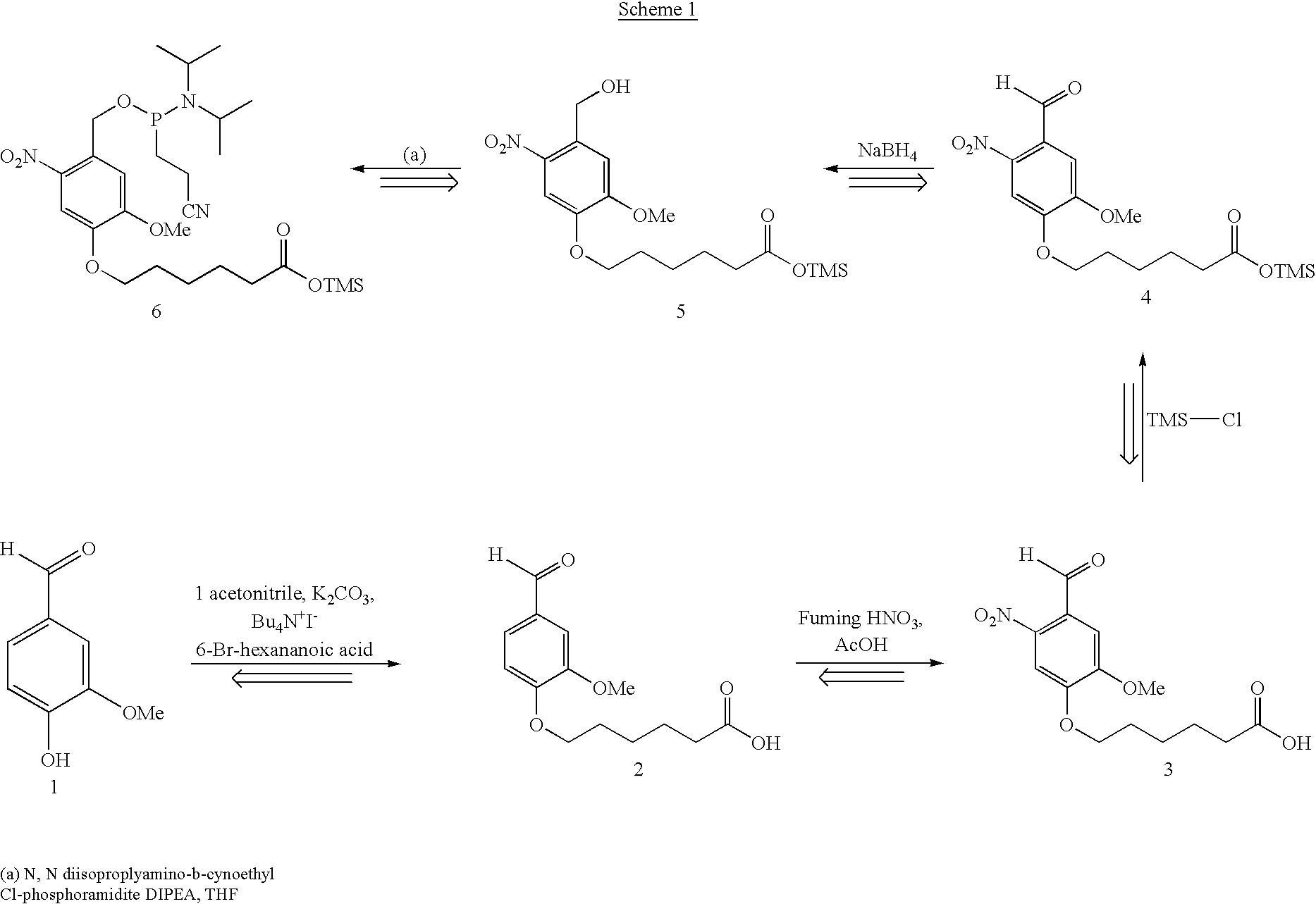
Photolabile linkers based on vanillin are synthesized as follows. Treatment of vanillin 1 with K.sub.2 CO.sub.3, provides alkylation of the phenol with 6-Bromohexanoic acid, yielding 2. 2 is then nitrated with fuming HNO.sub.3 and acetic acid to yield 3. 3 may be protected at the carboxylic acid with TMS depending on the stability of the final product. This step may or may not be necessary. 4 will be reduced to 5 with NaBH.sub.4. At which point 5 will be phosphitilated to the corresponding phosphoramidite 6. 6 Can be used in a synthesizer to couple the linker to an oligo, or in solution phase coupling. Below, a proposed synthesis is presented in scheme 1. ##STR2##
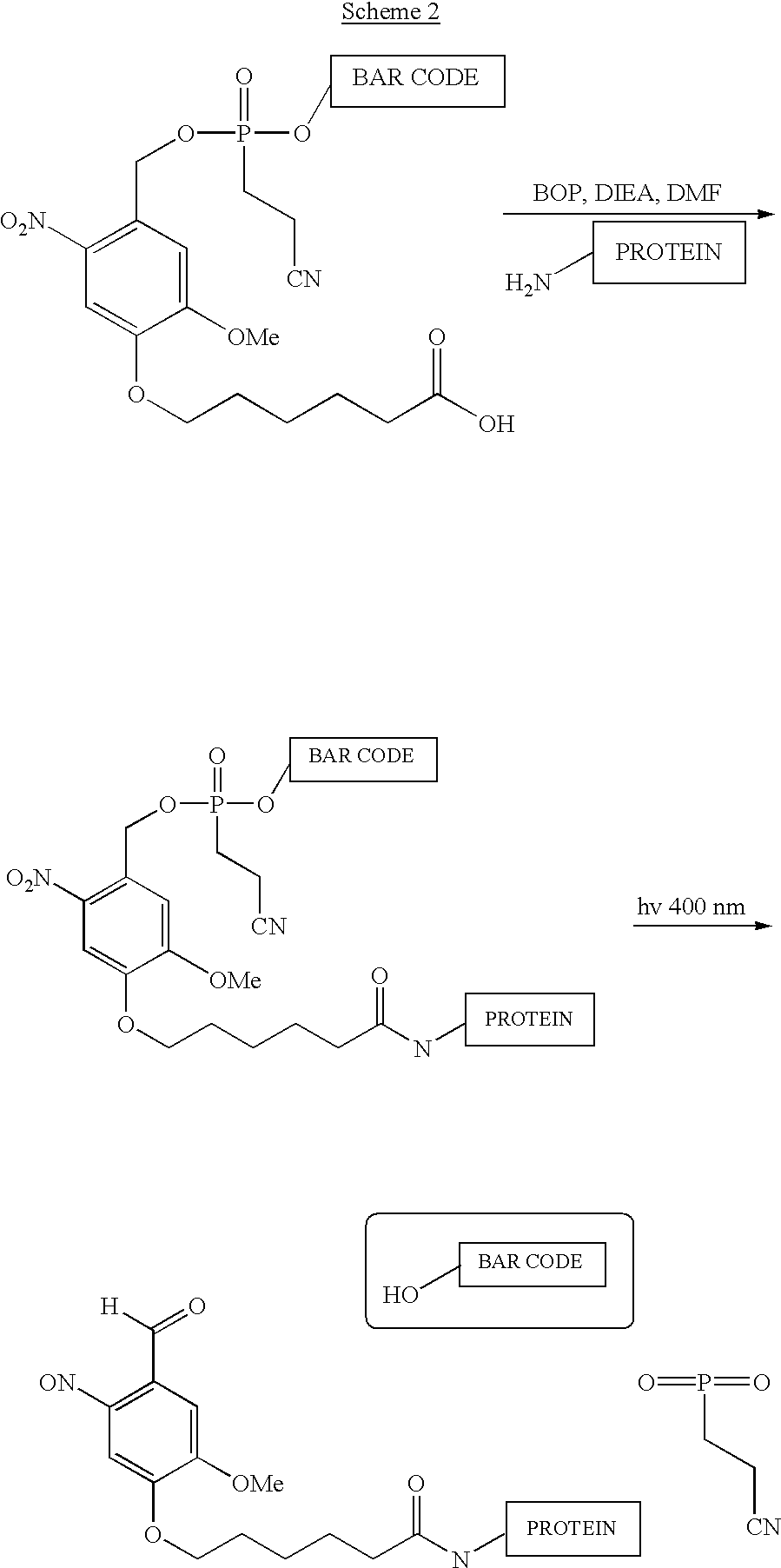
Standard peptide coupling using BOP reagent to activate the carboxyl group, will allow the peptide to be attached under mild conditions. To cleave and release the bar code at the appropriate time, UV light at 365-400 nm will be illuminated on the compound. The reaction scheme is presented below in scheme 2 ##STR3##
The proposed mechanism for photolysis of o-nitrobenzyl carbonate ester is present in the literature, but no specific mechanism for the photolysis of the corresponding phosphodiester is presented. Therefore, in scheme 3, the mechanism for a carbonate ester is shown. The mechanism for the phosphodiester should resemble the carbonate. ##STR4##
Dimethoxytrityl Linker
Dimethoxytrityl (DMT) is a common protecting group for the use of solid phase synthesis of DNA and RNA. Dimethoxytrityl is generally used to selectively protect primary alcohols. It is easily cleaved under mildly acidic conditions, but stable to basic work-up. A dimethoxytrityl group is incorporated into the cleavable linker for conjugation of an oligo to a protein as follows. The linker has a carboxylic acid functional group side chain to couple with an amine functionality 10. Scheme 4 describes the synthesis of this linker. ##STR5##
6-Bromohexanoic acid may be protected, where desired, prior to coupling with the phenol. If it is protected with TMS, it yields 7 the silyl ester of 6-bromohexanoic acid. 7 then alkylates p-hydroxy methylbenzoate to yield 8. 7 behaves like the methyl ester during Grignard reaction. If alkylation at the silyl ester occurs, it is necessary to reverse the addition order and form the tertiary alcohol before alkylating with the 6-Bromohexanoic acid. The next step involves a double Grignard reaction at the ester, yielding alcohol 9. If the TMS protection of the caboxylate is not necessary, three equivalents of the Grignard will be necessary. The tertiary alcohol is then substituted with Cl using acetyl-Cl, converting the compound to the linker/protecting group 10. This halide is then coupled to a primary alcohol using the same procedure as DMT protection. Once coupled to an oligo, the DMT linker is then attached to an amine functionality using standard peptide coupling techniques. Treatment with trichloroacetic acid is the standard deprotection method for DMT. The linker is more labile than the DMT because it has three electron donating groups vs. the two on DMT, so a milder acid treatment cleaves it readily. The general scheme is listed below. ##STR6## ##STR7##
It is evident from the above results and discussion that the subject targeted molecular bar codes constitute a significant advance in the field of analyte detection. The subject bar codes can be read directly without amplification and labeling steps. Furthermore, the bar codes can be read rapidly in much shorter times than that required for methods in which other labels, such as fluorescent or radioactive labels, are employed. In addition, the labels can be read from a heterogeneous population and need not be isolated before being read.
All publications and patent applications cited in this specification are herein incorporated by reference as if each individual publication or patent application were specifically and individually indicated to be incorporated by reference. The citation of any publication is for its disclosure prior to the filing date and should not be construed as an admission that the present invention is not entitled to antedate such publication by virtue of prior invention.
Although the foregoing invention has been described in some detail by way of illustration and example for purposes of clarity of understanding, it is readily apparent to those of ordinary skill in the art in light of the teachings of this invention that certain changes and modifications may be made thereto without departing from the spirit or scope of the appended claims.
PUM
Property | Measurement | Unit |
Length | 1.5E-8 ~ 2.5E-8 | m |
Nanoscale particle size | 20.0 | nm |
Description & Claims & Application Information
We can also present the details of the Description, Claims and Application information to help users get a comprehensive understanding of the technical details of the patent, such as background art, summary of invention, brief description of drawings, description of embodiments, and other original content. On the other hand, users can also determine the specific scope of protection of the technology through the list of claims; as well as understand the changes in the life cycle of the technology with the presentation of the patent timeline. Login to view more.