
Computed tomography system and method

a computed tomography and computed tomography technology, applied in tomography, instruments, applications, etc., can solve the problems of insufficient ebct, and insufficient longitudinal spatial resolution,

Inactive Publication Date: 2002-10-15
TOSHIBA MEDICAL SYST CORP
View PDF15 Cites 79 Cited by
  • Summary
  • Abstract
  • Description
  • Claims
  • Application Information

AI Technical Summary

Problems solved by technology

However, some projections are measured twice in the projection data and might cause artifacts in the absence of additional process.
However, EBCT has the disadvantages of insufficient longitudinal spatial resolution, system cost, and limited X-ray exposure.
The two biggest problems of this method are that it can be a high dose examination with small helical pitch, and potential problems exist for studies with contrast agents.
Since they utilize data obtained at the same cardiac phase but at the different time, changes other than cardiac motion, such as the contrast concentration, can degrade image quality.
The temporal resolution of individual images obtained with methods such as described above are insufficient for rapidly moving organs such as the heart and adjacent pulmonary vessels.
This method of weighting may be easier to implement but it may be more expensive computation-wise.
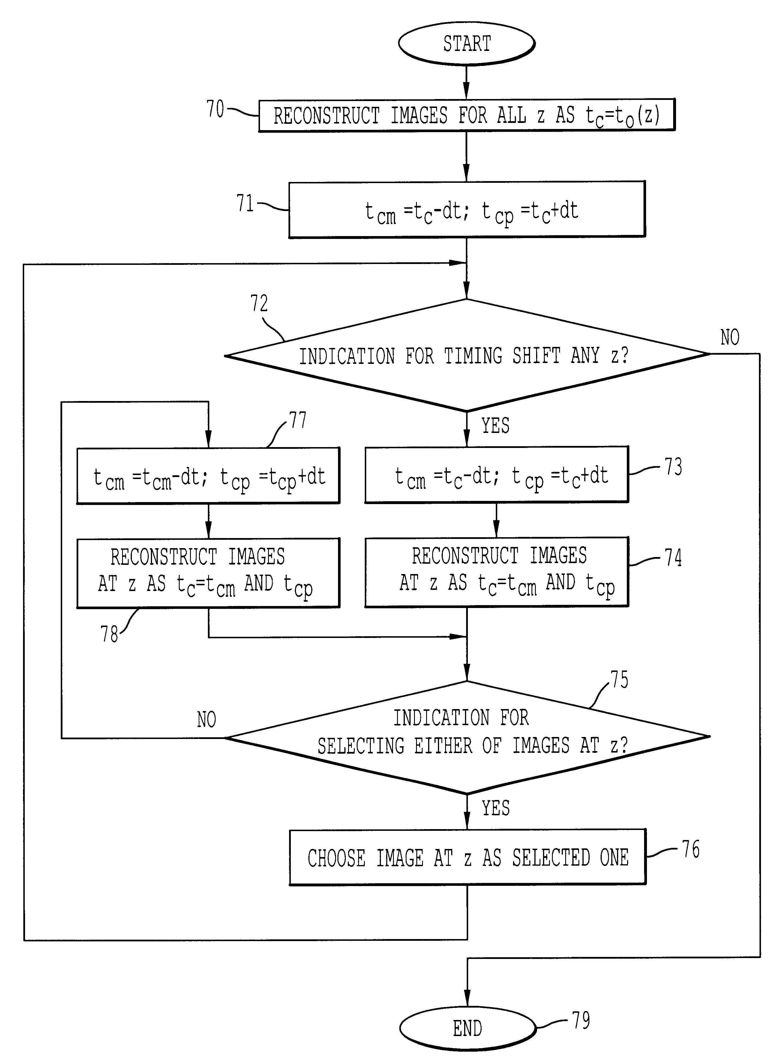
For example, a smaller time shift may be selected to increase the reconstructed image quality, but the computational expense is increased.

Method used


Image

  • Computed tomography system and method
  • Computed tomography system and method
  • Computed tomography system and method

Examples


Embodiment Construction

An example of the method according to the invention will now be described. Physical evaluations of the method were performed using computer simulations in order to evaluate the temporal and the spatial (z) resolution, the image noise, and the accuracy of the in-plane image of a moving phantom. The methods performed and compared were HHS with and without TS, and HFI for multi-slice CT, and 180LI for single-slice CT. The geometry and the X-ray tube rotation time were identical to those of a four-slice helical CT scanner (Aquilion; Toshiba Medical Systems Company, Tokyo, Japan), which acquires a full 360.degree. scan with 900 projections in 0.5 sec.

The temporal and spatial (z) resolution, and the image noise were evaluated varying the helical pitch and the filter width for HHS and HFI. In other evaluations, the helical multi-slice pitch was fixed at 2.5 for multi-slice CT and at 1.0 for single-slice CT, respectively, as will be discussed below. The nominal slice thickness was 2.0 mm fo...


PUM


Abstract

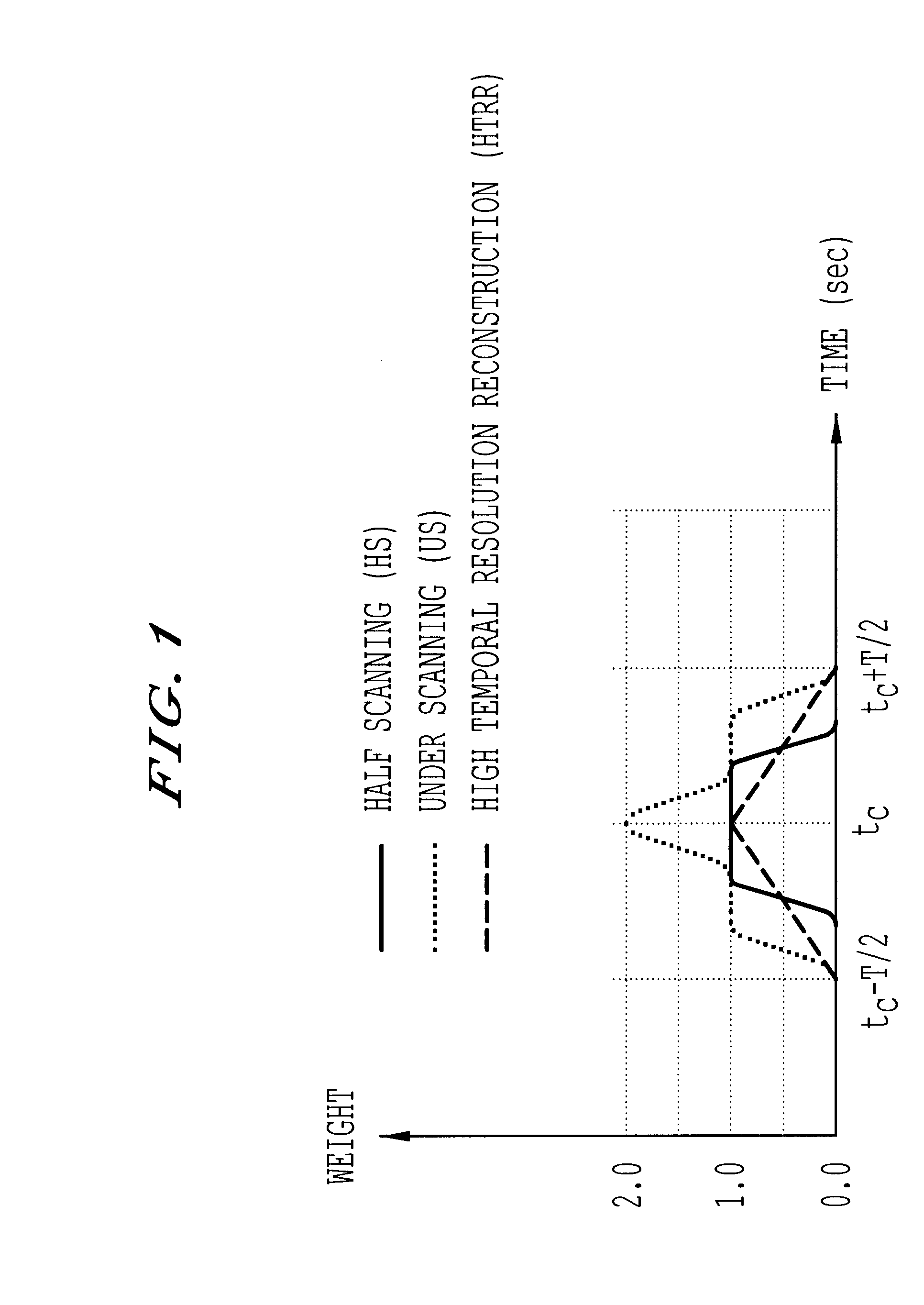
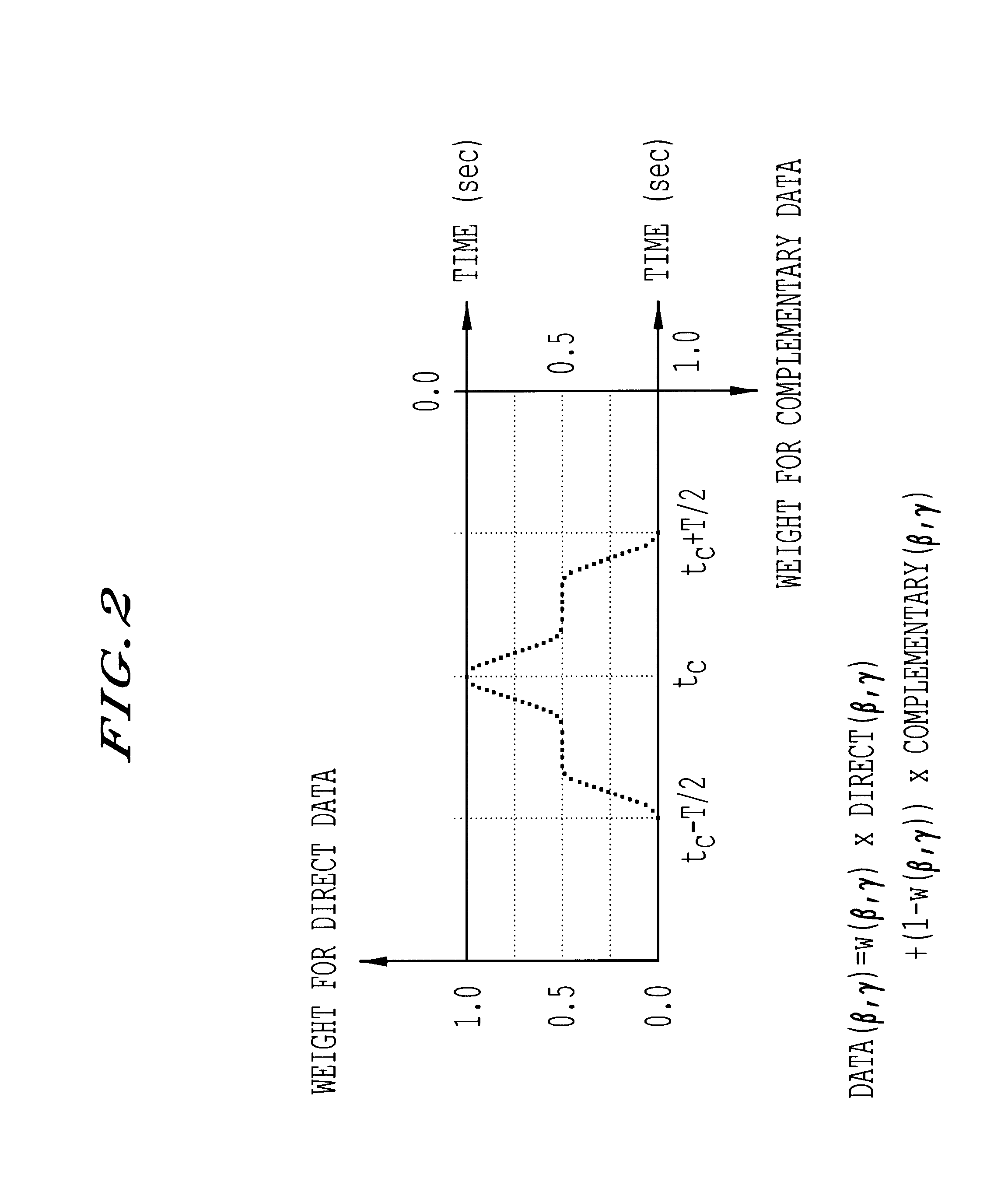
Method and system for computed tomography. Data is collected by irradiating a subject with x-rays. The data is interpolated and then weighted based upon the data collection time. The interpolated and weighted data is then used to reconstruct the image using fan beam reconstruction. The weighting function may by shifted in time. Signals may be obtained from the subject and used to control exposure, reduce the dose and provide automatic volumetric cardiac reconstruction. The method and system can increase the temporal and spatial resolution, and reconstruct images at different timing.

Description

1. Field of the InventionThe present invention is related to helical scan computed tomography and, in particular, to helical scan computed tomography with improved temporal resolution. The temporal resolution may be improved in each slice image by using an image reconstruction algorithm for multi-slice helical CT which does not need any special signals obtained from a patient.2. Discussion of the BackgroundHelical scanning computed tomography (CT) is described, for example, in U.S. Pat. No. 4,630,202. Helical scan CT may incorporate a multi-row detector array to collect data at multiple slice positions, as described in U.S. Pat. No. 4,965,726. Multi-slice helical CT uses such an array. This enables one to improve spatial resolution in the longitudinal direction, to extend the scanning length and to shorten scanning time, which results both in better temporal resolution in volume and better contrast resolution. If the helical scanning is repeated for the same volume, it is possible t...

Claims


Application Information

Patent Timeline
IPC IPC(8): A61B6/00A61B6/03
CPCA61B6/032A61B6/541A61B6/583A61B6/027A61B6/542Y10S378/901
Inventor TAGUCHI, KATSUYUKI
Owner TOSHIBA MEDICAL SYST CORP
Learn More