Electrophysiological cardiac mapping system based on a non-contact non-expandable miniature multi-electrode catheter and method therefor

a multi-electrode, non-contact technology, applied in catheters, instruments, therapies, etc., can solve the problems of limited recording sites, time-consuming, and difficulty in current techniques for mapping potentials directly from the endocardium

Inactive Publication Date: 2005-01-04
CASE WESTERN RESERVE UNIV
View PDF20 Cites 177 Cited by
  • Summary
  • Abstract
  • Description
  • Claims
  • Application Information

AI Technical Summary

Benefits of technology

FIG. 23 includes isochrone maps showing the effects of twisting on the present invention;
FIG. 24 includes electrograms showing the effects of catheter probe shape on the present invention;
FIG. 25 includes isochrone maps showing the effects of catheter shape on the present invention;
FIG. 26 includes isochrone maps showing the effects of catheter shape on the present invention;
FIG. 27 includes isochrone maps showing the effects of catheter shape on the present invention;
FIG. 28 includes isochrone maps showing the effects of catheter shape on the present invention;

Problems solved by technology

Unfortunately, current techniques of mapping potentials directly from the endocardium present certain difficulties.
For example, the well-known “roving” probe approach is 1) limited in the number of recording sites, 2) too time consuming and 3) only operative to collect data over a plurality of heart beats, instead of a single beat.
Therefore, this approach is not useful on a beat-by-beat basis to study dynamic changes in the activation process.
Although capable of mapping the entire endocardium, these devices occlude the heart cavity and require open heart surgery, heart-lung bypass and other complicated and high risk procedures.
However, the basket is nonetheless limited in the number of electrodes so that spatial resolution is relatively low.
Moreover, it is difficult to insure that all electrodes make contact with the endocardium.
The fact that the basket must be collapsed prior to catheter withdrawal from the ventricle adds complexity and risk to this procedure.
However, this system still involves undesirable expansion of a device in the heart chamber.
The expanded element may interfere with intracavity structures and adds complexity to the system because it must be collapsed before removal.
Moreover, it is difficult to determine the location of the electrodes within the chamber.
Also, the array may not expand as desired, leading to inaccuracies in mapping.

Method used


Image

  • Electrophysiological cardiac mapping system based on a non-contact non-expandable miniature multi-electrode catheter and method therefor
  • Electrophysiological cardiac mapping system based on a non-contact non-expandable miniature multi-electrode catheter and method therefor
  • Electrophysiological cardiac mapping system based on a non-contact non-expandable miniature multi-electrode catheter and method therefor

Examples


Embodiment Construction

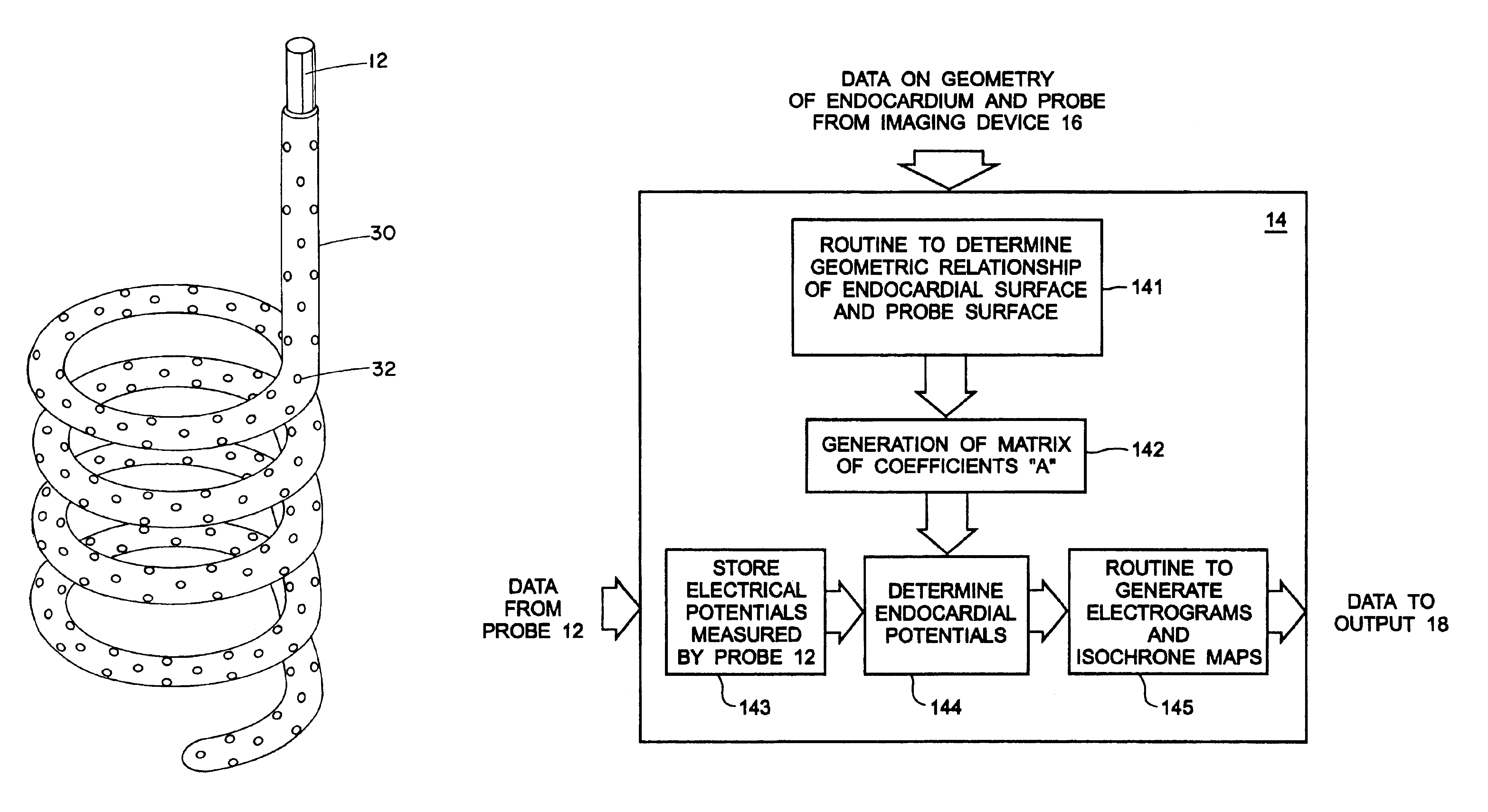
A mathematical inverse methodology for reconstruction of endocardial potentials, electrograms and isochrones from non-contact, intracavitary probe measurements has been developed and validated. A study in an isolated canine left ventricle (LV) demonstrated that the inversely computed endocardial potential maps reconstruct with good accuracy and resolve the major features of the measured endocardial potential maps, including maxima, minima and regions of negative and positive potentials. A more recent systematic evaluation demonstrated that computed temporal electrograms and isochrones also closely approximate their directly measured counterparts. The isochronal maps correctly captured the regions of early and late activation for a single pacing site and for two simultaneous pacing sites separated by 17 mm. Moreover, the entire activation sequence was closely approximated, including regions of nonuniform conduction (e.g. isochrone crowding indicating slow conduction). The size of the...


PUM


Abstract

A system (10) for determining electrical potentials on an endocardial surface of a heart is provided. The system includes a non-contact, non-expandable, miniature, multi-electrode catheter probe (12), a plurality of electrodes (32) disposed on an end portion (30) thereof, means for determining endocardial potentials (14) based on electrical potentials measured by the catheter probe, a matrix of coefficients that is generated based on a geometric relationship between the probe surface, and the endocardial surface. A method is also provided.

Description

BACKGROUND OF THE INVENTIONThis invention relates to an apparatus and method for electrophysiological cardiac mapping. More particularly, the invention is directed to a system based on a nonexpandable, noncontact, miniature, multielectrode catheter which is used to measure electrical potentials within a heart cavity. These measured potentials are then used, along with data on the geometric relationship between the catheter and the endocardial surface, to reconstruct maps representing endocardial electrical activity. In this regard, electrograms and isochrones are reconstructed.While the invention is particularly directed to the art of electrophysiological cardiac mapping, and will be thus described with specific reference thereto, it will be appreciated that the invention may have usefulness in other fields and applications.By way of background, endocardial potential mapping is a tool for studying cardiac excitation and repolarization processes. Mapping endocardial potential distrib...

Claims


Application Information

Patent Timeline
Patent Type & Authority Patents(United States)
IPC IPC(8): A61B17/22A61B5/0408A61B5/042G06F17/00A61B5/05A61B5/04A61B6/12A61B8/08
CPCA61B5/0422A61B5/6852A61B5/6855A61B5/6856A61B17/22012A61B5/6857A61B5/04011A61B2019/5253A61B6/12A61B6/503A61B8/0833A61B8/0883A61B2034/2053A61B5/341A61B5/287
Inventor RUDY, YORAM
Owner CASE WESTERN RESERVE UNIV
Why Eureka
Try Eureka