Apparatus And Method For Cardiac Ablation

a technology of cardiac ablation and apparatus, applied in the field of medical devices, can solve the problems of life-threatening arrhythmias, inability to treat dysrhythmias of the heart with drugs, and high cost of both procedures

Inactive Publication Date: 2007-07-12
CATHEFFECTS
View PDF15 Cites 115 Cited by
  • Summary
  • Abstract
  • Description
  • Claims
  • Application Information

AI Technical Summary

Benefits of technology

[0064] An important advantage of the present invention is the capability of allowing a medical practitioner to use a roving catheter to locate the site of origin of tachycardia in the endocardium quickly and accurately, without the need for open-chest and open-heart surgery. This is accomplished by the use of the multi-electrode catheter 70 in combination with real-time data-processing and interactive display by the system 10.
[0065] Essentially, the multi-electrode catheter 70 must be able to deploy at least a two-dimensional array of electrodes against a site of the endocardium to be mapped. The intracardiac signals detected by each of the electrodes provide data sampling of the electrical activity in the local site spanned by the array of electrodes. This data is processed by the computer to produce a real-time display including arrival times of intracardiac signals at each electrode, and a local isochrone map of the sampled site. By plotting contours of equal arrival time of the intracardiac signals, the local isochrone map is an expedient way of indicating how close and where the electrode array is from the site of origin. Also, at each sampled site, the computer computes and displays in real-time an estimated location of the site of origin relative to the electrodes, so that a medical practitioner can interactively and quickly move the electrodes towards the site of origin.
[0066] A suitable multi-electrode catheter for use in the present invention is a five-electrode orthogonal electrode catheter array (OECA) that has been disclosed in U.S. Pat. No. 4,940,064 to Desai. Relevant portions of aid disclosure are incorporated herein by reference.
[0067]FIG. 2A illustrates the proximal end of the orthogonal electrode catheter array (OECA) 70 in its fully retracted position or mode. Because the catheter material has a “set” or “memory” it will normally return to this retracted position. The OECA comprises an eight-french five-pole electrode catheter 70. It has a central stylet 102 with four peripheral or circumferential electrodes 112, 113, 114 and 115. A fifth electrode 111 is located centrally at the tip of the stylet 102. All five electrodes are hemispherical and have individual leads 116 connected thereto. Each peripheral electrode is 2 mm in diameter while the central electrode is 2.7 mm in diameter. Slits 120 are cut longitudinally near where the electrodes are located.
[0068]FIG. 2B illustrates the OECA in its fanned out mode. When the proximal end (not shown) of the catheter is pulled, the stylet's slits 120 allow four side arms 122 to open from the stylet body in an orthogonal configuration. Each of the four arms 122 extend a peripheral electrode radially from the stylet so that the four peripheral electrodes forms a cross with the fifth electrode 111 at its center. The inter-electrode distance from the central electrode to each peripheral electrode is 0.5 cm, and the distance between peripheral electrodes is 0.7 cm. The surface area of the catheter tip in an open position is 0.8 cm2.
[0069]FIG. 2C shows the footprints of the five-electrode OECA electrodes. The four peripheral electrodes 112, 113, 114 and 115 or (2)-(5) form a cross configuration. The fifth electrode 111 or (1) is located at the center of the cross. The orthogonal array of electrodes therefore provides five sampling points over the zone 130 in an endocardium site spanned by the electrodes. Isochrone Maps

Problems solved by technology

Another common type of cardiac dysrhythmias is ventricular tachycardia (VT), which is a complication of a heart attack or reduction of blood supply to an area of heart muscle, and is a life threatening arrhythmia.
However, some dysrhythmias of the heart are not treatable with drugs.
Both procedures have increased morbidity and mortality and are extremely expensive.
In addition, some patients of advanced age or illness cannot tolerate invasive surgery to excise tachycardia focus which causes dysrhythmias.
While the sock or balloon electrode arrays allow global mapping by acquiring electrogram signals over a wider area of the heart simultaneously, they can only be installed after open-chest surgery.
Locating the site of origin and tracking the whereabouts of the catheter are at best tricky and time-consuming, and often proved unsuccessful.

Method used


Image

  • Apparatus And Method For Cardiac Ablation
  • Apparatus And Method For Cardiac Ablation
  • Apparatus And Method For Cardiac Ablation

Examples


Embodiment Construction

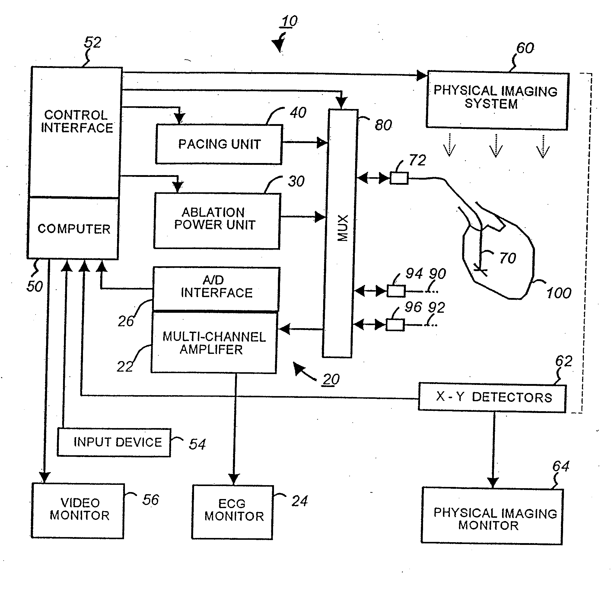
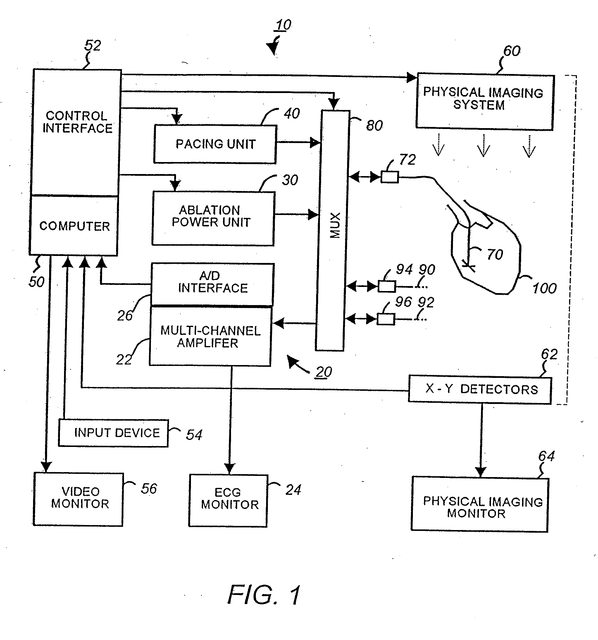
[0055]FIG. 1 is a schematic block diagram of a multi-electrode catheter mapping and ablation system 10 according to a preferred embodiment of the present invention.

[0056] The system 10 essentially comprises of three functioning units, namely a mapping unit 20, an ablation unit 30 and a pacing unit 40. A computer 50 controls the operation of each of the units and their cooperations via a control interface 52. The computer receives operator inputs from an input device 54 such as a keyboard, a mouse and a control panel. The output of the computer may be displayed on a video monitor 56 or other output devices (not shown).

[0057] In the preferred embodiment the system 10 also includes a physical imaging system 60. The physical imaging system 60 is preferably a 2-axis fluoroscope or an ultrasonic imaging system. The physical imaging system 60 is controllable by the computer 50 via the control interface 52. In one implementation, the computer triggers the physical imaging system to take “...


PUM


Abstract

A system and method for cardiac mapping and ablation include a multi-electrode catheter introduced percutaneously into a subject's heart and deployable adjacent to various endocardial sites. The electrodes are connectable to a mapping unit, an ablation power unit a pacing unit, all of which are under computer control. Intracardiac electrogram signals emanated from a tachycardia site of origin are detectable by the electrodes. Their arrival times are processed to generate various visual maps to provide real-time guidance for steering the catheter to the tachycardia site of origin. In another aspect, the system also include a physical imaging system which is capable of providing different imaged physical views of the catheter and the heart. These physical views are incorporated into the various visual maps to provide a more physical representation. Once the electrodes are on top of the tachycardia site of origin, electrical energy is supplied by the ablation power unit to effect ablation.

Description

[0001] This is a continuation-in-part of application Ser. No. 08 / 029,771, filed Mar. 11, 1993.BACKGROUND OF THE INVENTION [0002] This invention relates to medical devices and, in particular, a system and technique of employing multi-electrode catheters for cardiac mapping and ablation. [0003] Cardiac dysrhythmias are commonly known as irregular heart beats or racing heart. Two such heart rhythm irregularities are the Wolff-Parkinson-White syndrome and atrioventricular (AV) nodal reentrant tachycardia. These conditions are caused by an extraneous strand of conducting fibers in the heart that provides an abnormal short-circuit pathway for electric impulses normally conducting in the heart. For example, in one type of Wolff-Parkinson-White syndrome the accessory pathway causes the electric impulses that normally travel from the upper to the lower chamber of the heart to be fed back to the upper chamber. Another common type of cardiac dysrhythmias is ventricular tachycardia (VT), which ...

Claims


Application Information

Patent Timeline
Patent Type & Authority Applications(United States)
IPC IPC(8): A61B5/04A61B5/363A61B18/12A61B18/14
CPCA61B5/0422A61B5/7425A61B5/6852A61B5/6858A61B5/7445A61B6/464A61B6/5235A61B18/1206A61B18/1492A61B2018/0016A61B2018/00357A61B2018/00577A61B2018/0066A61B2018/00839A61B2018/1465A61B2019/5253A61B5/044A61B2034/2053A61B5/743A61B5/339A61B5/287
Inventor DESAI, JAWAHAR M.
Owner CATHEFFECTS
Why Eureka
Try Eureka